chapter 13. gene diagnosis - shandong university
TRANSCRIPT
Chapter 13. Gene Diagnosis
To identify the cause of a disease or condition based on gene analysis or test.
Methods of diagnosing a genetic disease:
1) Direct diagnosis: detect disease-causing mutation
2) Indirect diagnosis: tracking the disease gene by linkage analysis
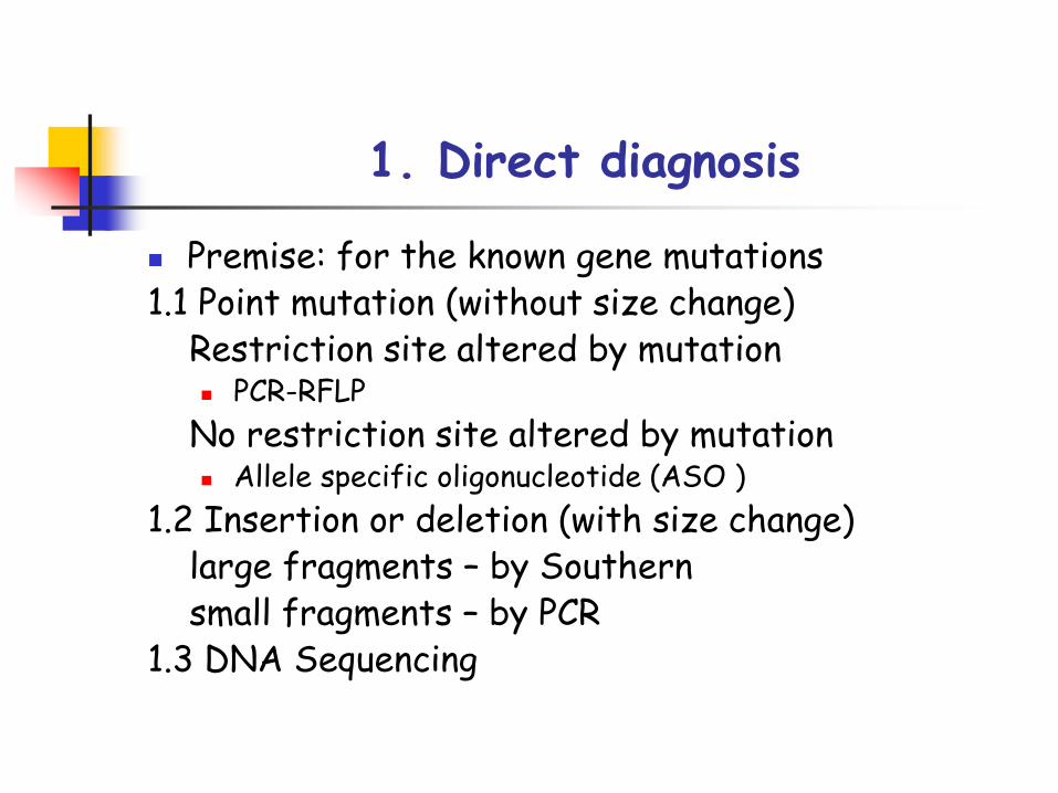
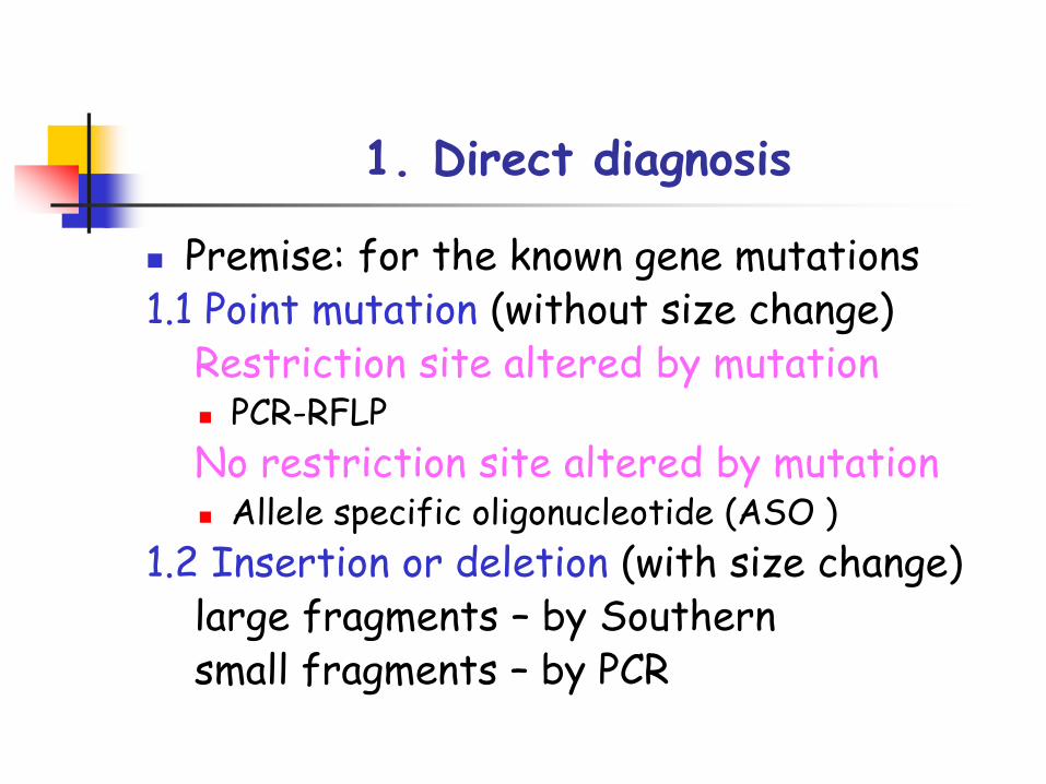
1. Direct diagnosis
Premise: for the known gene mutations 1.1 Point mutation (without size change)
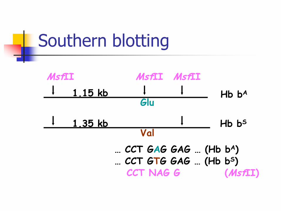
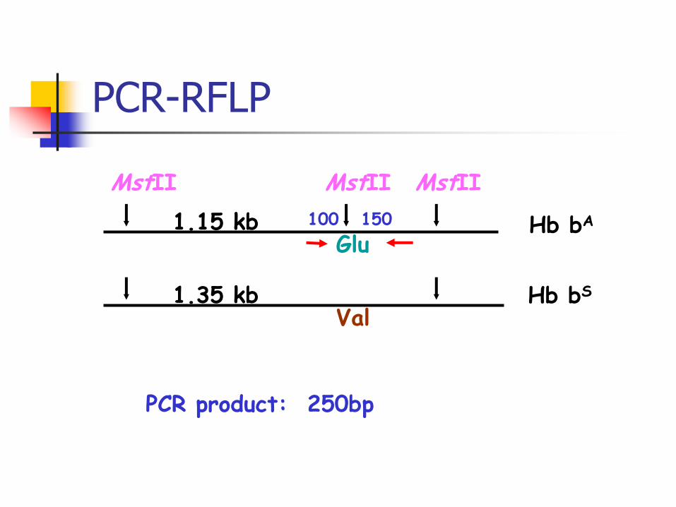
Restriction site altered by mutation PCR-RFLP
No restriction site altered by mutation Allele specific oligonucleotide (ASO )
1.2 Insertion or deletion (with size change)large fragments – by Southernsmall fragments – by PCR
1.3 DNA Sequencing
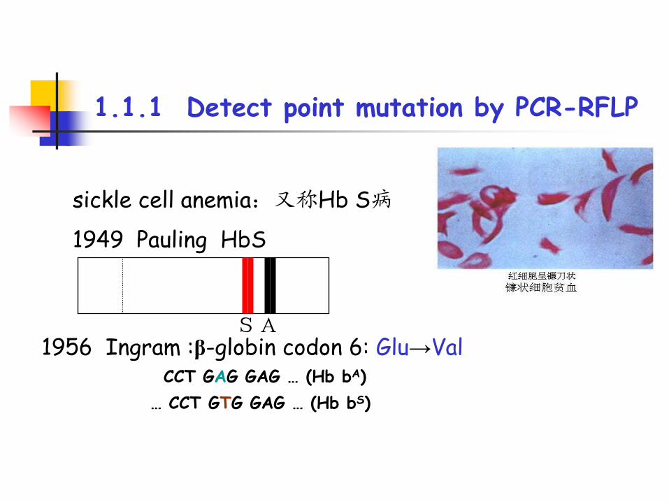
1.1.1 Detect point mutation by PCR-RFLP
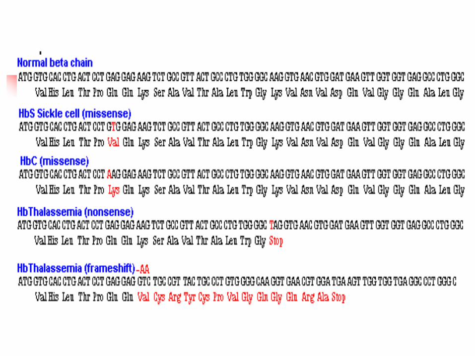
sickle cell anemia:又称Hb S病
1949 Pauling HbS
SA1956 Ingram :β-globin codon 6: Glu→Val
CCT GAG GAG … (Hb bA)
… CCT GTG GAG … (Hb bS)
Hb bA
Hb bS
MstII MstII MstII
Glu
Val
… CCT GAG GAG … (Hb bA)… CCT GTG GAG … (Hb bS)
CCT NAG G (MstII)
1.15 kb
1.35 kb
Southern blotting
Hb bA
Hb bS
MstII MstII MstII
Glu
Val
1.15 kb
1.35 kb
100 150
PCR product: 250bp
PCR-RFLP
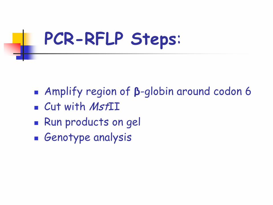
PCR-RFLP Steps:
Amplify region of β-globin around codon 6
Cut with MstII
Run products on gel
Genotype analysis
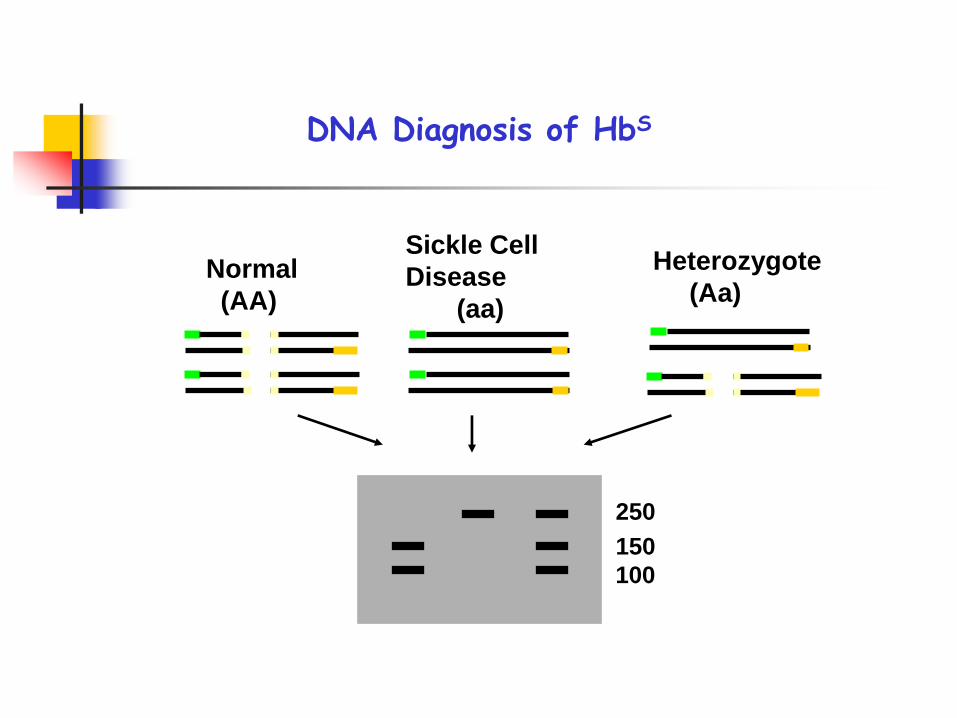
Sickle Cell
Disease
(aa)
Heterozygote
(Aa)Normal
(AA)
250
150
100
DNA Diagnosis of HbS

250150100
AA
DNA Diagnosis of HbS
Aa
?
aa
Aa Aa
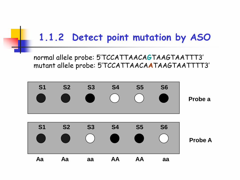
1.1.2 Detect point mutation by ASO
normal allele probe: 5′TCCATTAACAGTAAGTAATTT3′mutant allele probe: 5′TCCATTAACAATAAGTAATTTT3′
S1 S2 S3 S4 S5 S6
S1 S2 S3 S4 S5 S6
Probe a
Probe A
Aa Aa aa aaAA AA
1. Direct diagnosis
Premise: for the known gene mutations 1.1 Point mutation (without size change)
Restriction site altered by mutation PCR-RFLP
No restriction site altered by mutation Allele specific oligonucleotide (ASO )
1.2 Insertion or deletion (with size change)large fragments – by Southernsmall fragments – by PCR
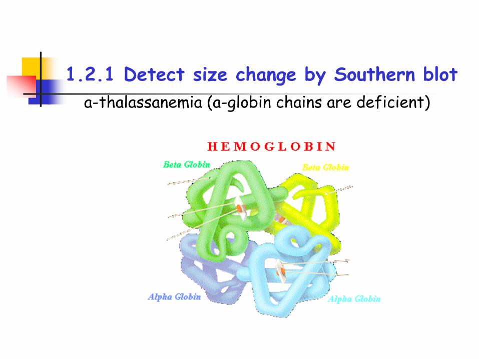
1.2.1 Detect size change by Southern blot
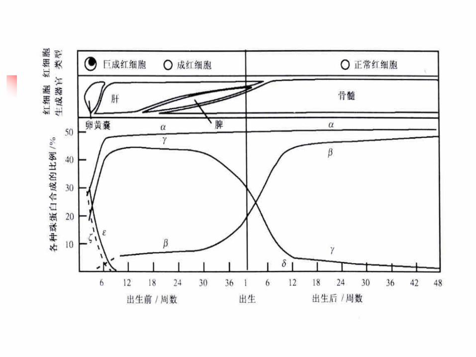
a-thalassanemia (a-globin chains are deficient)
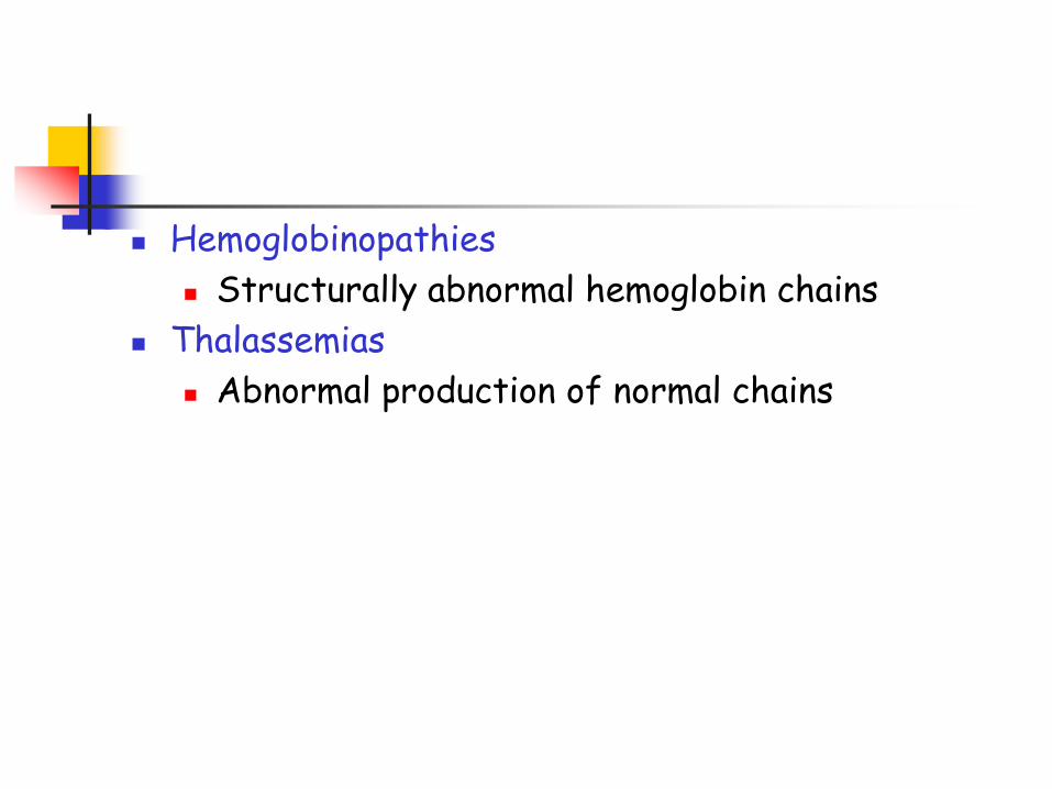
Hemoglobinopathies
Structurally abnormal hemoglobin chains
Thalassemias
Abnormal production of normal chains
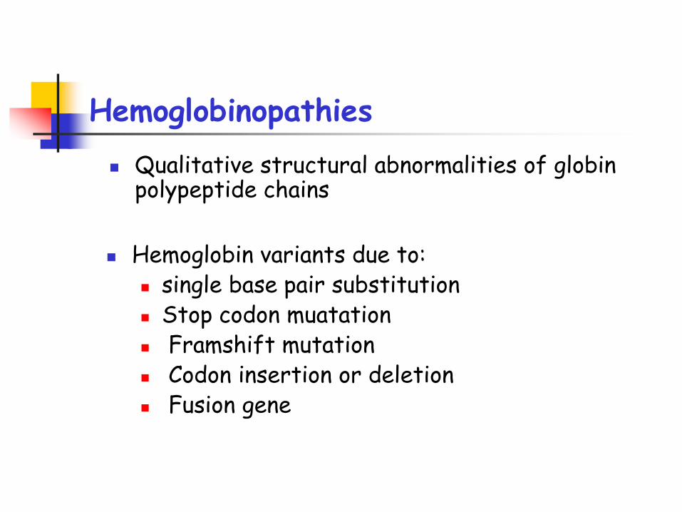
Hemoglobinopathies
Qualitative structural abnormalities of globin polypeptide chains
Hemoglobin variants due to: single base pair substitution Stop codon muatation Framshift mutation Codon insertion or deletion Fusion gene
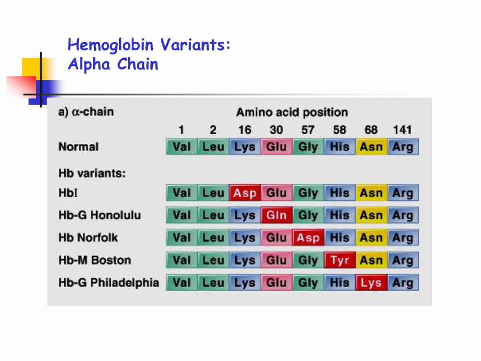
Hemoglobin Variants: Alpha Chain
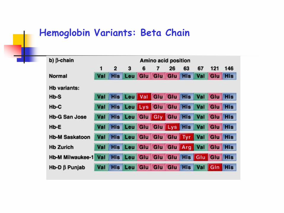
Hemoglobin Variants: Beta Chain
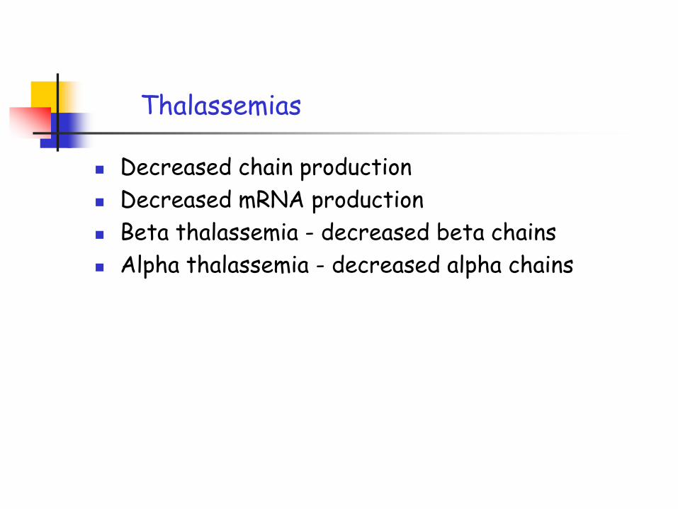
Thalassemias
Decreased chain production
Decreased mRNA production
Beta thalassemia - decreased beta chains
Alpha thalassemia - decreased alpha chains
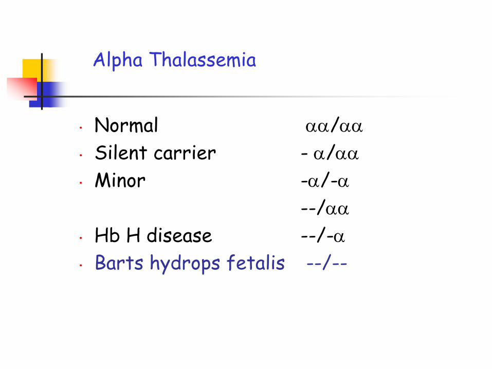
• Normal /
• Silent carrier - /
• Minor -/-
--/
• Hb H disease --/-
• Barts hydrops fetalis --/--
Alpha Thalassemia
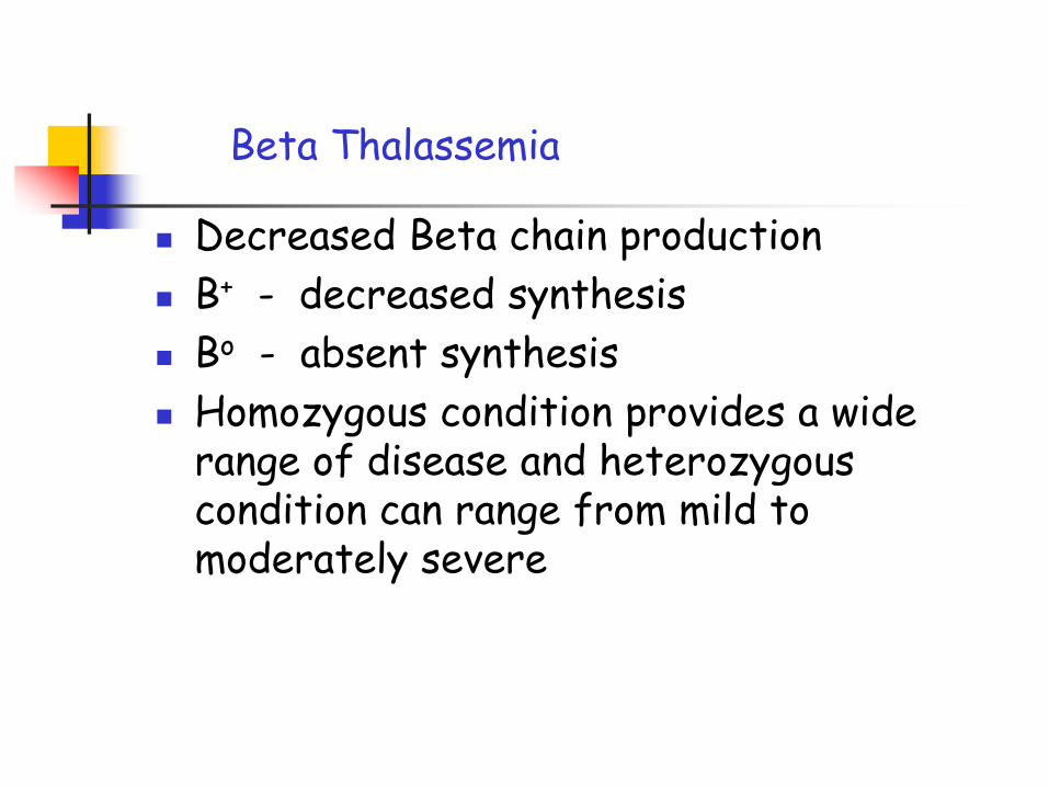
Beta Thalassemia
Decreased Beta chain production
B+ - decreased synthesis
Bo - absent synthesis
Homozygous condition provides a wide range of disease and heterozygous condition can range from mild to moderately severe
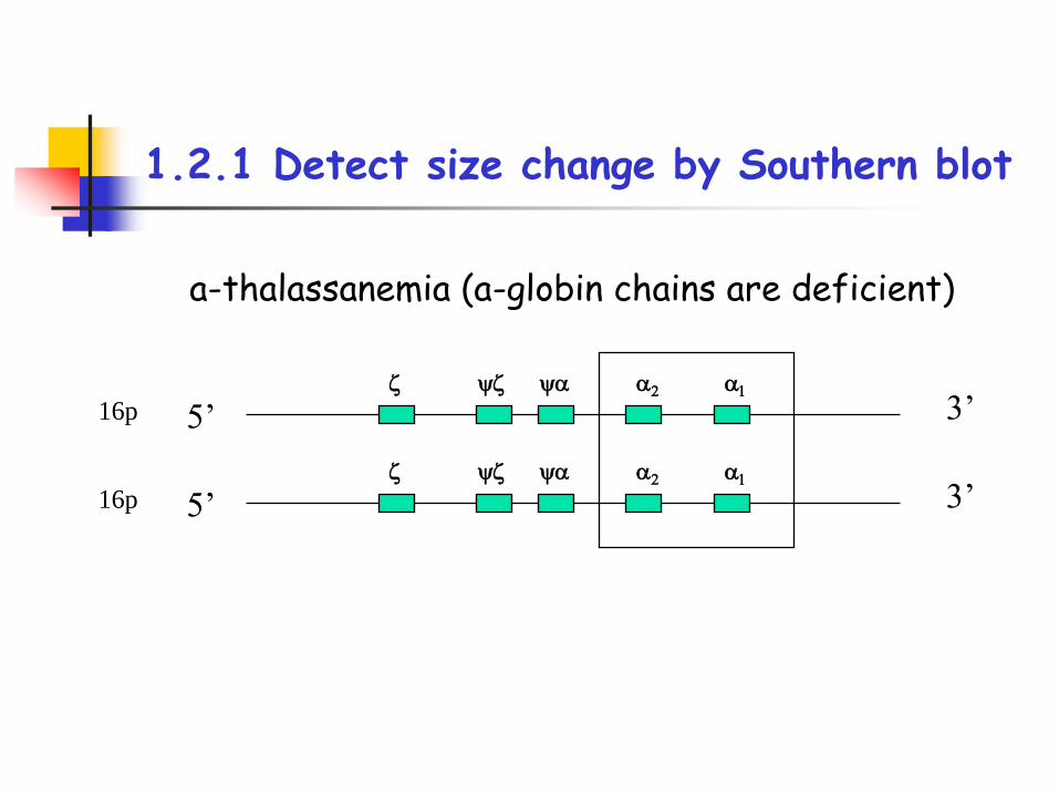
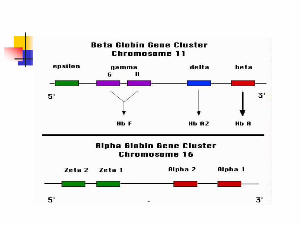
1.2.1 Detect size change by Southern blot
5’ 3’16pz yz y 2 1
5’ 3’16pz yz y 2 1
a-thalassanemia (a-globin chains are deficient)
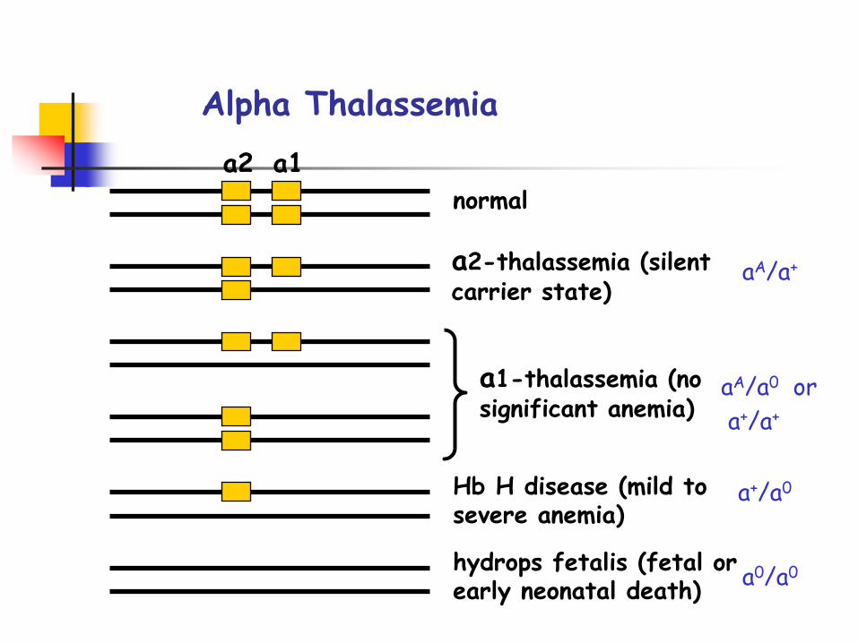
a2 a1
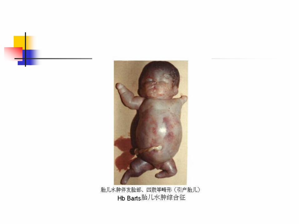
hydrops fetalis (fetal or early neonatal death)
Hb H disease (mild to severe anemia)
a1-thalassemia (no significant anemia)
a2-thalassemia (silent carrier state)
normal
Alpha Thalassemia
aA/a+
aA/a0 or
a+/a+
a+/a0
a0/a0
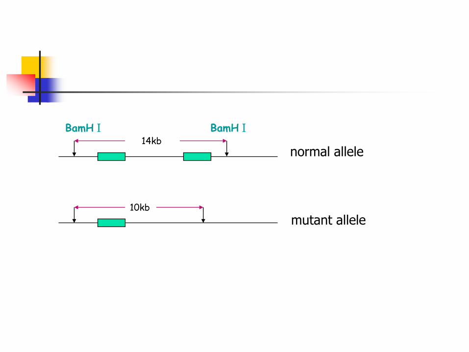
10kb
14kbnormal allele
mutant allele
BamHⅠ BamHⅠ
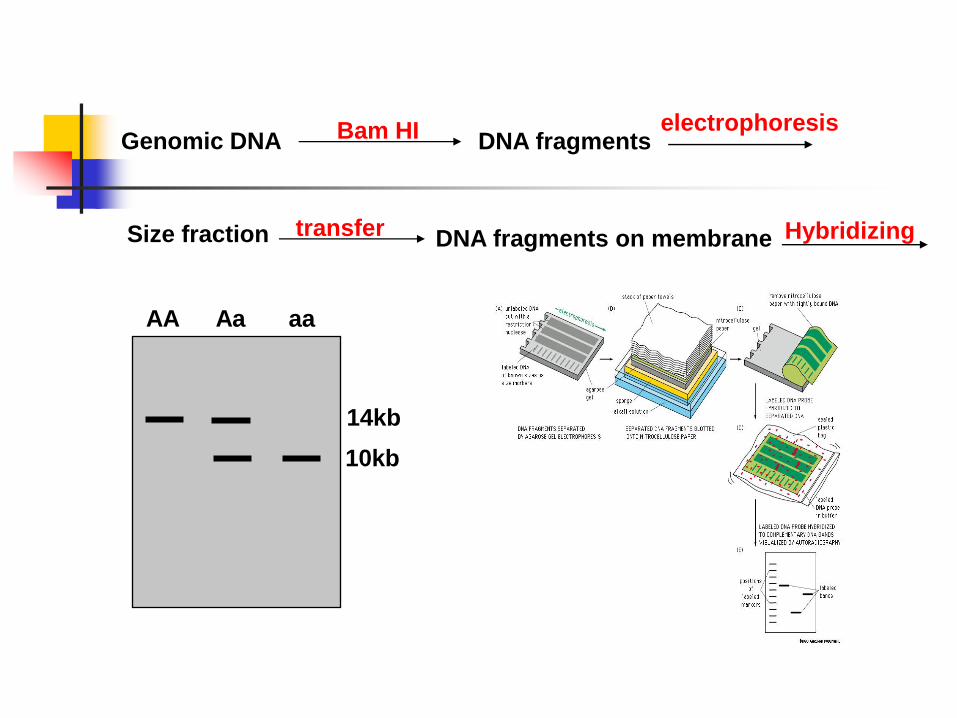
Genomic DNA Bam HI DNA fragmentselectrophoresis
Size fraction transfer DNA fragments on membrane Hybridizing
14kb
10kb
AA Aa aa
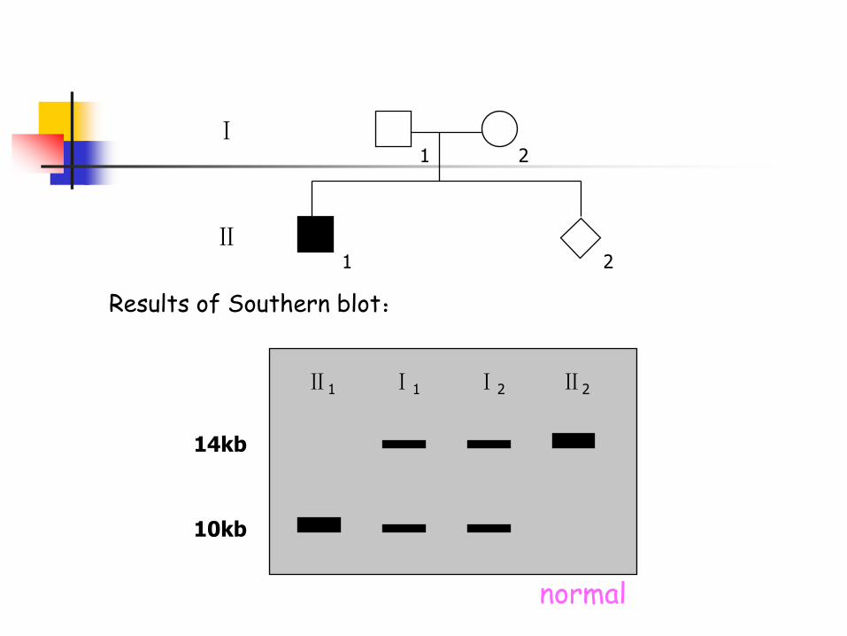
1 2
1 2
Ⅰ
Ⅱ
Results of Southern blot:
Ⅱ1 Ⅰ1 Ⅰ2 Ⅱ2
10kb
14kb
normal

Duchenne Muscular Dystrophy
17 18 19 44 45 46 47 48 49
17
19
44
45
48
C P1 P2 P3 P4Patient1: exon 44 deleted
Patient2: exon 17deleted
Patient4: exon 19 deleted
Patient3: exon 45 deleted
1.2.2 Detect size change by PCR
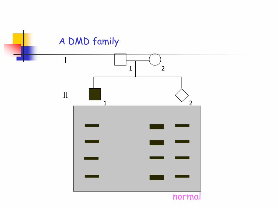
1 2
1 2
Ⅰ
Ⅱ
A DMD family
normal
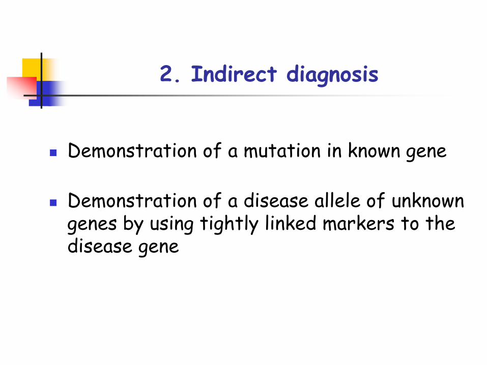
2. Indirect diagnosis
Demonstration of a mutation in known gene
Demonstration of a disease allele of unknown genes by using tightly linked markers to the disease gene
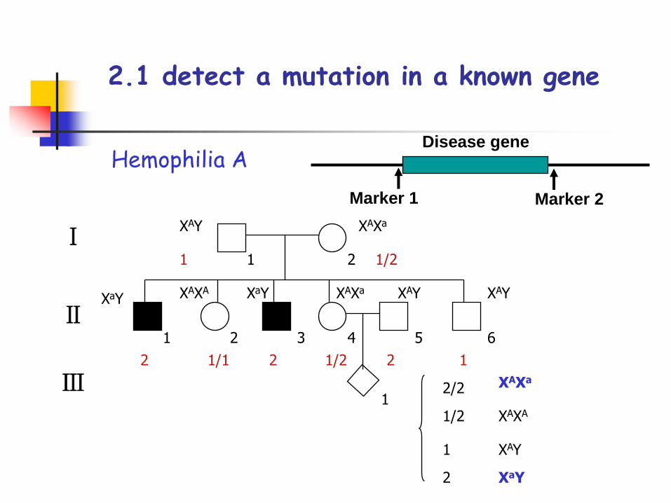
2.1 detect a mutation in a known gene
Hemophilia A
1 1/2
2 1/1 2 1/2 2 1
XAXa
XaY XaY
XAY
XAY XAYXAXA XAXa
Ⅰ
Ⅱ1
2
2
1
1
3 4 5 6
Ⅲ 2/2
1/2
1
2
XAY
XaY
XAXA
XAXa
Disease gene
Marker 1 Marker 2
2/5
P
Ⅰ
Ⅱ1
2
2
1
1
3 4 5 6
Ⅲ
2/4 3/5
2/3 4/5 2/5 3/4 4/52/5
2
2/4
P
3/5
N
3/4
N
2/2 P
2/5 ?
5/5 N
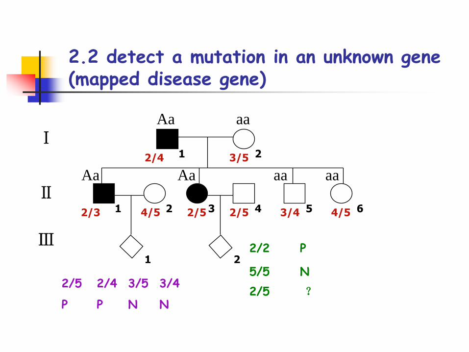
2.2 detect a mutation in an unknown gene (mapped disease gene)
Aa aa
Aa Aa aa aa
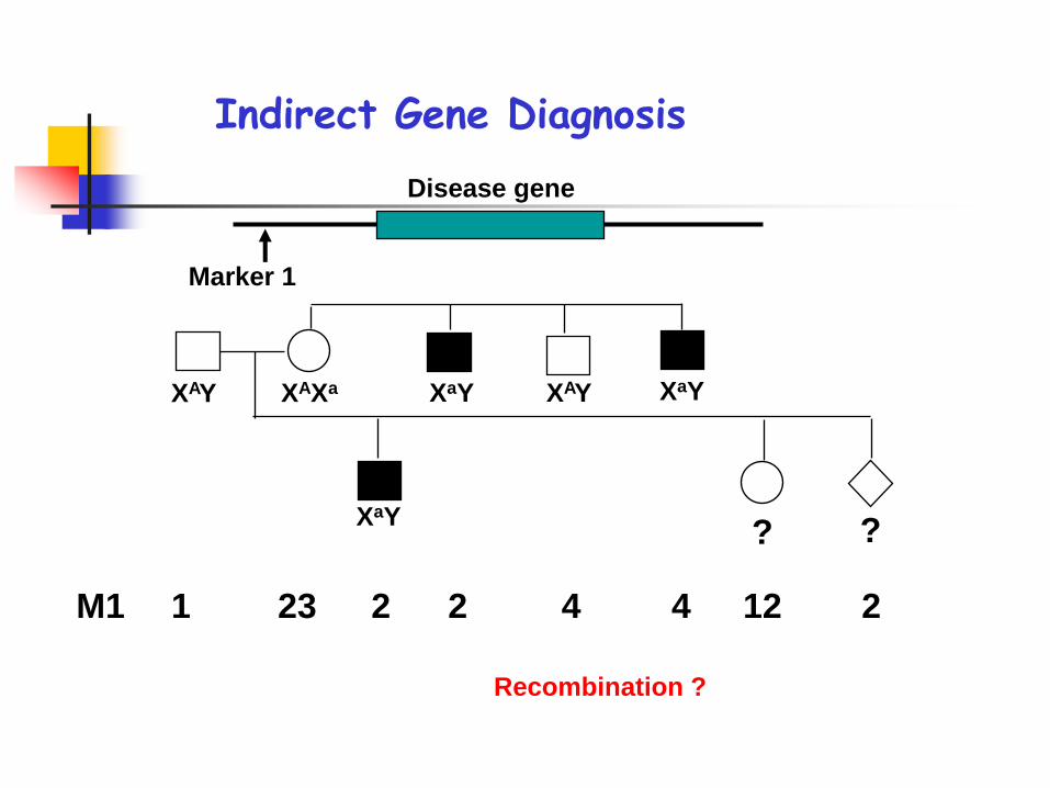
Indirect Gene Diagnosis
M1 1 23 2 2 4 4 12 2
XAY XAXa
XaY
XaY XAY XaY
? ?
Disease gene
Marker 1
Recombination ?
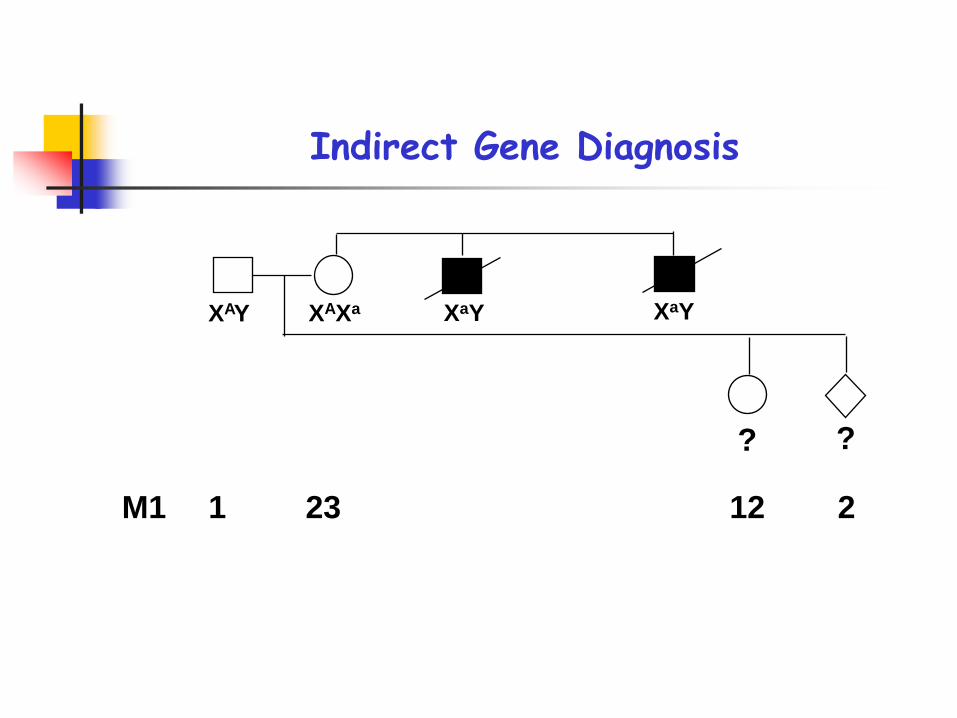
M1 1 23 12 2
XAY XAXa XaY XaY
? ?
Indirect Gene Diagnosis
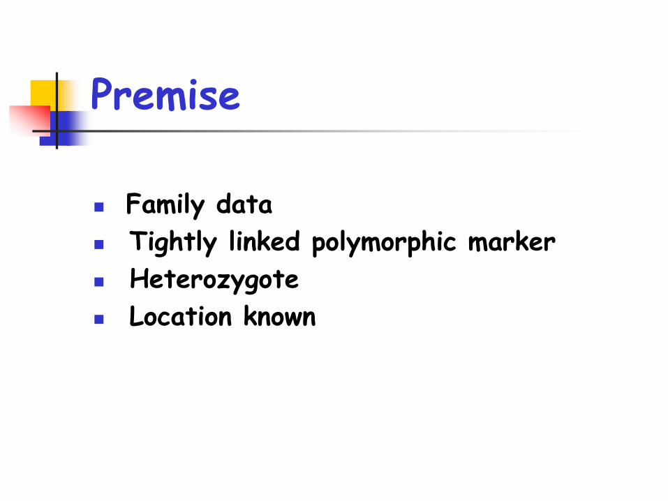
Premise
Family data
Tightly linked polymorphic marker
Heterozygote
Location known
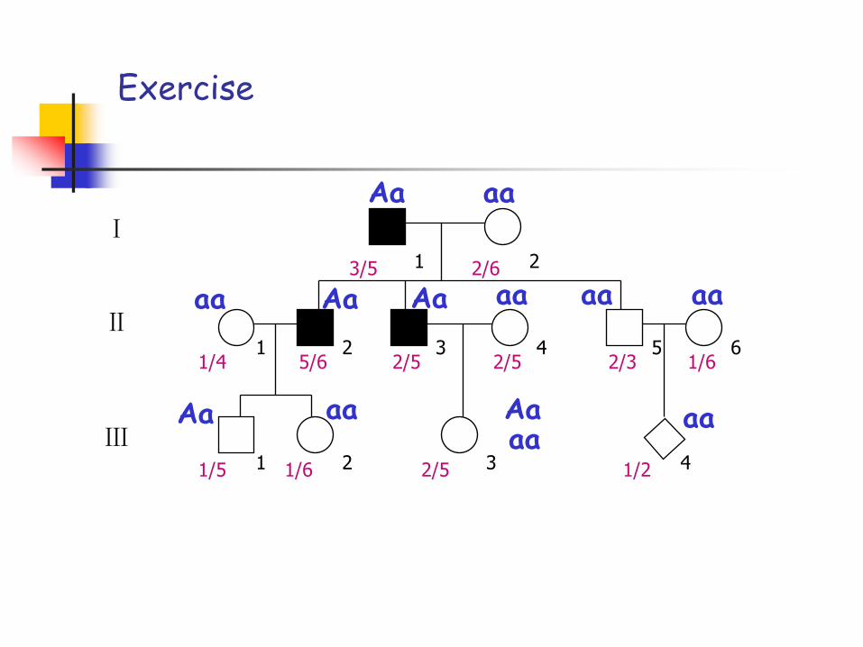
Exercise
Ⅰ
Ⅱ
Ⅲ1
1
1 2
2
2 3
3 4
4
5 6
3/5 2/6
5/6 2/5 2/5 2/3 1/61/4
1/5 1/6 2/5 1/2
Aa aa
aa Aa Aa aa aa aa
aaaaAaAa aa
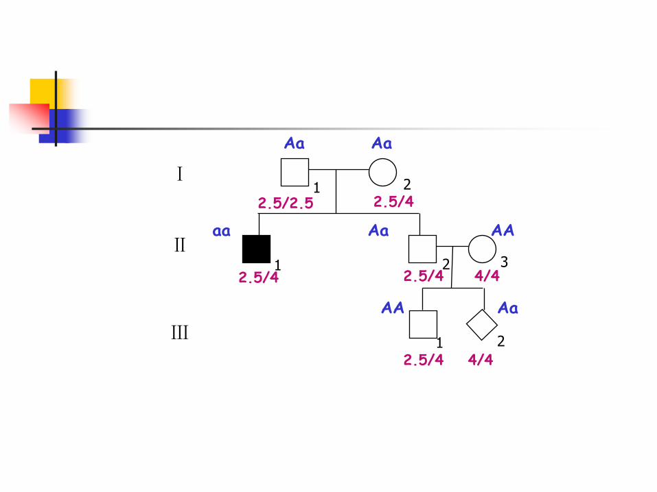
Ⅰ
Ⅱ
Ⅲ1
1 2
1
2
2 3
2.5/2.5 2.5/4
2.5/4 2.5/4 4/4
2.5/4 4/4
Aa
aa
Aa
Aa AA
AA Aa