description of the male genitalia of pristomyrmex ..._a.,_eguchi,_k._2016... · description of the...
TRANSCRIPT

ASIAN MYRMECOLOGY Volume 8, 1 – 8, 2016ISSN 1985-1944 © Aiki Yamada and Katsuyuki Eguchi
INTRODUCTION
The myrmicine genus Pristomyrmex is a mod-erate-sized taxon distributed in the Old World tropics and subtropics, primarily in the Oriental region (Wang 2003). Wang (2003) comprehen-sively revised the classification of the genus Pris-tomyrmex, and provided descriptions of males for 17 species. Currently, males are described for 19 of the 59 valid, extant species (Bolton et al. 2015), or one-third of the total known diversity. Pristomyrmex punctatus was described by Smith (1860) based on the material from Batjan Island, Indonesia, and is widely distributed in Southeast and East Asia, including Japan (Wang 2003). Pristomyrmex punctatus is a parthenogenetic spe-cies, i.e., colonies are usually composed of work-ers only, or often of workers and a few ergatoid queens (Itow et al. 1984). Although workers and ergatoids reproduce via thelytokous partheno-genesis, colonies containing alate males may be found in Japanese populations in early summer, although they are rare and no evidence of sexual reproduction has been found (Itow et al. 1984;
Description of the male genitalia of Pristomyrmex punctatus(Smith, 1860) (Hymenoptera, Formicidae, Myrmicinae)
Aiki Yamada1, Katsuyuki Eguchi2
1Department of Biological Sciences, Faculty of Science and Engineering, Tokyo Metropolitan University, Tokyo 192-0397, Japan.
2Department of Biological Sciences, Graduate School of Science and Engineering, Tokyo Metropolitan University, Tokyo 192-0397, Japan.
E-mail: [email protected]
Corresponding author’s email: [email protected]
ABSTRACT. Pristomyrmex punctatus (Smith, 1860) is a parthenogenetic ant species. Colonies are usually composed of workers only, or rarely of workers and a few ergatoid queens. Workers and ergatoids reproduce thelytokously, however colonies containing alate males are often found. The habitus of the body of P. punctatus has already been observed and described by previous authors. In the present study, a detailed description of the male genitalia is provided.
Keywords: Formicidae, Myrmicinae, Pristomyrmex punctatus, male genitalia, external morphology, taxonomy.
Tsuji 1988; Dobata et al. 2009). The external morphology of the male of this species was de-scribed by Ogata (1991) and Wang (2003). Itow et al. (1984) provided an illustration of the dorsal outline of entire male genitalia and mentioned “the copulatory organ is also normal”, but they did not provide a comparative description of the genital capsule, which is necessary for taxonomic purposes. Excluding Itow et al. (1984), the male genitalia of the genus Pristomyrmex, including P. punctatus, have not yet been described. In the present study, a detailed description of the male genitalia of P. punctatus is provided.
MATERIALS AND METHODS
Materials
A Pristomyrmex punctatus colony containing more than 100 alate males (Fig. 1) was found under a concrete block on the ground in a green tract of the Minami-Osawa Campus of Tokyo Metropolitan University. Twenty males of the
DOI: 10.20362/am.008010Published online ahead of print

2 Aiki Yamada & Katsuyuki Eguchi
colony were examined in the present study. The detailed collection information of the material is as follows: Japan: Tokyo: Hachioji-shi: Minami-Osawa: Tokyo Metropolitan University: Minami-Osawa Campus, N35˚37’10”, E139˚22’57”, ca. 135 m alt., 1.VIII.2015, K. Eguchi leg., Colony code: Eg01viii15-01.
Methods
Genitalia of 20 males preserved in 80% ethanol were slide-mounted following the steps below. The apical part of the gaster, including the geni-talia, was cut off and washed with ca. 500 µL TE
(pH 8.0) in a sterile disposable dish. The gastral apex was then transferred into 105 µL of dissolv-ing buffer [100 µL of 10% Chelex-TE solution and 5 µL Qiagen Proteinase K (Qiagen, Germany)], and incubated at 56˚C for ca. 24 h, then heated at 99˚C for 10 minutes to inactivate the Proteinase K. The apical part of the gaster was then picked up using a sterile disposable inoculating loop and transferred into a 1.5 mL microcentrifuge tube filled with in 99% ethanol for dehydration, and then male genitalia was separated from unneces-sary parts and cleaned in a small dish filled with 99% ethanol. The genitalia were dissected into several main components using forceps in a small amount of Euparal (WALDECK GmbH & Co. KG) on a slide glass, and covered with a cover-
Fig. 1. Male of P. punctatus , Scale bar = 1.0 mm.

3Description of the male genitalia of Pristomyrmex punctatus(Smith, 1860) (Hymenoptera, Formicidae, Myrmicinae)
Figs 2 – 3. Pygostyle and abdominal sternite IX: (2) pygostyle, left side, dorsal view; (3) abdominal sternite IX, ventral view. Scale bars = 0.1 mm. Abbreviations: Sp spiculum.
4 Aiki Yamada & Katsuyuki Eguchi
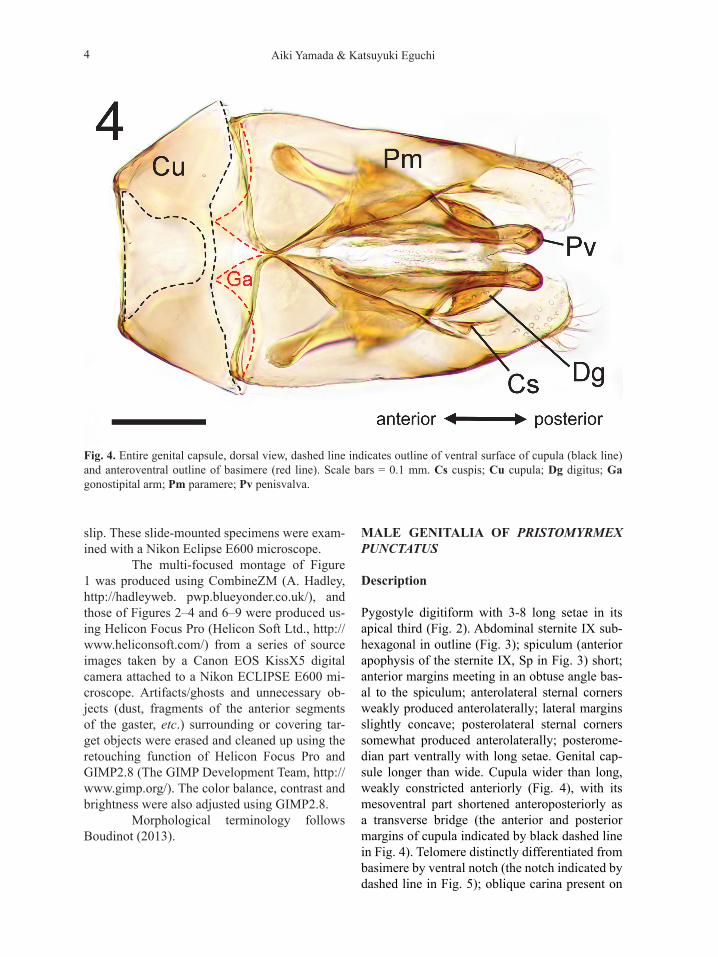
Fig. 4. Entire genital capsule, dorsal view, dashed line indicates outline of ventral surface of cupula (black line) and anteroventral outline of basimere (red line). Scale bars = 0.1 mm. Cs cuspis; Cu cupula; Dg digitus; Ga gonostipital arm; Pm paramere; Pv penisvalva.
slip. These slide-mounted specimens were exam-ined with a Nikon Eclipse E600 microscope. The multi-focused montage of Figure 1 was produced using CombineZM (A. Hadley, http://hadleyweb. pwp.blueyonder.co.uk/), and those of Figures 2–4 and 6–9 were produced us-ing Helicon Focus Pro (Helicon Soft Ltd., http://www.heliconsoft.com/) from a series of source images taken by a Canon EOS KissX5 digital camera attached to a Nikon ECLIPSE E600 mi-croscope. Artifacts/ghosts and unnecessary ob-jects (dust, fragments of the anterior segments of the gaster, etc.) surrounding or covering tar-get objects were erased and cleaned up using the retouching function of Helicon Focus Pro and GIMP2.8 (The GIMP Development Team, http://www.gimp.org/). The color balance, contrast and brightness were also adjusted using GIMP2.8. Morphological terminology follows Boudinot (2013).
MALE GENITALIA OF PRISTOMYRMEX PUNCTATUS
Description
Pygostyle digitiform with 3-8 long setae in its apical third (Fig. 2). Abdominal sternite IX sub-hexagonal in outline (Fig. 3); spiculum (anterior apophysis of the sternite IX, Sp in Fig. 3) short; anterior margins meeting in an obtuse angle bas-al to the spiculum; anterolateral sternal corners weakly produced anterolaterally; lateral margins slightly concave; posterolateral sternal corners somewhat produced anterolaterally; posterome-dian part ventrally with long setae. Genital cap-sule longer than wide. Cupula wider than long, weakly constricted anteriorly (Fig. 4), with its mesoventral part shortened anteroposteriorly as a transverse bridge (the anterior and posterior margins of cupula indicated by black dashed line in Fig. 4). Telomere distinctly differentiated from basimere by ventral notch (the notch indicated by dashed line in Fig. 5); oblique carina present on

5Description of the male genitalia of Pristomyrmex punctatus(Smith, 1860) (Hymenoptera, Formicidae, Myrmicinae)
Figs 5 – 7. Paramere and volsella. (5) paramere and volsella, right side, inner view; (6) anterior part of paramere, right side, inner view; (7) volsella separated from paramere, right side, inner view. Scale bars = 0.1 mm. Bm basimere; BmC carina of lower basimere; Cs cuspis; Dg digitus; Ga gonostipital arm;Tm telomere; Vo volsella.

6 Aiki Yamada & Katsuyuki Eguchi
Figs 8 – 9. Penisvalva. (8) penisvalva, left side, outer view. (9) pair of penisvalvae, dorsal view. Scale bars = 0.1 mm. Lp lateral apodeme of penisvalva; Pvm penisvalva membrane; Va valvura.
lower of basimere (BmC in Fig. 5); gonostipital arm (median anteroventral extension of the basi-mere indicated by red dashed line in Fig. 4 and Ga in Fig. 4 and 5) in ventral view almost as long as basal width, with its lateral and mesal margins
converging and forming an acute apex; telomere in lateral view almost as long as high, with se-tae on the outer surface of its posterior part (Fig. 6); ventral ridge of volsella with setae (Fig. 5); cuspis in lateral view roundly lobate and short,
7Description of the male genitalia of Pristomyrmex punctatus(Smith, 1860) (Hymenoptera, Formicidae, Myrmicinae)
not reaching posterior margin of digitus (Fig. 7); digitus in lateral view claw-shaped, entirely hooked ventrad (Fig. 7); very short setae with dis-tinct sockets scattered ventrally on digitus. Valvi-ceps in lateral view with posterior apex hooked ventrad, anteroventral corner not produced; ven-tral margin with 9–13 denticles (Fig. 8); foveae (recognized as tunnels running inside valviceps) sparsely present on apical and ventral area of valviceps (some of apical foveae probably with short setae but obscure in optical microscope ob-servation); valvura directed dorsoanterolaterally (Va in Fig. 9); lateral apodeme produced ventro-laterally (Lp in Fig. 9), forming wing-like struc-ture which hold basal part of volsella in genital complex; penisvalva membrane densely spinate (Pvm in Fig. 9).
Remarks
General constitution of formicid male genitalia as shown by previous authors (Krafchick 1959; Ogata 1987; Ogata 1991) was preserved in ir-regularly produced males of Pristomyrmex punc-tatus, and conspicuous deletion, deformation, or ornamentation of the main components was not recognized. Gonostipital arm which was claimed as a synapomorphy of the Myrmicinae by Ogata (1991) is present in the male genitalia of Pris-tomyrmex punctatus. But this structure is not a synapomorphy of the Myrmicinae because of its presence in other ant subfamilies and in other hy-menopteran lineages (Schulmeister 2003; Boudi-not 2013). Based on a comparison of the male genitalia of P. punctatus with those of other myr-micine genera described and illustrated by Ogata (1991), the general features of male genitalia of Myrmicinae can be summarized as follows: (1) cupula wider than long; (2) gonostipital arm pres-ent, but variable in shape; (3) digitus strongly curved ventrad (except for Solenopsis japonica: both of digitus and cuspis reduced); (4) cuspis simple and reduced, shorter than digitus and nev-er protruding dorsad above digitus in lateral view, or absent. In order to understand morphological synapomorphies of Myrmicinae in male genita-lia, more detailed and comprehensive compari-sion across taxa is needed.
In agreement with previous observa-tions by Itow et al. (1984), the male genitalia of P. punctatus seem to be functional. Further com-parison of the male genitalia between P. puncta-tus and amphigonic sister species, e.g., members of P. punctatus group (sensu Wang 2003), might provide valuable information for our better un-derstanding of the origin of parthenogenesis in the genus.
ACKNOWLEDGMENTS
We wish to thank Mr. Rijal Satria (Tokyo Metro-politan Univ., Japan) for his valuable suggestions. The present study was supported by the Japan So-ciety for the Promotion of Science (JSPS) Grant-in-Aid for scientific Research (C, no. 15K07193).
REFERENCES
Bolton B. 2015. AntCat.org: An online catalog of the ants of the world. Available from: http://ant-cat.org (accessed 8 December 2015).
Boudinot B. 2013. The male genitalia of ants: muscula-ture, homology, and functional morphology (Hymenoptera, Aculeata, Formicidae). Jour-nal of Hymenoptera Research. 30: 29–49.
Dobata S, Sasaki T, Mori H, Hasegawa E, Shimada M and Tsuji K. 2009. Cheater genotypes in the parthenogenetic ant Pristomyrmex punc-tatus. Proceedings of the Royal Society of London B 276(1656): 567–574.
Itow T, Kobayashi K, Kubota M, Ogata K, Imai HT and Crozier RH. 1984. The reproductive cy-cle of the queenless ant Pristomyrmex pun-gens. Insectes Sociaux 31(1):87–102.
Krafchick B. 1959. A comparative study of the male genitalia of North American ants (Formi-cidae) with emphasis on generic differences. Thesis submitted to the Faculty of the Grad-uate School of the University of Maryland. 79 pp.
Ogata K. 1987. A Generic Synopsis of the Poneroid Complex of the Family Formicidae in Japan (Hymenoptera). Part 1. Subfamilies Poneri-nae and Cerapachyinae. Esakia : occasional papers of the Hikosan Biological Laboratory in Entomology 25: 97-132.

8 Aiki Yamada & Katsuyuki Eguchi
Ogata K. 1991. A Generic Synopsis of the Poneroid Complex of the Family Formicidae in Japan (Hymenoptera). Part 2. Subfamily Myrmi-cinae. Bulletin of the Institute of Tropical Agriculture Kyushu University. 14: 61–149.
Schulmeister S. 2003. Genitalia and terminal abdomi-nal segments of male basal Hymenoptera (Insecta): morphology and evolution. Organ-isms Diversity and Evolution 3: 253–279.
Smith F. 1860. Catalogue of hymenopterous insects collected by Mr. A. R. Wallace in the islands of Bachian, Kaisaa, Amboyna, Gilolo, and at Dory in New Guinea. Journal of the Proceed-ings of the Linnean Society of London. Zool-ogy. 5(s1): 93-143.
Tsuji K. 1988. Obligate parthenogenesis and reproduc-tive division of labor in the Japanese queen-less ant Pristomyrmex pungens: comparison of intranidal and extranidal workers. Be-havioral Ecology and Sociobiology. 23(4): 247–255.
Wang M, 2003. A monographic revision of the ant genus Pristomyrmex (Hymenoptera: Formi-cidae). Bulletin of the Museum of Compara-tive Zoology 157(6): 383–542.
ASIAN MYRMECOLOGYA Journal of the International Network for the Study of Asian Ants
Communicating Editor: Himmender Bharti