dr. b ch 06_lecture_presentation
TRANSCRIPT
© 2012 Pearson Education, Inc.
6The Skeletal System: Axial Division
PowerPoint® Lecture Presentations prepared bySteven BassettSoutheast Community College Lincoln, Nebraska
© 2012 Pearson Education, Inc.
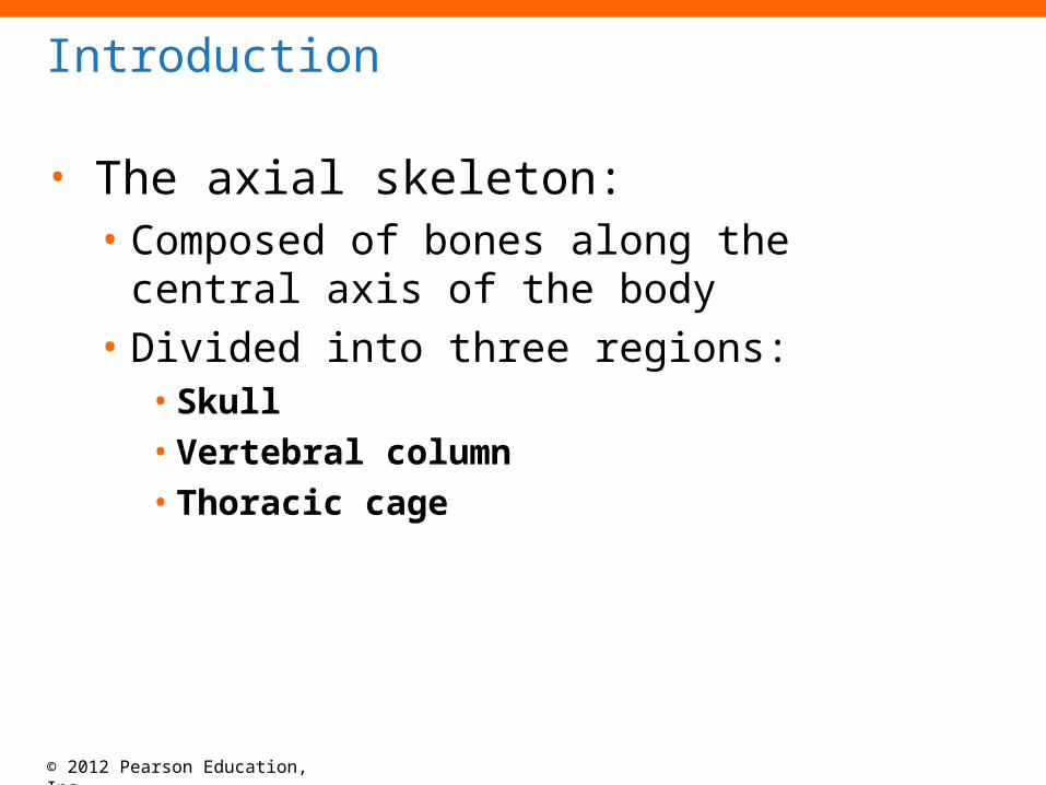
Introduction
• The axial skeleton:• Composed of bones along the central axis of
the body• Divided into three regions:
• Skull• Vertebral column• Thoracic cage
© 2012 Pearson Education, Inc.
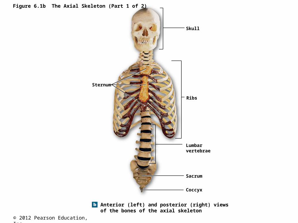
Figure 6.1b The Axial Skeleton (Part 1 of 2)
Anterior (left) and posterior (right) views of the bones of the axial skeleton
Skull
Ribs
Lumbarvertebrae
Sacrum
Coccyx
Sternum

© 2012 Pearson Education, Inc.
Introduction
• Functional anatomy of the axial skeleton: • Framework that supports and protects organs in the
dorsal and ventral body cavities• Protects special sense organs for taste, smell,
hearing, balance, and vision• Attachment sites for muscles that:
• Adjust the posture of the head, neck, and trunk• Move the thoracic cage for respiration• Stabilize the appendicular skeleton• The connection between axial and appendicular skeleton in
the upper body is sternum-clavicle.
© 2012 Pearson Education, Inc.
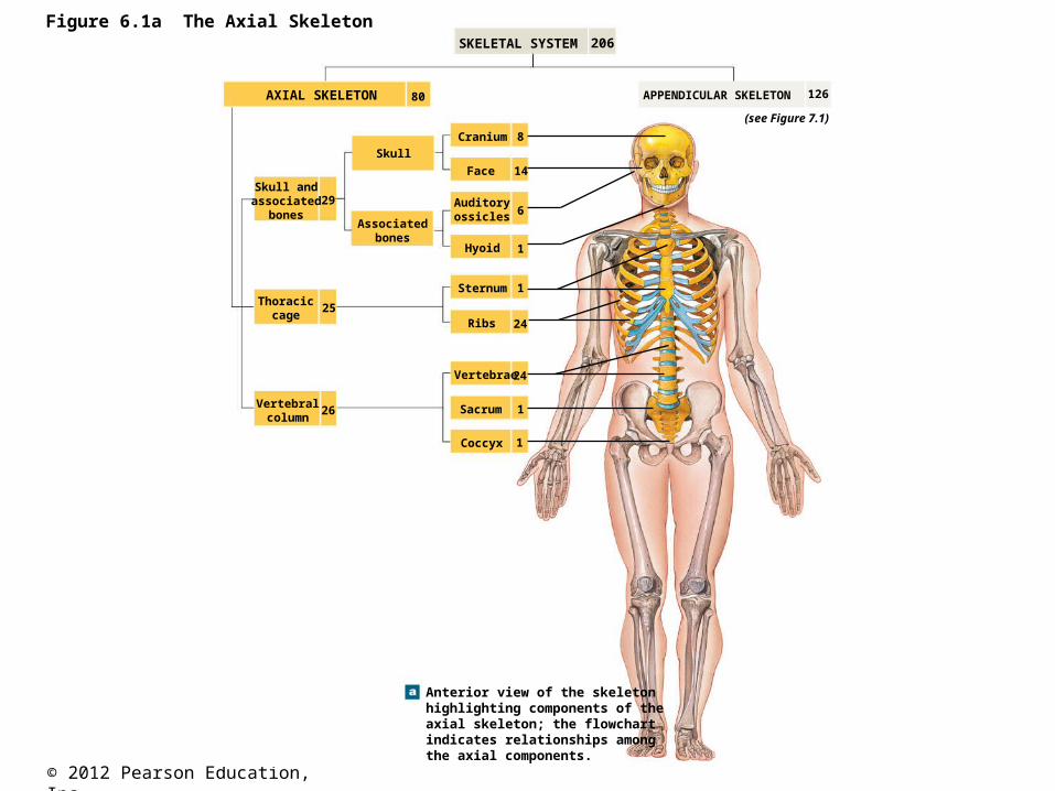
Figure 6.1a The Axial SkeletonSKELETAL SYSTEM
APPENDICULAR SKELETON
(see Figure 7.1)
AXIAL SKELETON 80
Skull andassociated
bones
Thoraciccage
Vertebralcolumn
Skull
Associatedbones
Auditoryossicles
Cranium
Face
Hyoid
Sternum
Ribs
Vertebrae
Sacrum
Coccyx
29
25
26
14
24
24
8
6
1
1
1
1
Anterior view of the skeletonhighlighting components of theaxial skeleton; the flowchartindicates relationships amongthe axial components.
126
206
© 2012 Pearson Education, Inc.
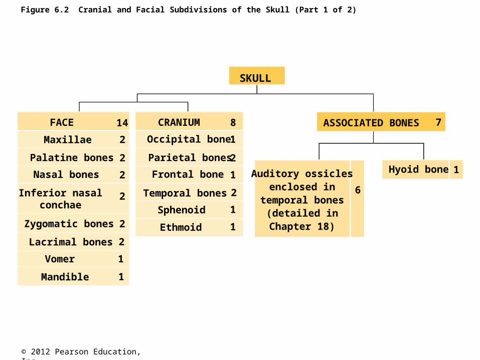
Figure 6.2 Cranial and Facial Subdivisions of the Skull (Part 1 of 2)
SKULL
ASSOCIATED BONES
Hyoid bone 1
7
6
Auditory ossiclesenclosed in
temporal bones(detailed inChapter 18)
FACE CRANIUM14 8
2
2
2
2
2
2
1
1
2
1
2
1
1
Maxillae
Palatine bones
Nasal bones
Inferior nasalconchae
Zygomatic bones
Lacrimal bones
Vomer
Mandible
Occipital bone
Parietal bones
Frontal bone
Temporal bones
Sphenoid
Ethmoid
1
© 2012 Pearson Education, Inc.
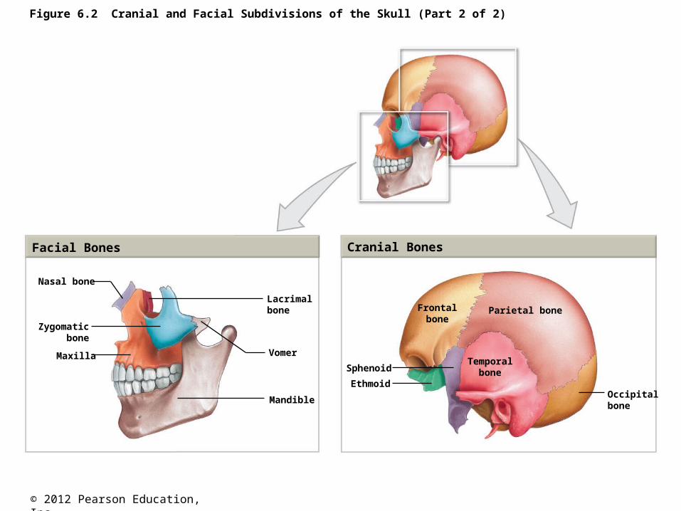
Figure 6.2 Cranial and Facial Subdivisions of the Skull (Part 2 of 2)
Facial Bones Cranial Bones
Nasal bone
Zygomaticbone
Maxilla
Lacrimalbone
Vomer
Mandible
Frontalbone
Sphenoid
Ethmoid
Parietal bone
Temporalbone
Occipitalbone
© 2012 Pearson Education, Inc.
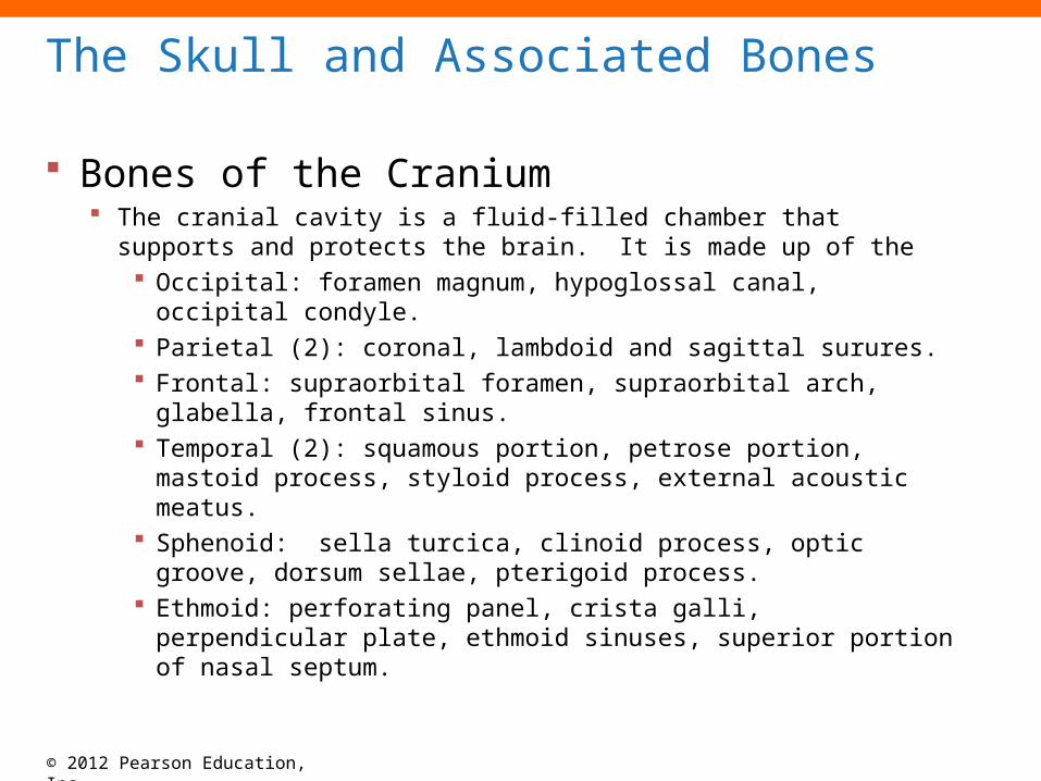
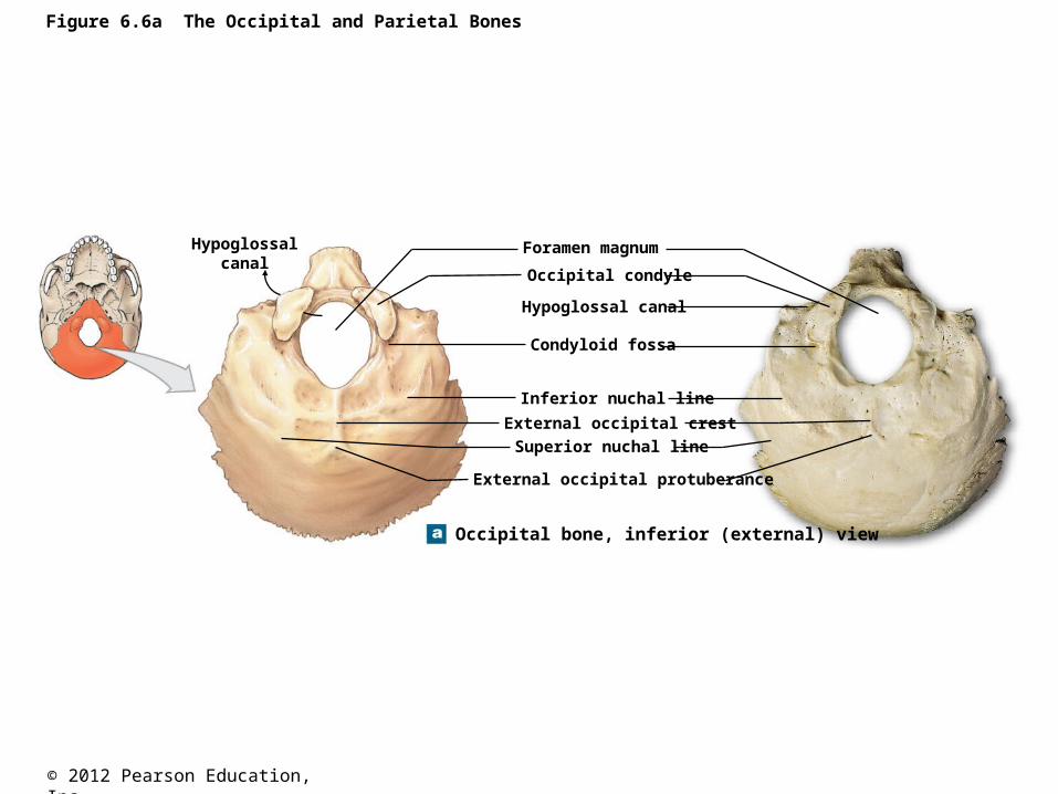
Bones of the Cranium The cranial cavity is a fluid-filled chamber that supports and
protects the brain. It is made up of the Occipital: foramen magnum, hypoglossal canal, occipital
condyle. Parietal (2): coronal, lambdoid and sagittal surures. Frontal: supraorbital foramen, supraorbital arch, glabella,
frontal sinus. Temporal (2): squamous portion, petrose portion, mastoid
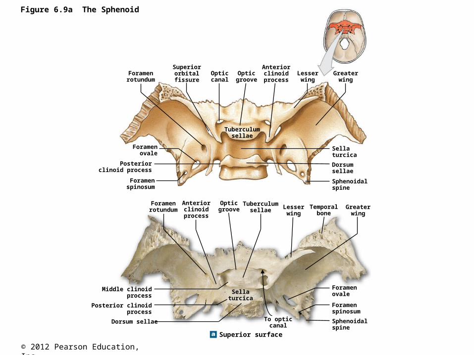
process, styloid process, external acoustic meatus. Sphenoid: sella turcica, clinoid process, optic groove,
dorsum sellae, pterigoid process. Ethmoid: perforating panel, crista galli, perpendicular plate,
ethmoid sinuses, superior portion of nasal septum.
The Skull and Associated Bones
© 2012 Pearson Education, Inc.
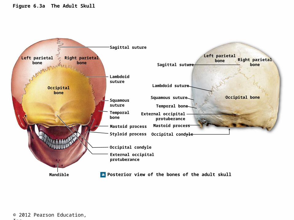
Figure 6.3a The Adult Skull
Posterior view of the bones of the adult skullMandible
External occipitalprotuberance
Occipital condyle
Styloid process
Mastoid process
Temporal bone
Squamoussuture
Lambdoidsuture
Sagittal suture
Left parietalbone
Right parietalbone
Occipitalbone
Sagittal suture
Lambdoid suture
Squamous suture
Temporal bone
External occipitalprotuberance
Mastoid process
Occipital condyle
Left parietalbone Right parietal
bone
Occipital bone
© 2012 Pearson Education, Inc.
Figure 6.6a The Occipital and Parietal Bones
Occipital bone, inferior (external) view
Hypoglossalcanal
Foramen magnum
Occipital condyle
Hypoglossal canal
Condyloid fossa
Inferior nuchal line
External occipital crest
Superior nuchal line
External occipital protuberance
© 2012 Pearson Education, Inc.
Figure 6.3b The Adult Skull
Superior view of the bonesof the adult skull
Nasal bones
Frontalbone
Rightparietal
bone
Leftparietal
bone
Occipitalbone
Occipitalbone
Frontalbone
Rightparietal
bone
Leftparietal
bone
Lambdoidsuture
Sagittalsuture
Coronalsuture
Zygomaticbone
© 2012 Pearson Education, Inc.
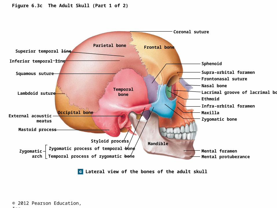
Figure 6.3c The Adult Skull (Part 1 of 2)
Lateral view of the bones of the adult skull
Parietal bone Frontal bone
Temporalbone
Occipital bone
Superior temporal line
Inferior temporal line
Squamous suture
Lambdoid suture
External acousticmeatus
Mastoid process
Zygomaticarch
Styloid process
Zygomatic process of temporal bone
Temporal process of zygomatic bone
Mandible
Coronal suture
Sphenoid
Supra-orbital foramen
Frontonasal suture
Nasal bone
Lacrimal groove of lacrimal bone
Ethmoid
Infra-orbital foramen
Maxilla
Zygomatic bone
Mental foramenMental protuberance
© 2012 Pearson Education, Inc.
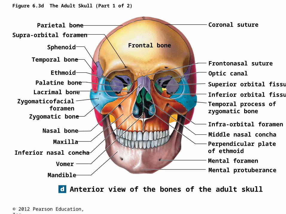
Figure 6.3d The Adult Skull (Part 1 of 2)
Anterior view of the bones of the adult skull
Parietal bone
Supra-orbital foramen
Sphenoid
Temporal bone
Ethmoid
Palatine bone
Lacrimal bone
Zygomaticofacialforamen
Zygomatic bone
Nasal bone
Maxilla
Inferior nasal concha
Vomer
Mandible
Frontal bone
Coronal suture
Frontonasal suture
Optic canal
Superior orbital fissure
Inferior orbital fissure
Temporal process ofzygomatic bone
Infra-orbital foramen
Middle nasal concha
Perpendicular plateof ethmoid
Mental foramen
Mental protuberance
© 2012 Pearson Education, Inc.
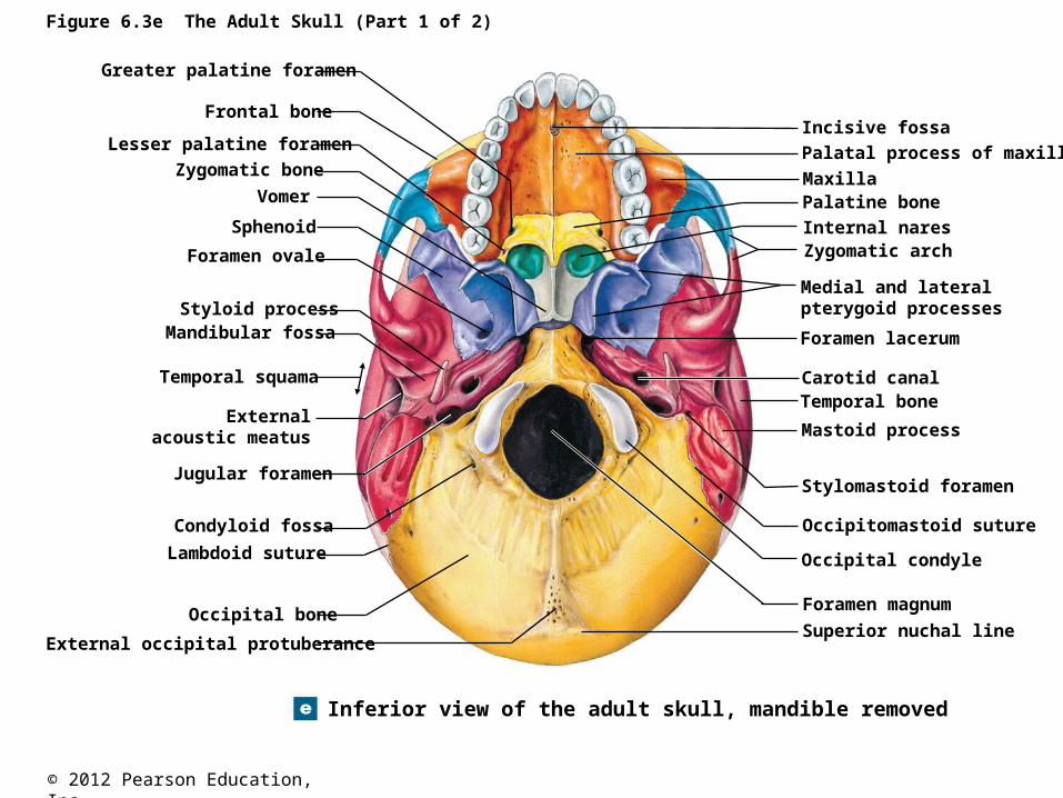
Figure 6.3e The Adult Skull (Part 1 of 2)
Inferior view of the adult skull, mandible removed
Greater palatine foramen
Frontal bone
Lesser palatine foramen
Zygomatic bone
Vomer
Sphenoid
Foramen ovale
Styloid processMandibular fossa
Temporal squama
Externalacoustic meatus
Jugular foramen
Condyloid fossa
Lambdoid suture
Occipital bone
External occipital protuberance
Incisive fossa
Palatal process of maxilla
Maxilla
Internal nares
Palatine bone
Zygomatic arch
Medial and lateralpterygoid processes
Foramen lacerum
Carotid canalTemporal bone
Mastoid process
Stylomastoid foramen
Occipitomastoid suture
Occipital condyle
Foramen magnum
Superior nuchal line
© 2012 Pearson Education, Inc.
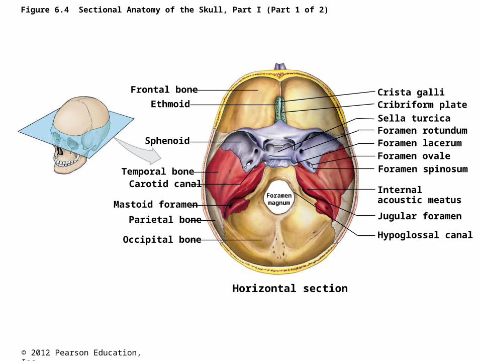
Figure 6.4 Sectional Anatomy of the Skull, Part I (Part 1 of 2)
Frontal bone
Ethmoid
Sphenoid
Temporal boneCarotid canal
Mastoid foramen
Parietal bone
Occipital bone
Crista galliCribriform plate
Sella turcicaForamen rotundumForamen lacerumForamen ovaleForamen spinosum
Internalacoustic meatus
Jugular foramen
Hypoglossal canal
Foramenmagnum
Horizontal section
© 2012 Pearson Education, Inc.
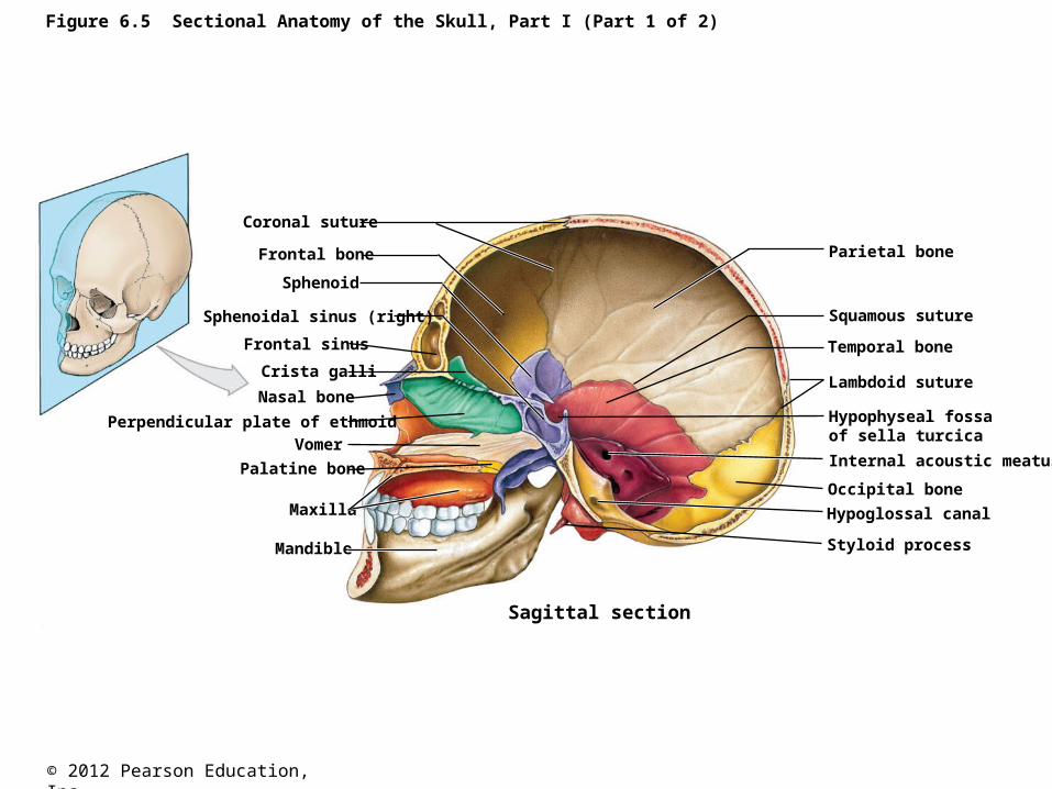
Figure 6.5 Sectional Anatomy of the Skull, Part I (Part 1 of 2)
Sagittal section
Coronal suture
Frontal bone
Sphenoid
Sphenoidal sinus (right)
Frontal sinus
Crista galli
Nasal bone
Perpendicular plate of ethmoid
Vomer
Palatine bone
Maxilla
Mandible Styloid process
Hypoglossal canal
Occipital bone
Internal acoustic meatus
Hypophyseal fossaof sella turcica
Lambdoid suture
Temporal bone
Squamous suture
Parietal bone
© 2012 Pearson Education, Inc.
Figure 6.9a The Sphenoid
Superior surface
Tuberculumsellae
Foramenrotundum
Superiororbitalfissure
Opticcanal
Opticgroove
Anteriorclinoid
processLesserwing
Greaterwing
Foramenovale
Posteriorclinoid process
Foramenspinosum
Sellaturcica
Dorsumsellae
Sphenoidalspine
Foramenrotundum
Anteriorclinoid
process
Opticgroove
Tuberculumsellae Lesser
wingGreater
wingTemporal
bone
Foramenovale
Foramenspinosum
Sphenoidalspine
Sellaturcica
Dorsum sellae
Posterior clinoidprocess
Middle clinoidprocess
To opticcanal
© 2012 Pearson Education, Inc.
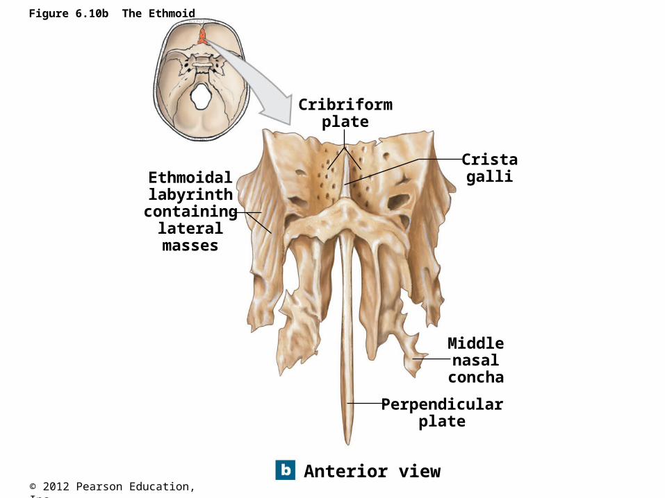
Figure 6.10b The Ethmoid
Anterior view
Ethmoidallabyrinth
containinglateral
masses
Cribriformplate
Cristagalli
Middlenasal
concha
Perpendicularplate
© 2012 Pearson Education, Inc.
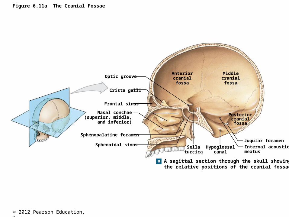
Figure 6.11a The Cranial Fossae
A sagittal section through the skull showingthe relative positions of the cranial fossae
Optic groove
Crista galli
Frontal sinus
Sphenopalatine foramen
Sphenoidal sinus
Nasal conchae(superior, middle,
and inferior)
Anteriorcranialfossa
Middlecranialfossa
Posteriorcranialfossa
Sellaturcica
Hypoglossalcanal
Jugular foramenInternal acousticmeatus
© 2012 Pearson Education, Inc.
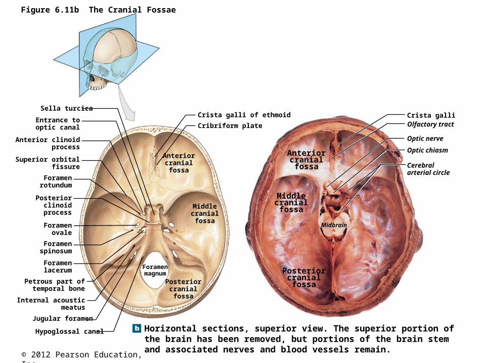
Figure 6.11b The Cranial Fossae
Horizontal sections, superior view. The superior portion ofthe brain has been removed, but portions of the brain stemand associated nerves and blood vessels remain.
Sella turcica
Entrance tooptic canal
Anterior clinoidprocess
Superior orbitalfissure
Foramenrotundum
Posteriorclinoid
process
Foramenovale
Foramenspinosum
Foramenlacerum
Petrous part oftemporal bone
Internal acousticmeatus
Jugular foramen
Hypoglossal canal
Posteriorcranialfossa
Middlecranialfossa
Anteriorcranialfossa
Foramenmagnum
Crista galli of ethmoid
Cribriform plate
Crista galliOlfactory tract
Optic nerve
Optic chiasm
Cerebralarterial circle
Midbrain
Anteriorcranialfossa
Middlecranialfossa
Posteriorcranialfossa
© 2012 Pearson Education, Inc.
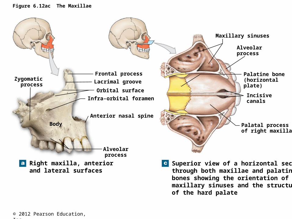
Bones of the Face The skull contains 14 total facial bones.
The facial bones included the paired bones named the Maxillae: alveolar process, maxillary sinus (the largest
paranasal sinus), infraorbital foramen, front part of hard palate. Palatine: dorsal part of hard palate. Nasal Zygomatic: cheek bones Lacrimal Inferior nasal conchae
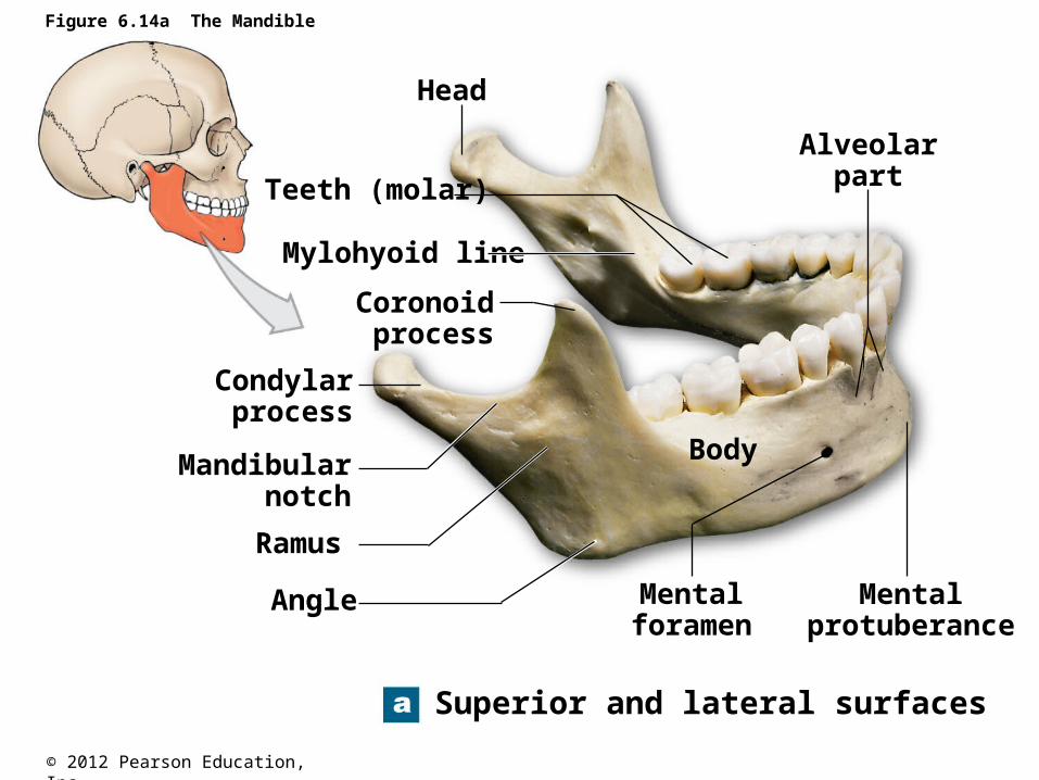
Single bones of the face are the Vomer: makes the inferior portion of nasal septum. Mandible: Alveolar process, ramus, condylar process, coronoid
process.
The Skull and Associated Bones
© 2012 Pearson Education, Inc.
Figure 6.12ac The Maxillae
Right maxilla, anteriorand lateral surfaces
Body
Zygomaticprocess
Frontal process
Lacrimal groove
Orbital surface
Infra-orbital foramen
Anterior nasal spine
Alveolarprocess
Superior view of a horizontal sectionthrough both maxillae and palatinebones showing the orientation of themaxillary sinuses and the structureof the hard palate
Alveolar process
Maxillary sinuses
Palatine bone(horizontalplate)
Incisivecanals
Palatal processof right maxilla
© 2012 Pearson Education, Inc.
Figure 6.14a The Mandible
Superior and lateral surfaces
Head
Teeth (molar)
Mylohyoid line
Coronoidprocess
Condylarprocess
Mandibularnotch
Ramus
Angle
Body
Mentalforamen
Mentalprotuberance
Alveolarpart
© 2012 Pearson Education, Inc.
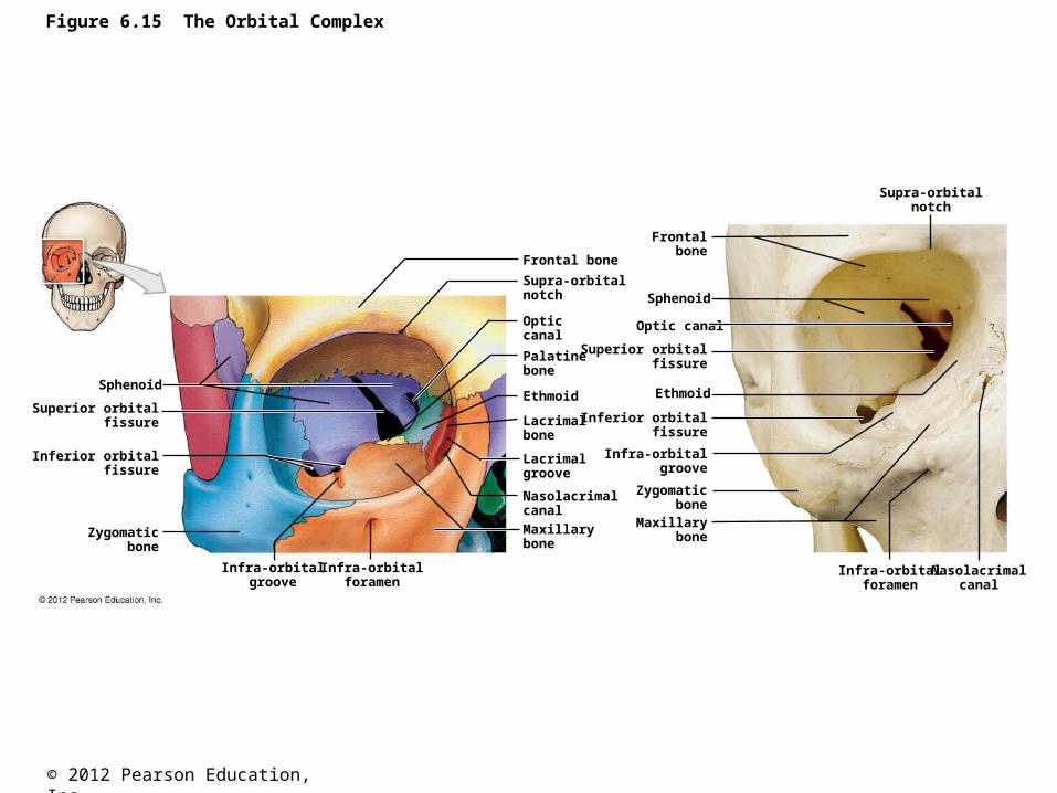
Figure 6.15 The Orbital Complex
Sphenoid
Superior orbitalfissure
Inferior orbitalfissure
Zygomaticbone
Infra-orbitalgroove
Infra-orbitalforamen
Frontal bone
Supra-orbitalnotch
Opticcanal
Palatinebone
Ethmoid
Lacrimalbone
Lacrimalgroove
Nasolacrimalcanal
Maxillarybone
Supra-orbitalnotch
Frontalbone
Sphenoid
Optic canal
Superior orbitalfissure
Ethmoid
Inferior orbitalfissure
Infra-orbitalgroove
Zygomaticbone
Maxillarybone
Infra-orbitalforamen
Nasolacrimalcanal
© 2012 Pearson Education, Inc.
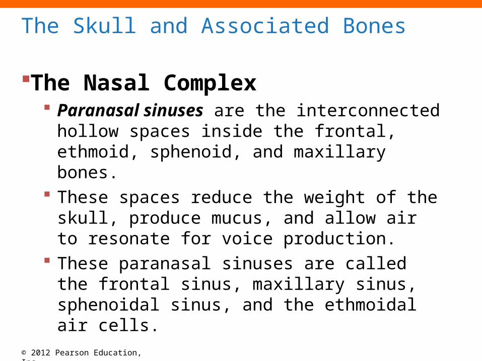
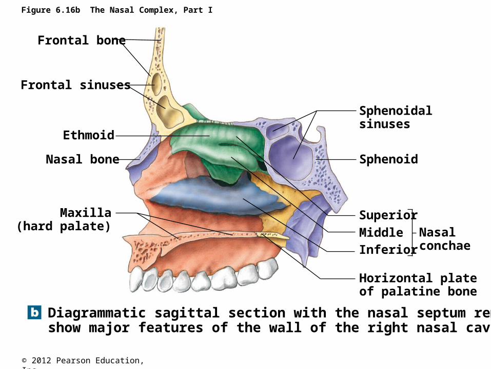
The Nasal Complex Paranasal sinuses are the interconnected
hollow spaces inside the frontal, ethmoid, sphenoid, and maxillary bones.
These spaces reduce the weight of the skull, produce mucus, and allow air to resonate for voice production.
These paranasal sinuses are called the frontal sinus, maxillary sinus, sphenoidal sinus, and the ethmoidal air cells.
The Skull and Associated Bones
© 2012 Pearson Education, Inc.
Figure 6.16b The Nasal Complex, Part I
Diagrammatic sagittal section with the nasal septum removed toshow major features of the wall of the right nasal cavity
Nasal bone
Ethmoid
Frontal sinuses
Frontal bone
Maxilla(hard palate)
Sphenoidalsinuses
Sphenoid
Superior
MiddleInferior
Nasalconchae
Horizontal plateof palatine bone
© 2012 Pearson Education, Inc.
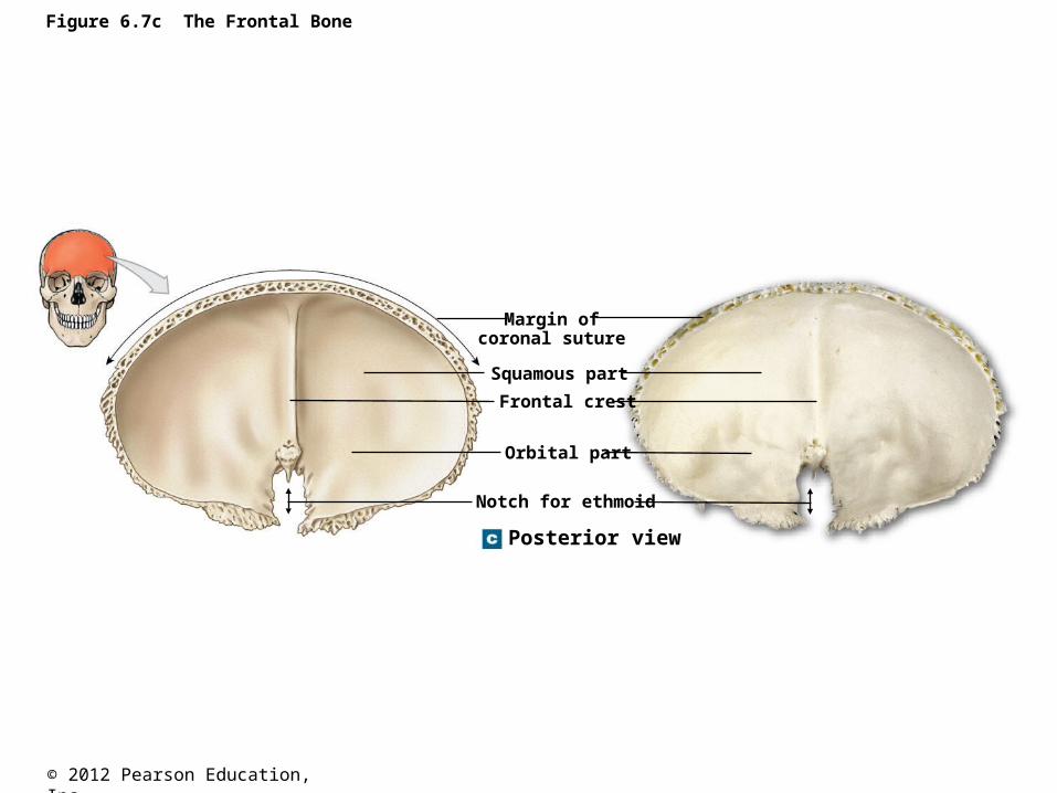
Figure 6.7c The Frontal Bone
Posterior view
Margin ofcoronal suture
Squamous part
Frontal crest
Orbital part
Notch for ethmoid
© 2012 Pearson Education, Inc.
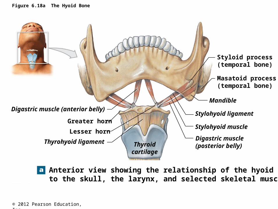
Figure 6.18a The Hyoid Bone
Anterior view showing the relationship of the hyoid boneto the skull, the larynx, and selected skeletal muscles
Digastric muscle (anterior belly)
Greater horn
Lesser horn
Thyrohyoid ligament Thyroidcartilage
Digastric muscle(posterior belly)
Stylohyoid muscle
Stylohyoid ligament
Mandible
Masatoid process(temporal bone)
Styloid process(temporal bone)
© 2012 Pearson Education, Inc.
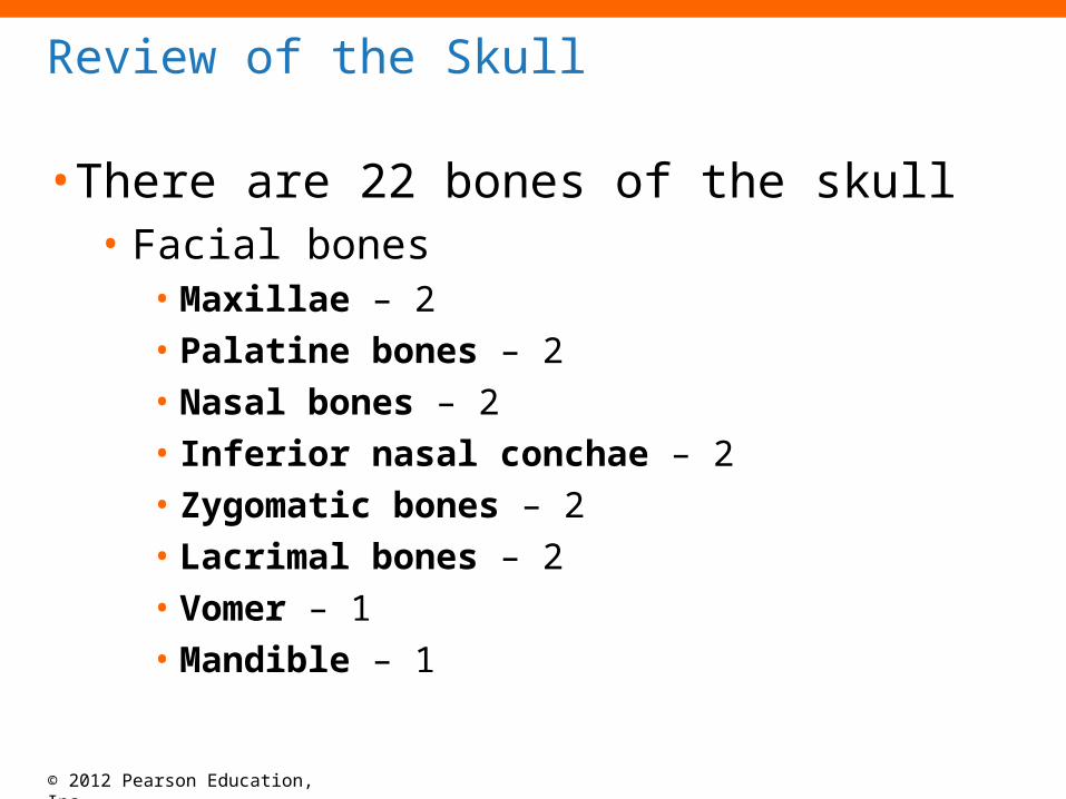
• There are 22 bones of the skull• Facial bones
• Maxillae – 2• Palatine bones – 2• Nasal bones – 2• Inferior nasal conchae – 2• Zygomatic bones – 2• Lacrimal bones – 2• Vomer – 1• Mandible – 1
Review of the Skull

© 2012 Pearson Education, Inc.
• There are 22 bones of the skull• Cranial bones
• Occipital bone – 1• Parietal bones – 2• Frontal bone – 1• Temporal bones – 2• Sphenoid bone – 1• Ethmoid bone – 1
Review of the Skull (continued)
© 2012 Pearson Education, Inc.
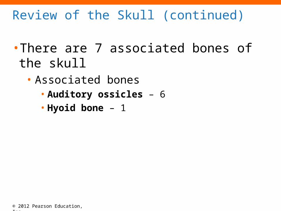
• There are 7 associated bones of the skull• Associated bones
• Auditory ossicles – 6• Hyoid bone – 1
Review of the Skull (continued)
© 2012 Pearson Education, Inc.
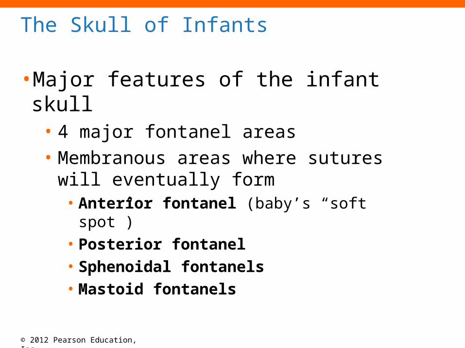
• Major features of the infant skull• 4 major fontanel areas• Membranous areas where sutures will
eventually form• Anterior fontanel (baby’s “soft spot”)• Posterior fontanel• Sphenoidal fontanels• Mastoid fontanels
The Skull of Infants
© 2012 Pearson Education, Inc.
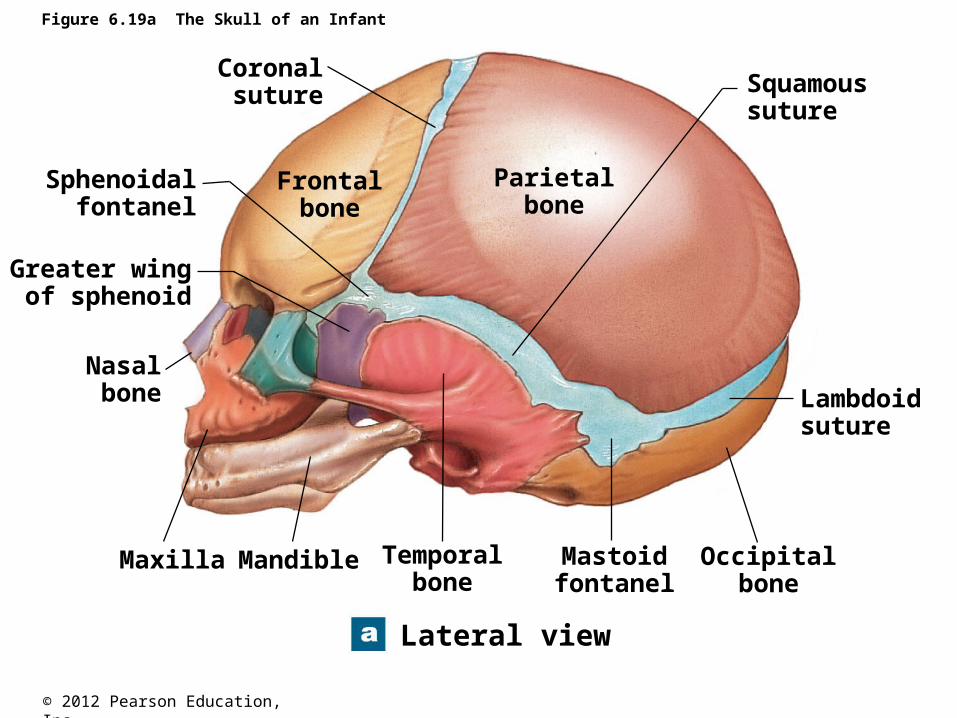
Figure 6.19a The Skull of an Infant
Lateral view
Maxilla Mandible Temporalbone
Mastoidfontanel
Occipitalbone
Lambdoidsuture
Squamoussuture
Parietalbone
Frontalbone
Coronalsuture
Sphenoidalfontanel
Greater wingof sphenoid
Nasalbone
© 2012 Pearson Education, Inc.
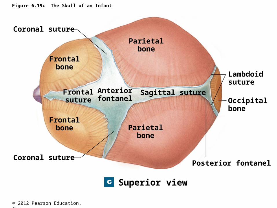
Figure 6.19c The Skull of an Infant
Superior view
Coronal suture
Coronal suturePosterior fontanel
Lambdoidsuture
Occipital bone
Frontalbone
Frontalbone
Parietalbone
Parietalbone
Frontalsuture
Anteriorfontanel
Sagittal suture
© 2012 Pearson Education, Inc.
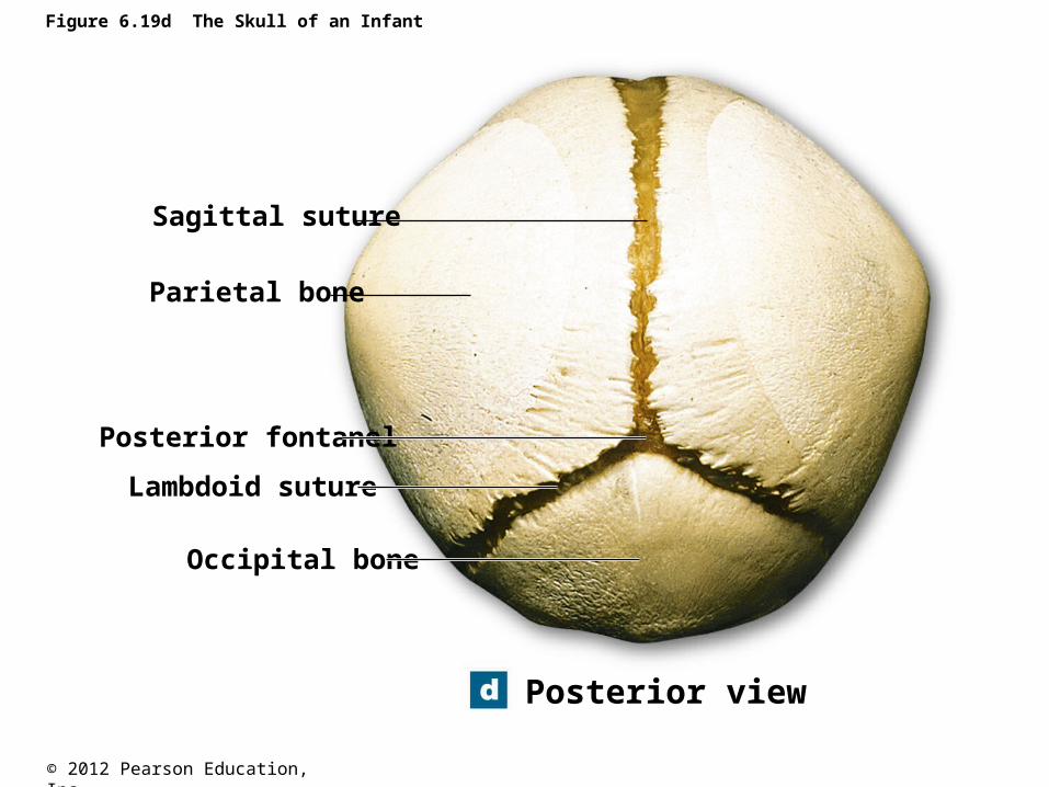
Figure 6.19d The Skull of an Infant
Posterior view
Sagittal suture
Parietal bone
Posterior fontanel
Lambdoid suture
Occipital bone
© 2012 Pearson Education, Inc.
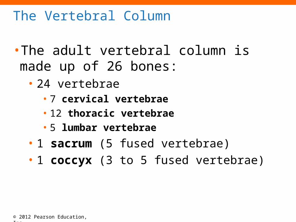
• The adult vertebral column is made up of 26 bones:
• 24 vertebrae• 7 cervical vertebrae• 12 thoracic vertebrae• 5 lumbar vertebrae
• 1 sacrum (5 fused vertebrae)• 1 coccyx (3 to 5 fused vertebrae)
The Vertebral Column
© 2012 Pearson Education, Inc.
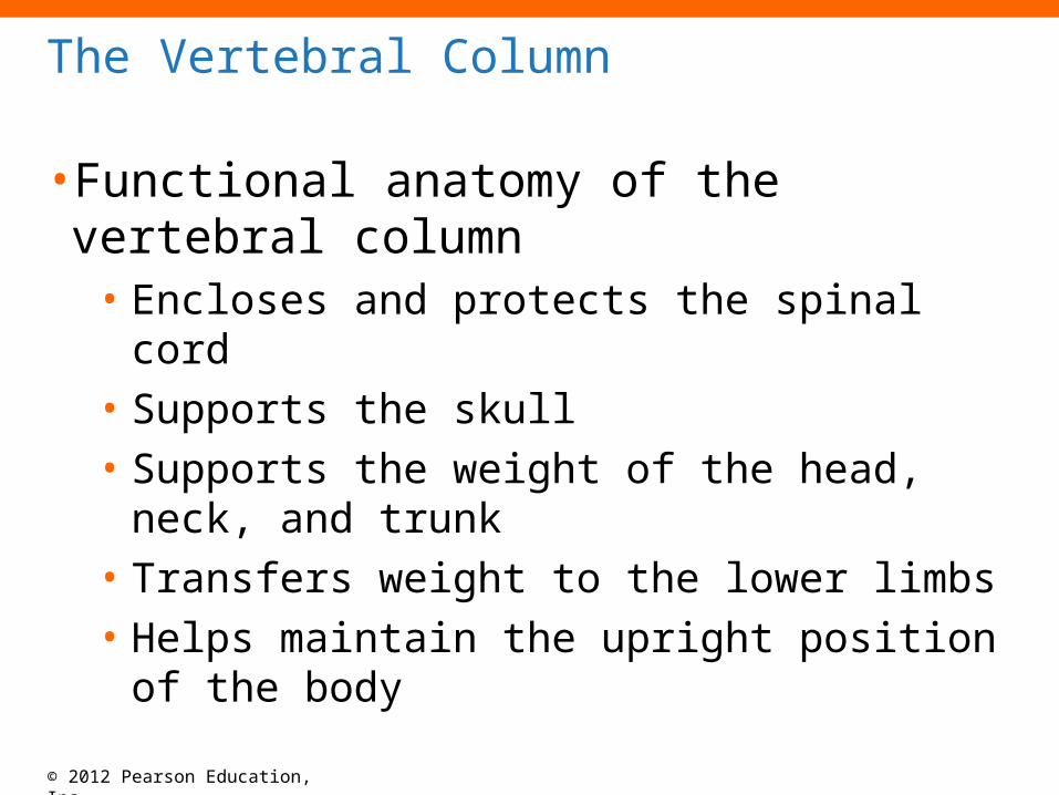
The Vertebral Column
• Functional anatomy of the vertebral column• Encloses and protects the spinal cord• Supports the skull• Supports the weight of the head, neck, and trunk• Transfers weight to the lower limbs• Helps maintain the upright position of the body
© 2012 Pearson Education, Inc.
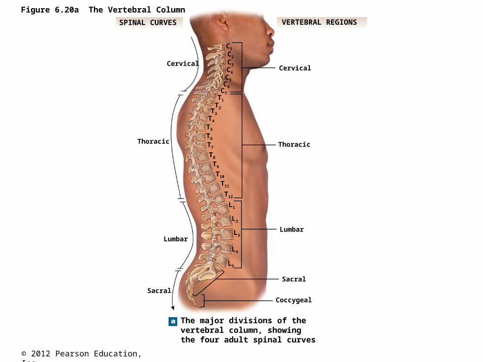
Figure 6.20a The Vertebral Column
The major divisions of thevertebral column, showingthe four adult spinal curves
Sacral
Lumbar
Thoracic
CervicalCervical
Sacral
Lumbar
Thoracic
Coccygeal
C1
C2
C3
C4
C5C6
C7T1
T2T3
T4
T5
T6
T7
T8
T9
T10
T11
T12
L1
L2
L3
L4
L5
SPINAL CURVES VERTEBRAL REGIONS
© 2012 Pearson Education, Inc.
The Vertebral Column
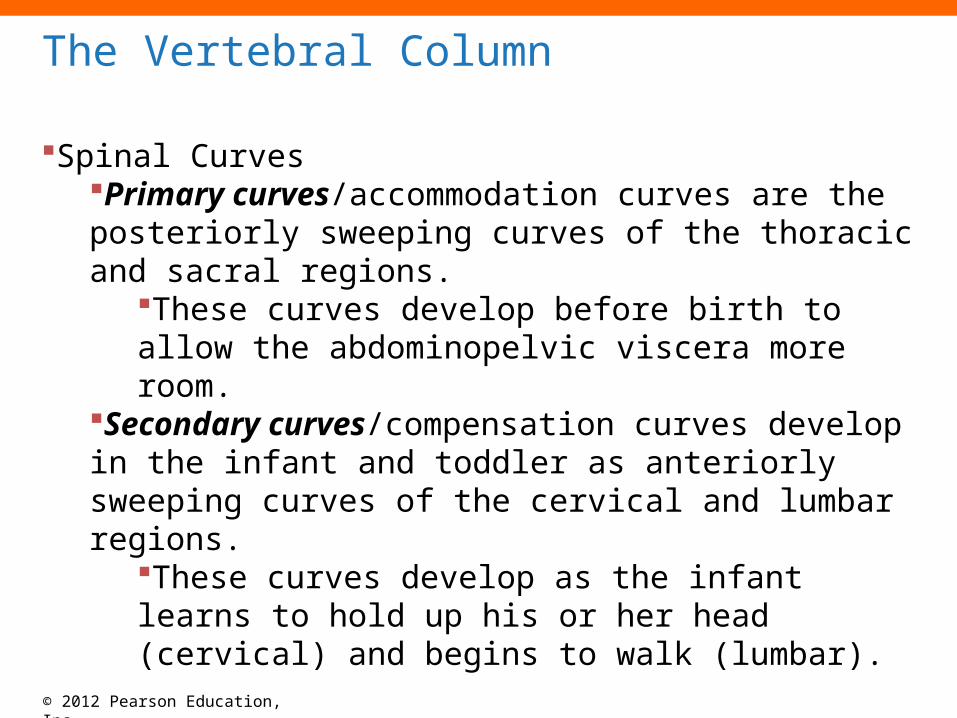
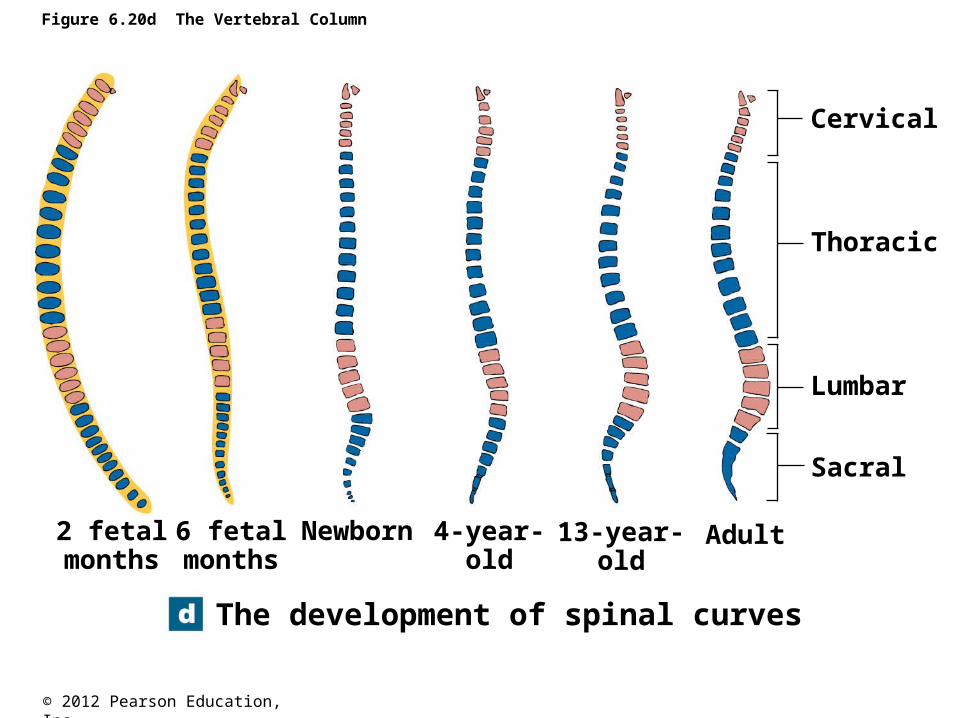
Spinal CurvesPrimary curves/accommodation curves are the posteriorly sweeping curves of the thoracic and sacral regions.
These curves develop before birth to allow the abdominopelvic viscera more room.
Secondary curves/compensation curves develop in the infant and toddler as anteriorly sweeping curves of the cervical and lumbar regions.
These curves develop as the infant learns to hold up his or her head (cervical) and begins to walk (lumbar).
© 2012 Pearson Education, Inc.
Figure 6.20d The Vertebral Column
The development of spinal curves
Cervical
Thoracic
Lumbar
Sacral
2 fetalmonths
6 fetalmonths
Newborn 4-year-old
13-year-old
Adult
© 2012 Pearson Education, Inc.
The Vertebral Column
• Abnormal curvatures of the vertebral column
• Scoliosis• Abnormal lateral curvature
• Kyphosis• Exaggerated posterior curvature of the thoracic
region
• Lordosis• Exaggerated anterior curvature of the lumbar
region

© 2012 Pearson Education, Inc.
Spinal curve deformities
© 2012 Pearson Education, Inc.
The Vertebral Column
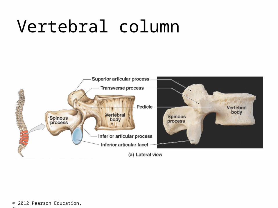
• Vertebral Processes (Cervical Vertebrae)• Vertebral body• Vertebral foramen• Spinous process• Transverse process
• Transverse foramen
• Lamina• Pedicle
© 2012 Pearson Education, Inc.
The Vertebral Column
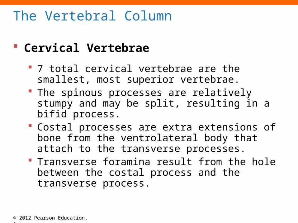
Cervical Vertebrae
7 total cervical vertebrae are the smallest, most superior vertebrae.
The spinous processes are relatively stumpy and may be split, resulting in a bifid process.
Costal processes are extra extensions of bone from the ventrolateral body that attach to the transverse processes.
Transverse foramina result from the hole between the costal process and the transverse process.
© 2012 Pearson Education, Inc.
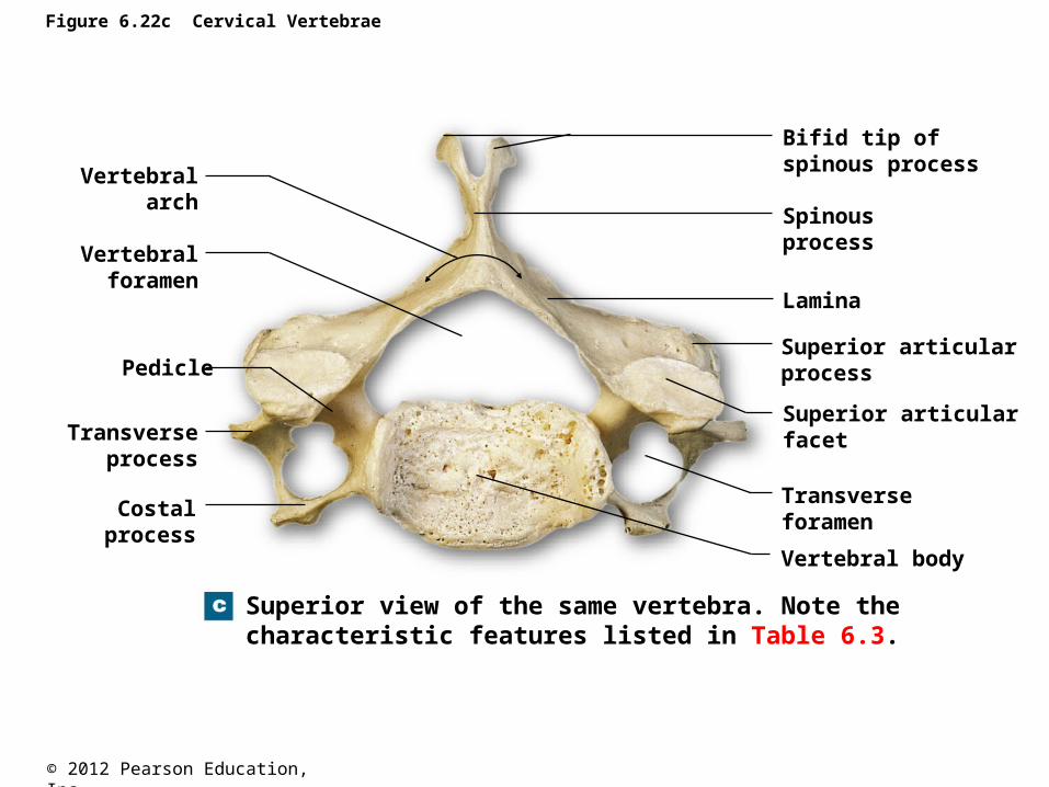
Figure 6.22c Cervical Vertebrae
Superior view of the same vertebra. Note thecharacteristic features listed in Table 6.3.
Vertebralarch
Vertebralforamen
Pedicle
Transverseprocess
Costalprocess
Bifid tip ofspinous process
Spinous process
Lamina
Superior articularprocess
Superior articularfacet
Transverseforamen
Vertebral body
© 2012 Pearson Education, Inc.
The Vertebral Column
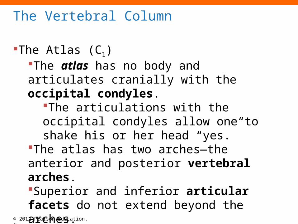
The Atlas (C1)The atlas has no body and articulates cranially with the occipital condyles.
The articulations with the occipital condyles allow one to shake his or her head “yes.”
The atlas has two arches—the anterior and posterior vertebral arches.Superior and inferior articular facets do not extend beyond the arches.
© 2012 Pearson Education, Inc.
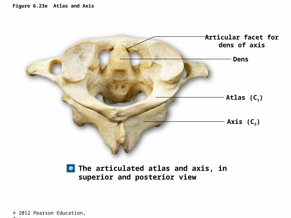
Figure 6.23e Atlas and Axis
The articulated atlas and axis, insuperior and posterior view
Articular facet fordens of axis
Dens
Atlas (C1)
Axis (C2)
© 2012 Pearson Education, Inc.
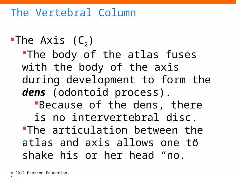
The Vertebral Column
The Axis (C2)The body of the atlas fuses with the body of the axis during development to form the dens (odontoid process).
Because of the dens, there is no intervertebral disc.
The articulation between the atlas and axis allows one to shake his or her head “no.”
© 2012 Pearson Education, Inc.
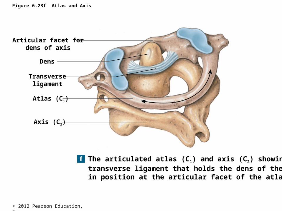
Figure 6.23f Atlas and Axis
Articular facet fordens of axis
Dens
Atlas (C1)
Axis (C2)
The articulated atlas (C1) and axis (C2) showing thetransverse ligament that holds the dens of the axisin position at the articular facet of the atlas
Transverseligament
© 2012 Pearson Education, Inc.
The vertebral column
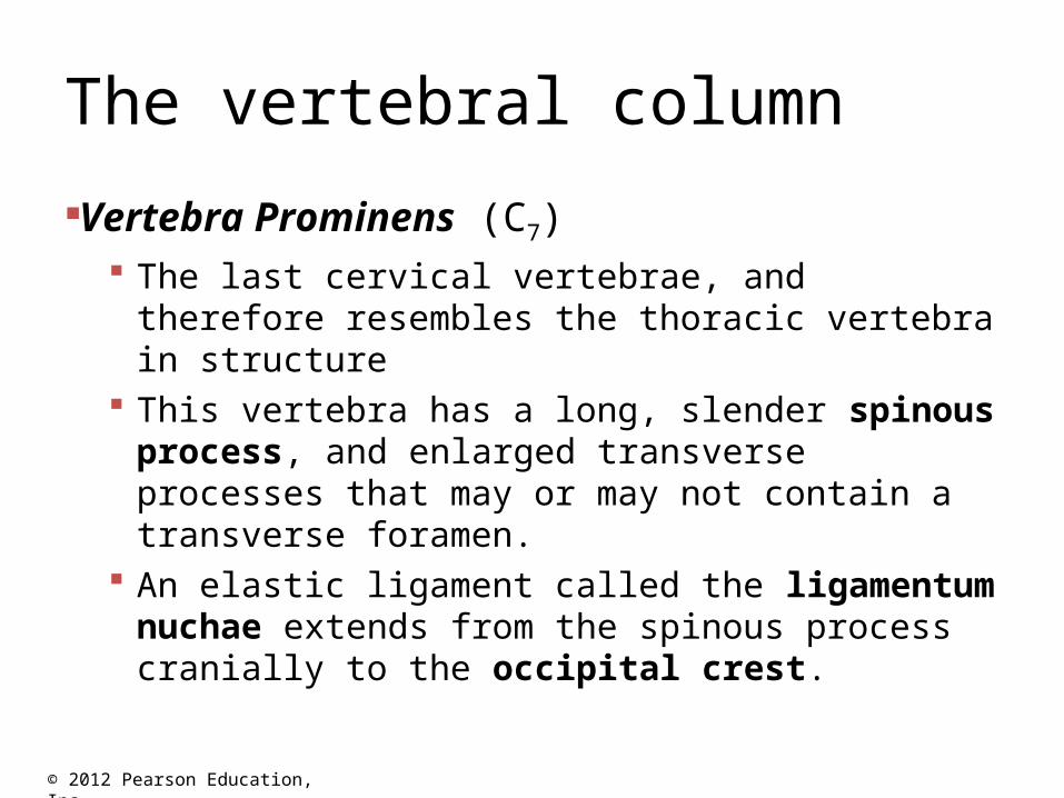
Vertebra Prominens (C7) The last cervical vertebrae, and therefore
resembles the thoracic vertebra in structure This vertebra has a long, slender spinous
process, and enlarged transverse processes that may or may not contain a transverse foramen.
An elastic ligament called the ligamentum nuchae extends from the spinous process cranially to the occipital crest.
© 2012 Pearson Education, Inc.
Vertebral column
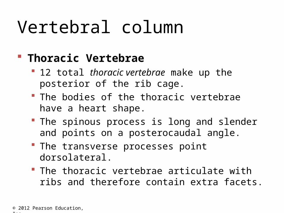
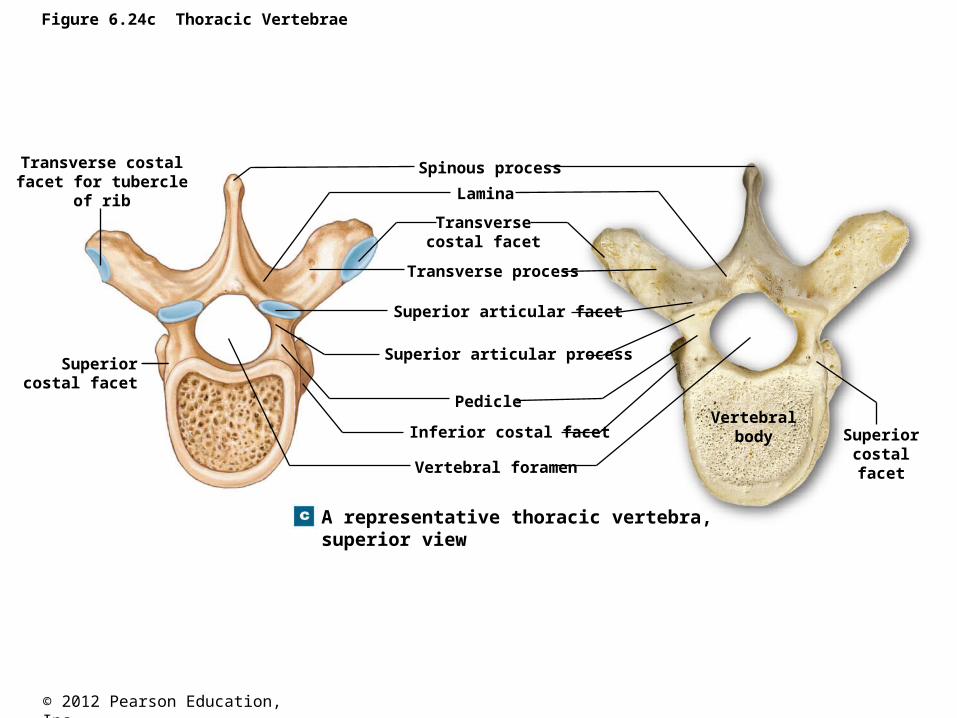
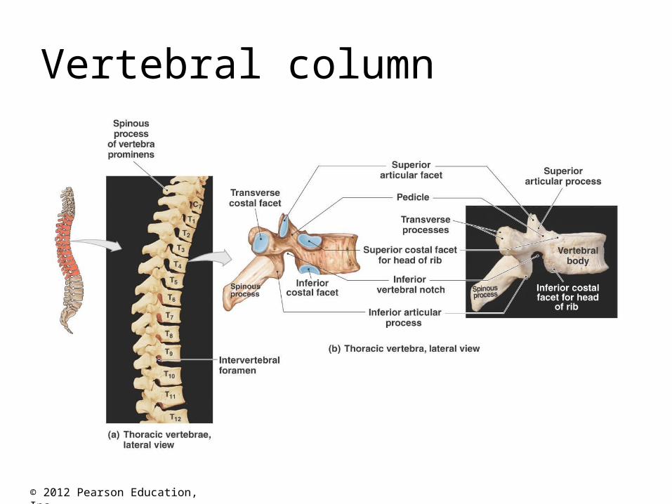
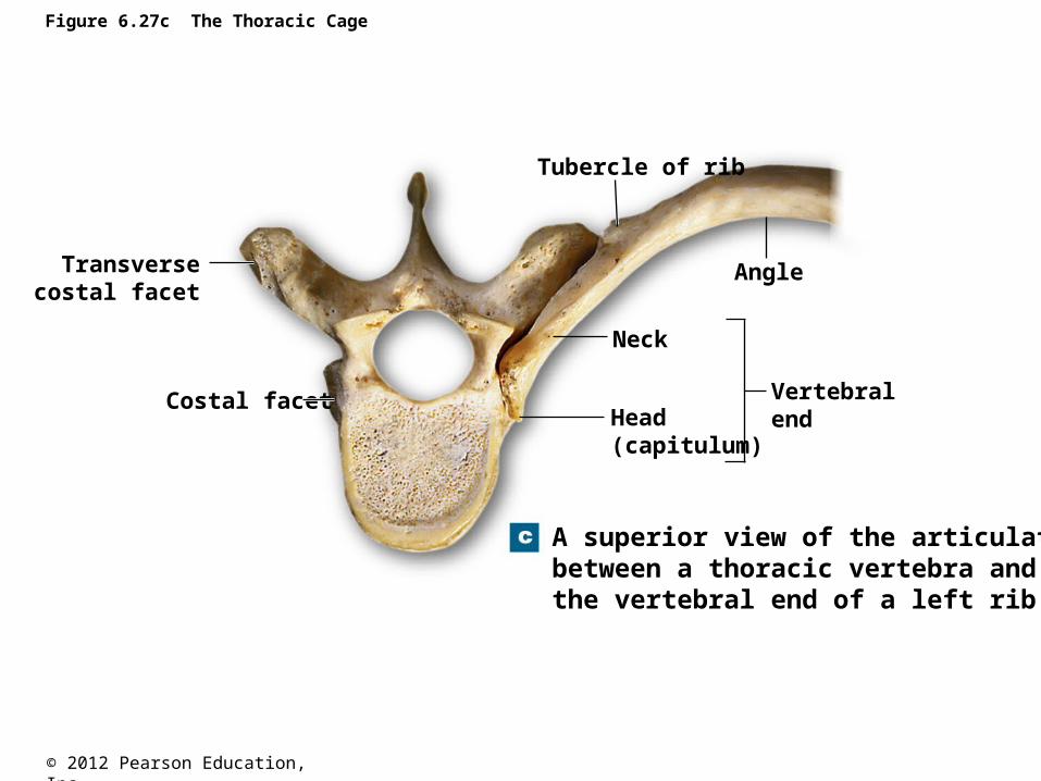
Thoracic Vertebrae 12 total thoracic vertebrae make up the posterior of
the rib cage. The bodies of the thoracic vertebrae have a heart
shape. The spinous process is long and slender and points
on a posterocaudal angle. The transverse processes point dorsolateral. The thoracic vertebrae articulate with ribs and
therefore contain extra facets.
© 2012 Pearson Education, Inc.
Figure 6.24c Thoracic Vertebrae
A representative thoracic vertebra,superior view
Transverse costalfacet for tubercle
of rib
Superiorcostal facet
Spinous process
Lamina
Transversecostal facet
Transverse process
Superior articular facet
Superior articular process
Pedicle
Inferior costal facet
Vertebral foramen
Vertebralbody Superior
costalfacet
© 2012 Pearson Education, Inc.
Vertebral column
© 2012 Pearson Education, Inc.
Vertebral column
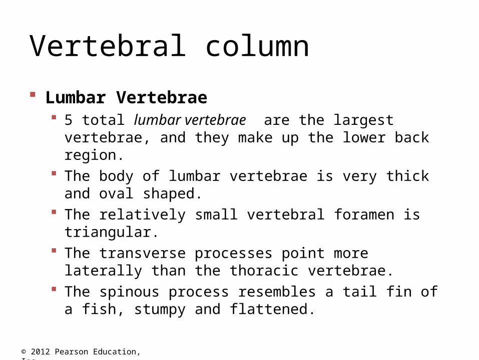
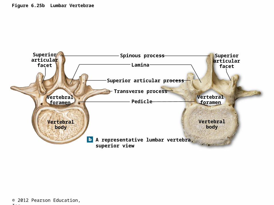
Lumbar Vertebrae 5 total lumbar vertebrae are the largest vertebrae, and they
make up the lower back region. The body of lumbar vertebrae is very thick and oval
shaped. The relatively small vertebral foramen is triangular. The transverse processes point more laterally than the
thoracic vertebrae. The spinous process resembles a tail fin of a fish, stumpy
and flattened.
© 2012 Pearson Education, Inc.
Figure 6.25b Lumbar Vertebrae
A representative lumbar vertebra,superior view
Vertebralbody
Vertebralbody
Vertebralforamen
Vertebralforamen
Superiorarticular
facet
Superiorarticular
facet
Pedicle
Superior articular process
Transverse process
Lamina
Spinous process
© 2012 Pearson Education, Inc.
Vertebral column
© 2012 Pearson Education, Inc.
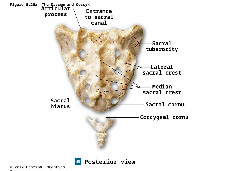
Figure 6.26a The Sacrum and Coccyx
Posterior view
Articularprocess
Entranceto sacral
canal
Sacralhiatus
Sacraltuberosity
Lateralsacral crest
Mediansacral crest
Sacral cornu
Coccygeal cornu
© 2012 Pearson Education, Inc.
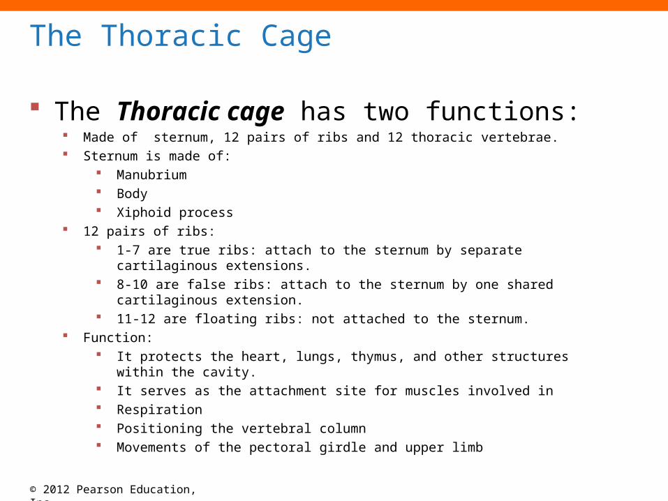
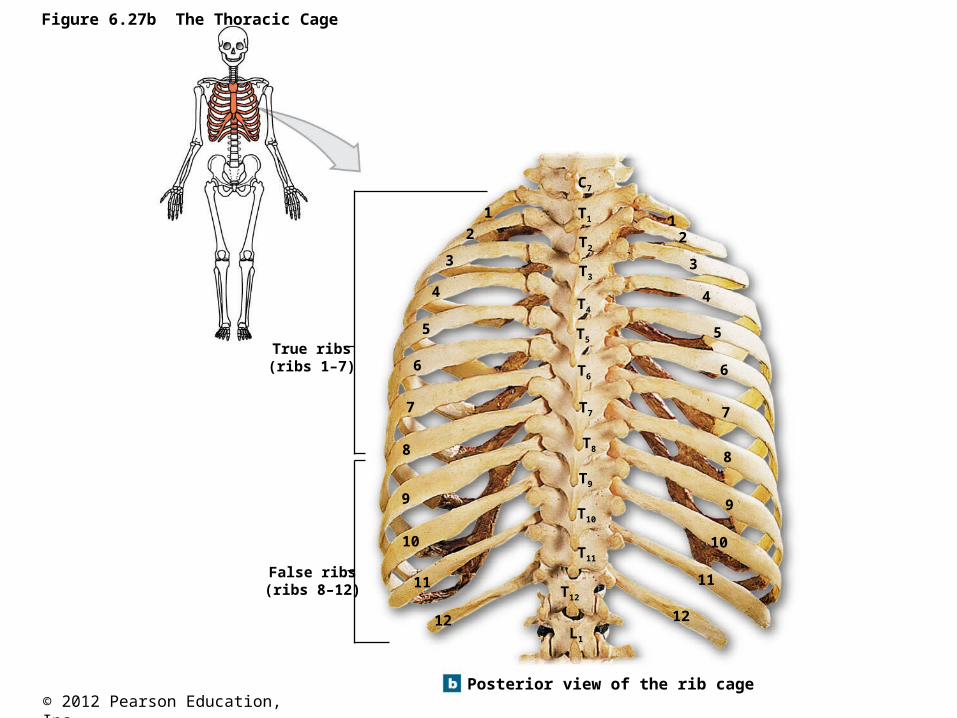
The Thoracic Cage
The Thoracic cage has two functions: Made of sternum, 12 pairs of ribs and 12 thoracic vertebrae. Sternum is made of:
Manubrium Body Xiphoid process
12 pairs of ribs: 1-7 are true ribs: attach to the sternum by separate cartilaginous extensions. 8-10 are false ribs: attach to the sternum by one shared cartilaginous extension. 11-12 are floating ribs: not attached to the sternum.
Function: It protects the heart, lungs, thymus, and other structures within the cavity. It serves as the attachment site for muscles involved in Respiration Positioning the vertebral column Movements of the pectoral girdle and upper limb
© 2012 Pearson Education, Inc.
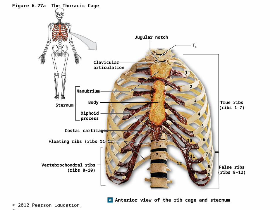
Figure 6.27a The Thoracic Cage
Anterior view of the rib cage and sternum
T11
T12
T1
11
12
10
1
2
3
4
5
6
7
8
9
Jugular notch
Claviculararticulation
Sternum
Manubrium
Body
Xiphoidprocess
Costal cartilages
Floating ribs (ribs 11–12)
Vertebrochondral ribs(ribs 8–10)
True ribs(ribs 1–7)
False ribs(ribs 8–12)
© 2012 Pearson Education, Inc.
Figure 6.27b The Thoracic Cage
Posterior view of the rib cage
True ribs(ribs 1–7)
False ribs(ribs 8–12) T12
T11
T10
T9
T8
T7
T6
T5
T4
T3
T2
T1
C7
1 12 2
3 3
4 4
5 5
6 6
7 7
8 8
9 9
10 10
11 11
12 12L1
© 2012 Pearson Education, Inc.
Figure 6.27c The Thoracic Cage
A superior view of the articulationbetween a thoracic vertebra andthe vertebral end of a left rib
Transversecostal facet
Costal facet
Tubercle of rib
Neck
Angle
Head(capitulum)
Vertebralend