faculdade de engenharia da universidade do porto · 1 departamento de engenharia metalúrgica e de...
TRANSCRIPT

FACULDADE DE ENGENHARIA DA UNIVERSIDADE DO PORTO
Departamento de Engenharia Metalúrgica e Materiais
In vitro biological assessment of silicon substituted hydroxyapatite:
Angiogenesis and Osteogenesis
Ana Catarina Gil Campos
Licenciada em Cardiopneumologia pela Escola Superior de Tecnologias da Saúde do
Porto
Dissertação submetida para satisfação parcial dos
requisitos do grau de mestre
em Engenharia Biomédica
(Área de especialização de Biomateriais)
Dissertação realizada sob a supervisão de
Professor Doutor José Domingos da Silva Santos1,2 e Doutora Cláudia Manuela da
Cunha Ferreira Botelho2,3
1 Departamento de Engenharia Metalúrgica e de Materiais, Faculdade de Engenharia da
Universidade do Porto (FEUP) 2 Laboratório de Biomateriais, Instituto de Engenharia Biomédica (INEB)
3 Life and Health Sciences Research Institute, Universidade do Minho
Porto, Setembro de 2007

Aos meus Pais, à Teresa e ao João
“Por vezes sentimos que aquilo que fazemos não é senão uma gota de água no mar.
Mas o mar seria menor se lhe faltasse uma gota.”
Madre Teresa de Calcutá

In vitro biological assessment of silicon substituted hydroxyapatite: Angiogenesis and Osteogenesis
Resumo
Um substituto ósseo deve não só promover a regeneração óssea, mas também
favorecer o desenvolvimento de uma rede microvascular. A hidroxiapatite modificada com
silício é um material bioactivo e osteocondutor, que permite a proliferação e a
diferenciação celular.
A primeira parte do trabalho consistiu na caracterização físico-química e estrutural
da Hidroxiapatite (HA) e da Hidroxiapatite modificada com Silício (SiHA) como materiais
densos e na forma de revestimentos. O titânio é usado como biomaterial em aplicações
biomédicas. Apesar das suas boas propriedades mecânicas e de resistência à corrosão, o
titânio não tem a capacidade de formar uma ligação ao tecido ósseo, sendo assim este deve
ser revestido com um cerâmico bioactivo, já que estes estimulam a ligação biológica entre
o implante e o tecido ósseo.
Realizaram-se estudos in vitro com culturas de células endoteliais cultivadas em
amostras densas de hidroxiapatite (HA) e de hidroxiapatite modificada com silício (SiHA),
assim como a avaliação dos parâmetros de crescimento celular e da actividade funcional
em determinadas fases do período de cultura, representativas da proliferação/diferenciação
celular. Verificou-se que a incorporação de Si4+ na malha de HA estimulou a adesão das
células endoteliais ao substrato comparativamente com a HA.
O processo de angiogénese pode ser definido como a formação de uma rede
vascular a partir de vasos sanguíneos pré-existentes e o processo de osteogénese como a
formação de novo tecido ósseo. Estes dois processos estão intimamente relacionados, de
modo estrutural, bioquímico e funcional. Sendo assim, após se verificar o efeito positivo
do silício na angiogénese em amostras densas, procedeu-se ao estudo da osteogénese da
hidroxiapatite modificada com silício, mas na forma de revestimentos da liga de titânio
(Ti-6Al-4V). Os resultados obtidos nestes estudos corroboram os resultados descritos na
literatura, os quais demonstram o efeito positivo do silício nas células osteoblásticas.
Estes estudos demonstram que a hidroxiapatite modificada com silício quer na
forma densa, quer na forma de revestimento pode estimular a regeneração do tecido ósseo.
Universidade do Porto, Faculdade de Engenharia, Departamento de Engenharia Metalúrgica e Materiais 3
In vitro biological assessment of silicon substituted hydroxyapatite: Angiogenesis and Osteogenesis
Abstract
A bone graft should promote bone regeneration and stimulate the development of a
vascular net. According to the literatures, the silicon substituted hydroxyapatite is a
bioactive and osteoconductive material that stimulates cellular proliferation and
differentiation.
In the first part of this thesis the silicon substituted hydroxyapatite (SiHA) and
hydroxyapatite (HA) in a dense and coating form were characterized.
Titanium and titanium alloys can be used as biomaterial in several biomedical
applications, although its good mechanical properties and corrosion resistance, titanium is
not a bioactive material, so it should be coated with a bioactive ceramics, that can form a
biological bond between the graft and bone tissue.
In vitro biological studies were performed by seeding endothelial cells in dense
samples of HA and SiHA. The colonized materials were evaluated for cellular growth
parameters and functional activity at different time points of culture period. The Si4+
incorporation in HA lattice induce a positive effect on the development of the endothelial
cells. The number of endothelial cells that adhered to SiHA substrate is higher than the
number of cells present on the surface of HA.
The angiogenesis process can be defined as the formation of vascular net from
preexisting vases and the osteogenic process as the formation of new bone tissue. These
processes are structurally, biochemically and functionally related. It has been shown that
the presence of silicon stimulates the adhesion of endothelial cells and that in accordance
to the literature the osteogenesis in the dense form. So, the second part of this thesis aimed
to performed in vitro studies with human osteoblastic cells cultured on the surface
Titanium alloy coated with HA and SiHA. The results obtained corroborate previous
results described in the literature, which showed the positive effect of silicon on bone cells.
Universidade do Porto, Faculdade de Engenharia, Departamento de Engenharia Metalúrgica e Materiais 4
In vitro biological assessment of silicon substituted hydroxyapatite: Angiogenesis and Osteogenesis
Objectives
The aim of this thesis was to study the behaviour of human umbilical vein
endothelial cells seeded on the HA and SiHA as dense form, and to study the behaviour of
human osteoblastic cells seeded on the HA and SiHA as coating material of titanium alloy,
to understand the importance of the silicon incorporated into HA lattice in the angiogenesis
and osteogenic process.
Universidade do Porto, Faculdade de Engenharia, Departamento de Engenharia Metalúrgica e Materiais 5
In vitro biological assessment of silicon substituted hydroxyapatite: Angiogenesis and Osteogenesis
Acknowledgements
I am very grateful to my supervisor Professor Doutor José Domingos Santos for the
opportunity to work in biomaterial research what allowed me to discover the taste of this
area.
I am deeply grateful to my co-supervisor Doutora Cláudia Botelho for the teachings and
constant support.
My specials thanks to Professora Helena Fernandes who help me in cell work as Dr. Pedro
and Dra. Lurdes of the Laboratório de Farmacologia da Faculdade de Medicina Dentária da
Universidade do Porto.
I would like to acknowledge to everyone at the INEB, especially the members of
Bioceramics and Glasses Group.
My thanks to the Faculdade de Engenharia da Universidade do Porto, Instituto Nacional de
Engenharia Biomédica, Faculdade de Medicina Dentária da Universidade do Porto and
Fundação para a Ciência e Tecnologia.
I am most grateful to my Parents, Teresa and João for all they represent and do for me.
Universidade do Porto, Faculdade de Engenharia, Departamento de Engenharia Metalúrgica e Materiais 6
In vitro biological assessment of silicon substituted hydroxyapatite: Angiogenesis and Osteogenesis
Contents
Chapter 1 – General introduction………………………………………... 11
1. The bone……………………………………………………………….. 12
1.1 Bone composition …………………………………………….. 12
1.2 Bone cells………………………………………………………. 12
1.3 Bone Structure………………………………………………….. 13
1.4 Bone Growth…………………………………………………… 14
1.5 Bone Remodelling……………………………………………… 15
1.6 Bone healing………………………………………… 15
2. Angiogenesis…………………………………………………………… 16
2.1 Vessel growth…………………………………………………... 16
2.2 Endothelial Progenitors………………………………………… 17
2.3 Vascular cell specification……………………………………... 18
2.4 Angiogenesis and arteriogenesis……………………………….. 18
2.5 Collateral growth……………………………………………….. 21
2.6 Leukocytes and angiogenesis…………………………………... 21
2.7 Coagulation and angiogenesis………………………………….. 21
2.8 Vessel regression……………………………………………….. 22
2.9 Types of angiogenesis………………………………………….. 23
2.9.1 Sprouting angiogenesis………………………………… 23
2.9.2 Intussusceptive angiogenesis…………………………... 23
2.10 Vascular endothelial growth factor…………………………… 23
2.11 Matrix Metalloproteinase……………………………………... 25
2.12 Angiogenesis and bone……………………………………….. 26
2.13 Osteogenesis and angiogenesis……………………………….. 26
3. Bone Tissue Engineering………………………………………………. 27
3.1 Biomaterials ............................................................................... 27
3.2 Interaction between bone tissue and bioceramics……………… 28
3.3 Hydroxyapatite……………………………………………….. 29
3.4 Silicon substituted Hydroxyapatite……………………………. 30
3.5 Coating Techniques ………………………………………….. 32
3.6 In vitro biological studies………………………………………. 33
Universidade do Porto, Faculdade de Engenharia, Departamento de Engenharia Metalúrgica e Materiais 7
In vitro biological assessment of silicon substituted hydroxyapatite: Angiogenesis and Osteogenesis
Chapter 2 – Experimental procedures…………………………………… 35
Introduction………………………………………………………………. 36
1. Preparation of HA and SiHA…………………………………………... 37
1.1 HA preparation………………………………………………… 37
1.2 SiHA preparation …………………………………………….. 37
1.3 Dense samples 38
1.4 Milling and sieving…………………………………………….. 38
1.5 Plasma-spray…………………………………………………… 38
2. Physico-chemical and structural characterization of HA and SiHA…… 38
3. In vitro biological studies of human vein endothelial
cells……………………………………………………………………….
39
3.1 Human Umbilical Vein Endothelial Cells …………………….. 39
3.2 Cell proliferation……………………………………………….. 39
3.3 Morphologic Evaluation ………………………………………. 39
4. In vitro biological studies of human osteoblastic cells………………… 40
4.1 Human osteoblastic cells …………………………………….. 40
4.2 Cell proliferation………………………………………………. 41
4.3 Morphologic evaluation……………………………………….. 41
Results……………………………………………………………………. 42
1. Hydroxyapatite and Silicon substituted apatite characterization… 43
2. In vitro biological studies of Human Umbilical Vein Endothelial
Cells ………………………………………………………………
46
3. In vitro biological studies of human osteoblastic cells ………….. 48
Discussion…………………………………………………………………
53
Conclusions……………………………………………………………….
57
References………………………………………………………………...
58
Universidade do Porto, Faculdade de Engenharia, Departamento de Engenharia Metalúrgica e Materiais 8
In vitro biological assessment of silicon substituted hydroxyapatite: Angiogenesis and Osteogenesis
List of figures
Figure 1 – A microscopic image of an osteocyte............................................... 13
Figure 2 - Compact and cancellous bone …………………………………….. 14
Figure 3 – HA structure ………………………………………………………. 30
Figure 4 - SiHA structure……………………………………………….......... 31
Figure 5 – XRD spectrum of phase pure SiHA.……………………………… 43
Figure 6 – FTIR spectrum of phase pure HA………………………………… 44
Figure 7 – FTIR spectrum of phase pure 0.8 wt% SiHA…………………….. 44
Figure 8 – SEM image of HA dense and SiHA dense ……………………… 45
Figure 9 - SEM appearance of the HA plasma-sprayed coated disc and SiHA
plasma-sprayed coated disc (original magnification, x 2000)…………………
45
Figure 10 – Cell viability/proliferation (MTT assay) of endothelial cells
cultured on the surface of dense samples of HA or SiHA.……………………
46
Figure 11 - Morphology of endothelial cells after 1 day and 3 days of culture;
actin staining and Pecam-1 staining..……….....................................................
47
Figure 12 – Morphology of endothelial cells after 1 day of culture; actin
staining of endothelial cells in HA and SiHA respectively and Pecam-1
staining of endothelial cells in HA and SiHA respectively..……………..........
48
Figure 13 - Cell viability/proliferation (MTT assay) of human osteoblastic
bone marrow cells cultured on the surface of Ti alloy coated with HA or
SiHA…………………………………………………………………………...
49
Figure 14 – SEM images of human osteoblastic bone marrow cells grown on
the surface of Ti alloy coated with SiHA, at different periods of the culture…
50
Figure 15 - Confocal laser scanning microscopy of the human osteoblastic
bone marrow cytoskeleton and nuclei, grown on the surface of Ti alloy
coated with SiHA, at different periods of the culture………………………....
51
Universidade do Porto, Faculdade de Engenharia, Departamento de Engenharia Metalúrgica e Materiais 9

In vitro biological assessment of silicon substituted hydroxyapatite: Angiogenesis and Osteogenesis
List of Abbreviations
Ang Angiopoietins
BMPs Bone morphogenetic proteins
CLSM confocal laser scanning microscopy
ECs Endothetial cells
ECM Extracellular matrix
EPCs Endothelial progenitor cells
FGF Fibroblast growth factor
FTIR Fourier transform infrared
HIFs Hypoxia-inducible transcription factors
HSCs Haematopoieitic stem cells
HSPGs Heparan sulfate proteoglycans
HUVECs Human Umbilical Vein Endothelial Cells
Id Inhibitor of differentiation
IMG Intussusceptive microvascular growth
KBr Potassium Bromide
MMPs Matrix metalloproteinases
NO Nitric oxide
PDGF Platelet-derived growth factor
PDGFR Platelet-derived growth factor receptor
PECAM-1 Platelet/endothelial cell adhesion molecule-1
PF Platelet factor
PIGF Placental growth factor
RTKs Receptor tyrosine kinases
SEM Scanning electron microscopy
SMCs Smooth muscle cells
TGF Transforming growth factor
TIMPs Tissue inhibitors of metalloproteinases
TSPs Thrombospondins
VEGF Vascular endothelial growth factor
VEGFRs VEGF signalling receptors
XRD X-ray powder diffraction
TCP Tricalcium phosphate
Universidade do Porto, Faculdade de Engenharia, Departamento de Engenharia Metalúrgica e Materiais 10

In vitro biological assessment of silicon substituted hydroxyapatite: Angiogenesis and Osteogenesis
CHAPTER 1
GENERAL INTRODUCTION
Universidade do Porto, Faculdade de Engenharia, Departamento de Engenharia Metalúrgica e Materiais 11
In vitro biological assessment of silicon substituted hydroxyapatite: Angiogenesis and Osteogenesis
CHAPTER 1
1. The bone
Bone is a dynamic connective tissue characterized by its hardness, growth
mechanisms and ability to regenerate. It has the functions of support and protection the
internal organs and provide attachment for muscles, facilitating the locomotion process.
Besides this, bone offers protection for blood-forming marrow and it is the reservoir of
mineral ions such as calcium and phosphorous [1].
1.1 Bone composition
Bone is a composite material consisting of an organic matrix that is strengthened by
deposits of calcium salts. Type I collagen constitutes approximately 95% of the organic
matrix. The remaining 5% is composed of numerous noncollageneous proteins such as:
proteoglycans, osteonectin, osteocalcin, osteopontin, sialoprotein, glycoproteins, enzymes
and cytokines. Bone apatite is a calcium and hydroxide deficient apatite containing
numerous ionic substitutions, such as carbonate, magnesium, potassium, fluoride, sodium,
phosphate and others [2-4].
1.2 Bone cells
The cells that are responsible for the structural and functional properties of bone
are: osteoprogenitors cells, osteoblasts, osteoclasts and osteocytes.
Osteogenics cells (osteoprogenitors)
This type of cells is mainly in the deepest layer of the periosteum and the
endosteum. They have a high mitotic potential and are recruited to repair bone defects [5].
Osteoblasts
Osteoblasts are fully differentiated cells responsible for the bone matrix production.
They are typical protein-producing cell, witch secrete type I collagen, noncolageneous
proteins of bone matrix and regulate the mineralization of bone matrix. Osteoblasts are
derived from mesenchymal stem cells. Factors such as bone morphogenetic proteins
Universidade do Porto, Faculdade de Engenharia, Departamento de Engenharia Metalúrgica e Materiais 12
In vitro biological assessment of silicon substituted hydroxyapatite: Angiogenesis and Osteogenesis
(BMPs) and transcription factors (Cbfa 1) mediate and regulate the induction of
mesenchymal stem cells into osteoblastic cells [5,6].
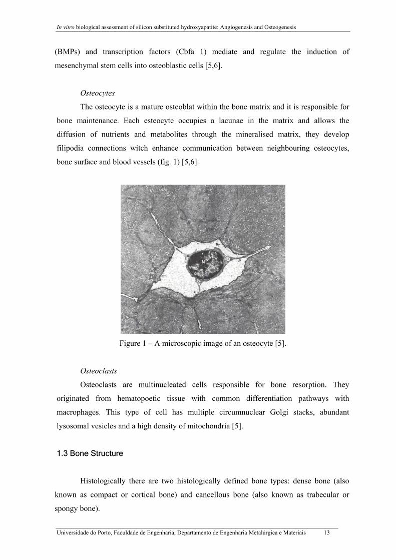
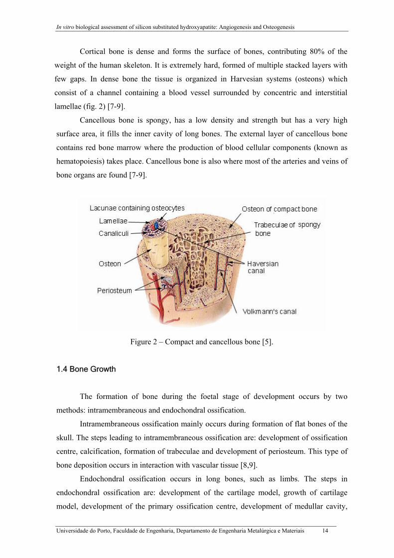
Osteocytes
The osteocyte is a mature osteoblat within the bone matrix and it is responsible for
bone maintenance. Each esteocyte occupies a lacunae in the matrix and allows the
diffusion of nutrients and metabolites through the mineralised matrix, they develop
filipodia connections witch enhance communication between neighbouring osteocytes,
bone surface and blood vessels (fig. 1) [5,6].
Figure 1 – A microscopic image of an osteocyte [5].
Osteoclasts
Osteoclasts are multinucleated cells responsible for bone resorption. They
originated from hematopoetic tissue with common differentiation pathways with
macrophages. This type of cell has multiple circumnuclear Golgi stacks, abundant
lysosomal vesicles and a high density of mitochondria [5].
1.3 Bone Structure
Histologically there are two histologically defined bone types: dense bone (also
known as compact or cortical bone) and cancellous bone (also known as trabecular or
spongy bone).
Universidade do Porto, Faculdade de Engenharia, Departamento de Engenharia Metalúrgica e Materiais 13
In vitro biological assessment of silicon substituted hydroxyapatite: Angiogenesis and Osteogenesis
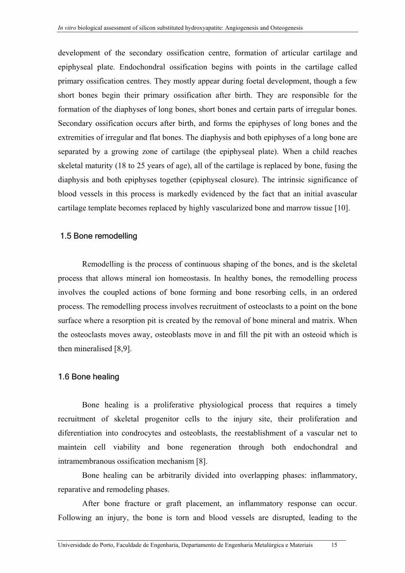
Cortical bone is dense and forms the surface of bones, contributing 80% of the
weight of the human skeleton. It is extremely hard, formed of multiple stacked layers with
few gaps. In dense bone the tissue is organized in Harvesian systems (osteons) which
consist of a channel containing a blood vessel surrounded by concentric and interstitial
lamellae (fig. 2) [7-9].
Cancellous bone is spongy, has a low density and strength but has a very high
surface area, it fills the inner cavity of long bones. The external layer of cancellous bone
contains red bone marrow where the production of blood cellular components (known as
hematopoiesis) takes place. Cancellous bone is also where most of the arteries and veins of
bone organs are found [7-9].
Figure 2 – Compact and cancellous bone [5].
1.4 Bone Growth
The formation of bone during the foetal stage of development occurs by two
methods: intramembraneous and endochondral ossification.
Intramembraneous ossification mainly occurs during formation of flat bones of the
skull. The steps leading to intramembraneous ossification are: development of ossification
centre, calcification, formation of trabeculae and development of periosteum. This type of
bone deposition occurs in interaction with vascular tissue [8,9].
Endochondral ossification occurs in long bones, such as limbs. The steps in
endochondral ossification are: development of the cartilage model, growth of cartilage
model, development of the primary ossification centre, development of medullar cavity,
Universidade do Porto, Faculdade de Engenharia, Departamento de Engenharia Metalúrgica e Materiais 14
In vitro biological assessment of silicon substituted hydroxyapatite: Angiogenesis and Osteogenesis
development of the secondary ossification centre, formation of articular cartilage and
epiphyseal plate. Endochondral ossification begins with points in the cartilage called
primary ossification centres. They mostly appear during foetal development, though a few
short bones begin their primary ossification after birth. They are responsible for the
formation of the diaphyses of long bones, short bones and certain parts of irregular bones.
Secondary ossification occurs after birth, and forms the epiphyses of long bones and the
extremities of irregular and flat bones. The diaphysis and both epiphyses of a long bone are
separated by a growing zone of cartilage (the epiphyseal plate). When a child reaches
skeletal maturity (18 to 25 years of age), all of the cartilage is replaced by bone, fusing the
diaphysis and both epiphyses together (epiphyseal closure). The intrinsic significance of
blood vessels in this process is markedly evidenced by the fact that an initial avascular
cartilage template becomes replaced by highly vascularized bone and marrow tissue [10].
1.5 Bone remodelling
Remodelling is the process of continuous shaping of the bones, and is the skeletal
process that allows mineral ion homeostasis. In healthy bones, the remodelling process
involves the coupled actions of bone forming and bone resorbing cells, in an ordered
process. The remodelling process involves recruitment of osteoclasts to a point on the bone
surface where a resorption pit is created by the removal of bone mineral and matrix. When
the osteoclasts moves away, osteoblasts move in and fill the pit with an osteoid which is
then mineralised [8,9].
1.6 Bone healing
Bone healing is a proliferative physiological process that requires a timely
recruitment of skeletal progenitor cells to the injury site, their proliferation and
diferentiation into condrocytes and osteoblasts, the reestablishment of a vascular net to
maintein cell viability and bone regeneration through both endochondral and
intramembranous ossification mechanism [8].
Bone healing can be arbitrarily divided into overlapping phases: inflammatory,
reparative and remodeling phases.
After bone fracture or graft placement, an inflammatory response can occur.
Following an injury, the bone is torn and blood vessels are disrupted, leading to the
Universidade do Porto, Faculdade de Engenharia, Departamento de Engenharia Metalúrgica e Materiais 15
In vitro biological assessment of silicon substituted hydroxyapatite: Angiogenesis and Osteogenesis
formation of a haematoma. Close to the fracture line, necrotic marrow and dead bone
accumulate and elicit an inflammatory response.
In the reparative phase, mesenchymal progenitors are recruited and differentiate
into chondrocytes or osteoblasts. The early reparative phase involves formation of (fibro)-
cartilage throughout the callus, particularly in domains distant from ingrowing capillaries
(internal callus), and of immature woven (spongy) bone predominantly in subperiosteal
areas with profound angiogenesis (external callus). Through endochondral ossification,
(fibro)-cartilage is replaced with mineralized bone, forming a hard callus bridging the
fracture gap.
In the remodeling phase, osteoclasts and osteoblasts continue to remodel the large
callus, ultimately restoring the former shape, strength and functioning of the bone [9-11].
2. Angiogenesis
Angiogenesis is a physiological process that leads to the formation of new blood
vessels from pre-existing vessels, by the migration and proliferation of endothelial cells
[12-14]. This process occurs during growth and development, wound healing, and in the
female reproductive system. Angiogenesis also occurs in different pathological processes
such as cancer (Carmeliet and Jain, 2000; Patan, 2004), rheumatoid arthritis, diabetes and
cardiovascular diseases. This process is complex and the mechanism behind angiogenesis
is not fully understood, although several stimulating factors are known.
2.1 Vessel growth
Small blood vessels are only composed by endothelial cells (ECs), while larger
vessels are composed by mural cells, pericytes in medium-sized and smooth muscle cells
(SMCs). Endothelial cells are oblong shaped cells that cover the lumen of all blood vessels
as a single epithelial cell layer, and they are derived from angioblasts and hemangioblasts
[15]. These cells play a major role in vascular biology under normal or pathological
conditions (Wiel et al, 2005), including the control of vasoconstriction and vasodilatation
(Cosentino and Volpe, 2005), thrombosis and fibrinolysis (Wiel et al, 2005; Chen and
Lopez, 2005), angiogenesis (Gerhardt and Betsholtz, 2005; Szekanecz and Koch, 2005),
leukocyte adhesion/trafficking and inflammatory processes (Cook-Mills and Deem, 2005;
Universidade do Porto, Faculdade de Engenharia, Departamento de Engenharia Metalúrgica e Materiais 16
In vitro biological assessment of silicon substituted hydroxyapatite: Angiogenesis and Osteogenesis
Aird, 2005; Szekanecz and Koch, 2005). Immature circulating endothelial cells derived
from the bone marrow are being referred to as endothelial progenitor cells (Khan et al,
2005).
Vessels can growth by different manners. Angiogenesis denotes the formation of
new blood vessels from pre-existing ones, while vasculogenesis is the term used for the
formation of new blood vessels when there are no pre-existing ones by endothelial
progenitors, during embryogenesis. Vasculogenesis is related to in situ differentiation and
growth of blood vessels from mesodermal derived hemangioblasts. Angiogenesis comprise
two different mechanisms: endothelial sprouting and intussusceptive microvascular growth
(IMG). Angiogenesis and arteriogenesis refer to the sprouting and subsequent stabilization
of these sprouts by mural cells, and collateral growth denotes the expansive growth of pre-
existing vessels, forming collateral bridges between arterial networks [14-16].
Lymphangiogenesis refers to the growth and formation of new lymphatic vessels, which
occurs in a normal development of tissues and also in pathological process (Al-Rawi et al,
2005).
2.2 Endothelial Progenitors
The endothelial progenitor cells (EPCs) contribute to vessel growth in embryo,
ischemic conditions, malignant or inflamed tissues in adults [16]. Haematopoietic and
other bone-marrow–derived stem cells might be recruited in the context of ischemia to
induce neovessel formation [17]. The EPCs have been investigated as therapeutic agents in
supply-side angiogenesis under pathological and physiological conditions [18]. ECs can be
differentiated from angioblasts in the embryo and from EPCs, mesoangioblasts,
multipotent adult progenitor cells, or side population cells in the adult bone marrow
[17,19]. EPCs can also contribute to vessel growth by releasing angiogenic growth factors,
like the vascular endothelial growth factor (VEGF), placental growth factor (PIGF),
angiopoietin (Ang)-1, inhibitor of differentiation (Id) proteins, and different cytokines [20].
EPCs, haematopoietic progenitors and their descendents share common markers and are
affected by common signals that will influence each other [21-23]. Identification of the
signals that recruit or differentiate these progenitors offers opportunities to manipulate
their contributions to vascular growth. The functional contribution of EPCs and
haematopoietic stem cells (HSCs) to pathological angiogenesis still undefined [15, 22-25].
Universidade do Porto, Faculdade de Engenharia, Departamento de Engenharia Metalúrgica e Materiais 17
In vitro biological assessment of silicon substituted hydroxyapatite: Angiogenesis and Osteogenesis
2.3 Vascular cell specification
Arteries and veins are distinct vessels anatomically and physiologically. They differ
in blood pressure, thickness of their smooth muscle cells (SMC) coat and, ECs and SMCs
have a distinct identity and origin [15]. Recent genetic studies show the signals that control
the arterial and venous identities of ECs. The Notch pathway, with its ligants and
receptors, promotes arterial destiny of ECs by repressing venous differentiation. Sonic
Hedgehog and VEGF act upstream, whereas Gridlock probably acts downstream of Notch
to determine arterial destiny, even before the onset of flow [26-28]. ECs have a phenotypic
plasticity because they can differentiate into either arterial or venous ECs in embryonic
development, in neonatal retina and even in adult heart. It means that a selective use of
arterial or venous ECs or their precursors may be useful for therapeutic vasculogenesis
[29].
Blood vessels in various tissues have specialized functions and ECs have distinct
properties, probably as many as the organs in the body. The expression and activity of
general angiogenic factors such as VEGF or Ang-1 varies greatly in different tissues.
Further more, organ-specific angiogenic factors determine the angiogenic switch, but in a
restricted manner in particular organs [30,31].
EPCs differentiate to arterial and venous ECs, which assemble in a primitive
capillary plexus. Vessels then sprout and become stabilized by SMCs, differentiating from
their progenitors. HSCs contribute to angiogenesis directly and indirectly, by
differentiating to leukocytes or platelets. A demarcation of arterial and venous boundaries
are required, as well as the establishment of vascular polarity. Ephrin-B2, an Eph family
transmembrane ligand, marks arterial ECs and SMCs, whereas EphB4, a receptor for
Ephrin-B2, marks only veins. Ephrin-B2-Eph4 participates in the formation of arterio-
venous anastomoses by arresting EC migration at the arterial-venous interface [32,33,34].
2.4 Angiogenesis and arteriogenesis
The regulation of angiogenesis by hypoxia is an important component of
homeostatic mechanisms that link vascular oxygen supply to metabolic demand. Initially,
cells are oxygenated by simple diffusion of oxygen, but when tissues grow beyond the
limit of oxygen diffusion, hypoxia triggers vessel growth by signalling through hypoxia-
inducible transcription factors (HIFs) [35].
Universidade do Porto, Faculdade de Engenharia, Departamento de Engenharia Metalúrgica e Materiais 18
In vitro biological assessment of silicon substituted hydroxyapatite: Angiogenesis and Osteogenesis
ECs built resistant channels and efficiently distribute blood to the various parts of
the body. They have long half-lives, several years, but when triggered are capable of
rapidly sending out sprouts in a coordinated and directional manner. Cells within the vessel
wall communicate with each other and with cells inside and outside of vessel lumen, they
sense changes in blood flow and pressure, and dynamically interact with the internal
cytoskeleton and surrounding ECM. When ECs migrate during vessel sprouting, these
contacts are transiently dissolved but later re-established, once ECs assemble a new sprout
[15].
Cellular interactions with the extracelular matrix (ECM) contribute to the
biochemical processes that regulate angiogenesis. The ECM provides necessary contacts
between ECs and the surrounding tissue, prevents vessels from collapsing, and regulates
the formation of new vessel sprouts. In quiescent vessels, the vascular cells are encased by
a basement membrane of collagen IV, laminin and other components, and pericytes and
ECs are also embedded in the same basement membrane. An interstitial matrix of collagen
I and elastin between vascular cells provides visco-elasticity and strength to the vessel
wall. When vascular cells migrate to form new sprouts, the matrix network is
proteolytically broken down and its composition is altered. The EC and SMC are induced
to migrate because proteinases expose new cryptic epitomes in ECM proteins (such as in
collagen IV) or change their structure (fibrillar versus monomer collagen). Further more, a
provisional matrix of fibronectin, fibrin and other components provides a support scaffold,
guiding ECs to their targets [36].
The cell-surface receptors of specific ECM molecules known as integrins are
crucial for vascular cells to build new vessels. Integrins are involved on the regulation of
proteolytic enzymes activity which degrades the basement membrane (the initial barrier to
surrounding tissue). Integrins are essential for cell migration and invasion, not only
because they directly mediate adhesion to the extracelular matrix, but also because they
regulate intracellular signalling pathways that control cytoskeleton organization, respond to
intracellular cues and modify the way they interact with the extracelular environment. The
binding to ligands in the extracelular matrix initiates several pro-survival mechanisms to
prevent apoptosis [37]. The αvβ3 and αvβ5 integrins have been considered to positively
regulate the angiogenic switch. However, genetic deletion studies suggest that vascular
integrins inhibit angiogenesis by suppressing VEGF and Flk-1 mediated EC survival, by
trans-dominantly blocking other integrins or by mediating the antiangiogenic activity of
Universidade do Porto, Faculdade de Engenharia, Departamento de Engenharia Metalúrgica e Materiais 19
In vitro biological assessment of silicon substituted hydroxyapatite: Angiogenesis and Osteogenesis
thrombospondins (TSPs) and other angiogenesis inhibitors (such as tumstatin, endostatin,
angiostatin and PEX) [38].
Remodelling of the ECM during vessel sprouting requires breakdown by
proteinases, including plasminogen activators, matrix metalloproteinases (MMPs and
tissue inhibitors of metalloproteinases (TIMPs)), heparinases, chymases, tryptases and
cathepsins [39-41]. Proteinases also facilitate EC sprouting by liberating matrix-bound
angiogenic activators (basic fibroblast growth factor (FGF), VEGF and transforming
growth factor (TGF) -β) and proteolytically activating angiogenic chemokines. In
proteolytic remodelling of the ECM, insufficient breakdown prevents vascular cells from
leaving their original position, but excessive breakdown removes critical support and
guidance cues for migrating ECs and, in fact, inhibits angiogenesis. Proteinases can also
have a role in the resolution of angiogenesis, as they liberate matrix-bound inhibitors (TSP-
1, canstatin, tumstatin, endostatin and platelet factor (PF)-4) and inactive angiogenic
cytokines [41,42].
Establishment of a functional vascular network further requires that nascent vessels
mature into durable vessels. Nascent vessels initially only consist of ECs and vessel
maturation requires a mix of angio- and arteriogenic factors for a sufficient duration, so
that ECs can tighten up and become covered by mural cells and ECM. Flow is a critical
determinant of vessel maintenance and durability. When insufficient angio- and
arteriogenic factors are present and angiogenesis inhibitors are present, EC channels
remain naked, leaky and fragile, are easily ruptured and bleed, what reduce flow and result
in vessel regression. The association of pericytes and SMCs with newly formed vessels
regulates EC proliferation, survival, migration, differentiation, vascular branching, blood
flow and vascular permeability. Platelet-derived growth factor (PDGF)-BB and its
receptor, PDGF-β, have essential roles in the stabilization of nascent blood vessels by
recruiting PDGF-β-positive mesenchymal progenitors [43,44].
Tie-2 receptor is involved in vessel maintenance, growth and stabilization, which
binds to angiopoietins (Ang-1 and Ang-2) [45,46]. Members of the TGF-β superfamily
contribute to the resolution and maturation phases of angiogenesis, but in a pleiotropic
manner [47].
Universidade do Porto, Faculdade de Engenharia, Departamento de Engenharia Metalúrgica e Materiais 20
In vitro biological assessment of silicon substituted hydroxyapatite: Angiogenesis and Osteogenesis
2.5 Collateral growth
When an artery is occluded its vascular territory becomes ischemic. Arterial
systems are often interconnected by pre-existing collateral vessels that can enlarge and
save the ischemic region [48]. The mechanisms of angiogenesis and collateral growth
differ significantly, due to the large pressure difference between the perfusion territories. In
collateral growth, the increased shear stress activates ECs, which then recruit monocytes.
These cells produce growth factors and proteinases, which enables SMCs to migrate and
divide [49, 50]. Cytokines that attract monocytes or prolong their life span, enhance
collateral growth, whereas anti-inflammatory cytokines are inhibitory [51]. PIGF also
enhances collateral growth, not only because it recruits monocytes, but also because it
stimulates EC and SMC growth [52]. Delivery of acidic FGF, FGF-4 or basic FGF
(together with PDGF-BB) stimulates collateral growth, in part by upregulating PDGFR
expression [53].
2.6 Leukocytes and angiogenesis
Inflammation and immune driven angiogenesis affect numerous disorders (cancer,
vascular malformations, obesity, psoriasis, diabetic retinopathy, arthritis, synovitis,
osteomyelitis, osteophyte formation, endometriosis, osteoporosis, Alzheimer disease,
atherosclerosis, and others) in part because most leukocyte subtypes produce a myriad of
angiogenic factors such as VEGF, PIGF, PDGF, basic FGF, Ang-2, epidermal growth
factor, TGF-β1, MCP-1 and various interleukins and proteinases (tryptase, chymase,
MMPs, heparanase and uPA) [54,55]. Leukocytes affect many angiogenic processes.
Monocytes are a source of EPCs and can differentiate into endothelial-like cells [56].
Leucocytes and vascular cells influence each other in other ways. Angiogenic factors
amplify the inflammatory process by recruiting leukocytes and affecting their function.
2.7 Coagulation and angiogenesis
Homeostasis and angiogenesis are closely linked. Fibrin-rich clot formation and
platelet aggregation precede infiltration of blood vessels into a wound. Platelets release
large stores of angiogenic factors such as VEGF, PDGF, TGF-β, IL-6, thrombin and
sphingosine-1-1phosphate, which stimulates the growth and stability of nascent vessels by
Universidade do Porto, Faculdade de Engenharia, Departamento de Engenharia Metalúrgica e Materiais 21
In vitro biological assessment of silicon substituted hydroxyapatite: Angiogenesis and Osteogenesis
tightening their junctions and recruit mural cells. Platelets also contain antiangiogenic
factors (TSP-1, PF-4 and others) that may have a role in the resolution of angiogenesis
once the wound has healed [57,58].
2.8 Vessel regression
Vessel regression occurs when the nascent vasculature consists of too many vessels,
and also constitutes the basis of many antiangiogenic therapeutic strategies. Abnormal
vessel regression also contributes to the pathogenesis of numerous disorders. Removal of
angiogenic stimulus causes vessels to regress, as in tumours and in heart, especially when
vessels have only been recently assembled and are still immature. When angiogenic
stimulus are provided for a sufficient length of time, new vessels mature and persist for
months, even after the angiogenic stimulus are withdrawn. Flow may have an important
role in determining whether neovessels regress or persist, by affecting several factors
(including MMPs, PDGF, basic FGF, integrins and nitric oxide (NO)). Flow stimulates
hyperplasia of ECs and SMCs, and induces the reorganization of endothelial junctions and
deposition of ECM, all of which contribute to vessel maturation. Thus, insufficient
perfusion may lead to regression, whereas sufficient perfusion promotes vessel persistence.
Pericytes also determine the susceptibility of vessels to regression, because once vessels
are surrounded by pericytes, they become resistant oxygen-induced regression. In contrast,
disruption of endothelial-pericyte associations results in regression of vessels [43,44,47].
Angiogenesis inhibitors also contribute to vessel regression. TSP-1 inhibits
angiogenesis through direct effects on ECs and indirect effects on growth factor
mobilization or activation. Upregulation of endogenous TSP-1 and TSP-2 contributes to
the resolution of angiogenesis and vessel stabilization after ischemia, and overexpression
of TSP-1 or TSP-2 in cancer cells results in reduced tumour vascularisation and tumour
growth. Macrophages contribute to vessel regression by releasing TGF-β1. Inhibitory PAS
dominate protein functions as a dominant-negative regulator of hypoxia-induced
angiogenesis to maintain an avascular phenotype in certain tissues. Additional inhibitors
include chemokines binding CXCR3, soluble receptors (Flt-1 and Tie-2), clotting
antagonists and others. It is being discovers inhibitors including cleavage products of
matrix components, proteinases, enzymes or plasma proteins [43,44].
Universidade do Porto, Faculdade de Engenharia, Departamento de Engenharia Metalúrgica e Materiais 22
In vitro biological assessment of silicon substituted hydroxyapatite: Angiogenesis and Osteogenesis
2.9 Types of angiogenesis
There are tow types of angiogenesis: sprouting angiogenesis and intussusceptive
angiogenesis [41,42].
2.9.1 Sprouting angiogenesis
In the sprouting angiogenesis the angiogenic growth factors activate receptors
present on endothelial cells in pre-existing venous blood vessels. The activated endothelial
cells begin to release enzymes called proteases that degrade the basement membrane in
order to allow endothelial cells to escape from the original vessel walls. After that the
endothelial cells proliferate into the surrounding matrix and form solid sprouts connecting
neighbouring vessels. As sprouts extend toward the source of the angiogenic stimulus,
endothelial cells migrate in tandem, using integrins. These sprouts then form loops to
become a full-fledged vessel lumen as cell migrate to the angiogenesis site. Sprouting
occurs at a rate of several millimetres per day, and enables new vessels to grow across gaps
in the vasculature [59].
2.9.2 Intussusceptive angiogenesis
In the intussusceptive angiogenesis the capillary wall extends into the lumen to split
a single vessel in two. The two opposing capillary walls establish a zone of contact, the
endothelial cell junctions are reorganized and the vessel bilayer is perforated to allow
growth factors and cells to penetrate into the lumen. A core is formed between the two new
vessels at the zone of contact that is filled with pericytes and myofibroblasts. These cells
begin laying collagen fibres into the core to provide an extracelular matrix for vessel lumen
growth. Finally, the core is fleshed out with no alterations in basic structure.
Intussusceptive angiogenesis allows a vast increase in capillaries number of without a
corresponding increase in endothelial cells number [59].
2.10 Vascular endothelial growth factor
Vascular endothelial growth factor (VEGF) is an important signalling protein
involved in both vasculogenesis and angiogenesis. It is a key regulator of physiological
Universidade do Porto, Faculdade de Engenharia, Departamento de Engenharia Metalúrgica e Materiais 23
In vitro biological assessment of silicon substituted hydroxyapatite: Angiogenesis and Osteogenesis
angiogenesis during embryogenesis, skeletal growth and reproductive functions. VEGF
activity is restricted mainly to cells of the vascular endothelium, although it has effects on
a limited number of other cell types (e.g. stimulation monocyte/macrophage migration). In
vitro VEGF has been shown to stimulate endothelial cell mitogenesis and cell migration.
VEGF has also been implicated in pathological angiogenesis associated with tumours,
intraocular neovascular disorders and other conditions. Upregulation of VEGF is a major
component of the physiological response to exercise and its role in angiogenesis is
suspected to be a possible treatment in vascular injuries. In vitro studies demonstrate that
VEGF is a potent stimulator of angiogenesis because in the presence of this growth factor,
plated endothelial cells will proliferate and migrate, eventually forming tube structures
resemble capillaries [60-64].
VEGF covers a number of proteins that result from alternative splicing of mRNA
from a single, 8 exon, VEGF gene. The different VEGF splice variants are referred to the
number of amino acids they contain (in human: VEGF121, VEGF145, VEGF165, VEGF189,
and VEGF206). These proteins differ by the presence or absence of short C-terminal
domains encoded by exons 6A, 6B and 7 of the VEGF gene. These domains have
important functional consequences for the VEGF splice variants as they mediate
interactions with heparan sulfate proteoglycans (HSPGs) and neuropilin co-receptors on
the cell surface, enhancing their ability to bind and activate the VEGF signalling receptors
(VEGFRs) [60-64].
The VEGF splice variants are released from cells as glycosylated disulfide-bonded
homodimers. Structurally VEGF belongs to the PDGF family of cystine-knot growth
factors. Subsequently, several closely-related proteins were discovered (Placenta growth
factor (PIGF), VEGF-B, VEGF-C and VEGF-D) which together comprises the VEGF sub-
family of growth factors. VEGF is sometimes referred to as VEGF-A to differentiate it
from these related growth factors [65].
All members of the VEGF family stimulate cellular responses by binding to
tyrosine kinase receptors (the VEGFRs) on the cell surface, causing them to dimerize and
become activated through transphosphorylation. The VEGF receptors have an extracelular
portion consisting of 7 immunoglobulin-like domains, a single transmembrane spanning
region and an intracellular portion containing a slit tyrosine-kinase domain. VEGF-A binds
to VEGFR-1 (Flt-1) and VEGFR-2 (KDR/Flk-1). VEGFR-2 appears to mediate almost all
of the known cellular responses to VEGF [66-69]. VEGF causes a massive signalling
cascade in endothelial cells. Binding to VEGFR-2 starts a tyrosine kinase signalling
Universidade do Porto, Faculdade de Engenharia, Departamento de Engenharia Metalúrgica e Materiais 24
In vitro biological assessment of silicon substituted hydroxyapatite: Angiogenesis and Osteogenesis
cascade that stimulates the production of factors that variously stimulate vessel
permeability (eNOS, production NO), proliferation/survival (bFGF), migration
(ICAMs/VCAMs/MMPs) and finally differentiation into mature blood vessels. The
increased flow also causes a large increase in the mRNA production of VEGF receptors 1
and 2 [60]. The function of VEGFR-1 is less well defined, although to modulate VEGFR-2
signalling. Another function of VEGFR-1 is to act as a dummy/decoy receptor,
sequestering VEGF from VEGFR-2 binding. A third receptor has been discovered
(VEGFR-3), however, VEGF-A is not a ligand for this receptor. VEGFR-3 mediates
lymphangiogenesis in response to VEGF-C and VEGF-D [66-69].
VEGF production can be induced in cells that are not receiving enough oxygen.
When a cell is deficient in oxygen, it produces HIF, Hypoxia Inducible Factor, a
transcription factor. HIF stimulates the release of VEGF, among other functions.
Circulating VEGF then binds to VEGF receptors on endothelial cells, triggering a Tyrosine
Kinase Pathway leading to angiogenesis [70,71].
VEGF has also effects on bone marrow-derived cells. It promotes monocyte
chemotaxis and induces colony formation by mature subsets of granulocyte-macrophage
progenitor cells. [72,73]. VEGF delivery to adult mice inhibits dendritic cell development
and increases production of B cells and generation of immature myeloid cells [74,75].
VEGF is known also as vascular permeability factor, based on its ability to induce vascular
leakage. It induces an increase in hydraulic conductivity of isolated microvessels, and this
effect is mediated by increased calcium influx [76-78].
2.11 Matrix Metalloproteinase
The matrix metalloproteinase (MMP) help to degrade the proteins that keep the
vessel walls solid. This proteolysis allows the endothelial cells to escape into the interstitial
matrix as seen in sprouting angiogenesis. Inhibition of MMPs prevents the formation of
new capillaries, and these enzymes are highly regulated during the vessel formation
process because wanton destruction of the extracellular matrix would destroy the integrity
of the microvasculature [60, 79].
Universidade do Porto, Faculdade de Engenharia, Departamento de Engenharia Metalúrgica e Materiais 25
In vitro biological assessment of silicon substituted hydroxyapatite: Angiogenesis and Osteogenesis
2.12 Angiogenesis and bone
The VEGF have the ability to induce neovascularisation. Studies had show that
neutralizing VEGF receptor decreased angiogenesis, bone formation, and callus
mineralization in femoral fractures. Exogenous VEGF enhanced blood vessel formation,
ossification, new bone (callus) maturation in mouse femur factures, and promoted bone
bridging of a rabbit radius segmental gap defect [80].
Fracture of bone disrupts its circulation and leads to necrosis and hypoxia of
adjacent bone. Under normal circumstances, fractured bone undergoes the orderly
regeneration of its component tissues with complete restoration of mechanical properties.
Reestablishment of the circulation is an early event in fracture healing. Bone repair is a
multistep process involving migration, proliferation, and activation of several cell types.
Expression of particular growth factors, such as FGFs, PDGFs, TGF-βs, VEGF, and bone
morphogenetic proteins (BMPs), during the course of healing suggests a possible role for
these secreted factors in bone repair [81,82]. VEGF is expressed in the fracture callus in
animal models in much the same temporal and spatial pattern as during long bone
development. Although VEGF can control hypertrophic cartilage structure and vascularity
within the development growth plate, the role of VEGF in bone repair has not yet been
determined [80, 83-85]. The fracture callus contains many factors that could promote bone
healing by coordinating angiogenesis with bone homeostasis [86].
2.13 Osteogenesis and angiogenesis
Bone formation is dependent on the signalling of BMPs, molecules initiating
vascularisation (e.g. VEGF) and osteogenic precursor cells capable of responding to these
cues and forming bone tissue [87]. Tissue development and regeneration are regulated by
interplay among various tissue inductive growth factors, formation of an appropriate
vascular bed to support the metabolic needs of the forming tissue mass, and a cell
population capable of responding to the chemical cues and creating the new tissue. BMPs
are responsible for initiating cartilage and bone progenitor cell differentiation and
sequencing new bone formation through endochondral ossification [88,89]. Angiogenesis
is also involved in the initiation of fracture healing and promotion of endochondral and
intramembranous ossification in bone growth. These factors must act on a population of
cells capable of responding to local factors and forming bone tissue. Multipotent stem cells
Universidade do Porto, Faculdade de Engenharia, Departamento de Engenharia Metalúrgica e Materiais 26
In vitro biological assessment of silicon substituted hydroxyapatite: Angiogenesis and Osteogenesis
originating from the bone marrow stroma, or bone marrow stromal cells (BMSCs), are a
particularly attractive source for osteogenic precursors for bone tissue engineering, because
they can be easily harvested and expanded in vitro and induced to differentiate into bone-
forming cells [87,90].
3. Bone Tissue Engineering
Biomaterials can be defined as a natural modified or synthetic material that
interacts with the biological system to improve, cure or substitute any tissue, organ or
function of the body (Williams, 1999) [2]. Biomaterials should be biocompatible, absence
of harmful or toxic effect for the organism and have biomechanics properties to support the
dynamic and static requests that will be subject during its useful life [91].
Medical problems can emerge from bone trauma, diseases and ageing. In order to
solve these problems, autografts (autogenous grafts), allografts (tissue harvested from a
different human patient) and xenografts (tissue harvested from an animal) have been used
in bone surgery [91]. The main advantage of an autograft is related to its osteogenic,
osteoinductive and osteoconductive properties. This graft contains cartilage matrix
minerals, osteogenic proteins and precursor cells [91]. Allografts can lead to the
transmission of viral diseases as HIV, hepatitis B and hepatitis C, and can induce
immunological reactions. Although, these risks can be diminished through sterilization
with gamma radiation, its structure can be affected and the bone graft may lose its
osteoinductive and osteogenic properties because most of the cells can be damaged during
the sterilization process. Xenografts can give rise to unfavourable immune response and
viral contamination. Due to the limitations described above related to autografts, allografts
and xenografts, researchers have been focus on the development of synthetic alternatives,
as metals, polymers, ceramic, and composites [91].
3.1 Biomaterials
Several materials can be used as biomaterials, namely metal, polymer, ceramic, and
composites.
Metal and metal alloys such as titanium, titanium alloys, aluminium, chromium and
cobalt alloys, are able to support mechanical loads. However, these materials do not have
Universidade do Porto, Faculdade de Engenharia, Departamento de Engenharia Metalúrgica e Materiais 27

In vitro biological assessment of silicon substituted hydroxyapatite: Angiogenesis and Osteogenesis
bioactive properties required for a proper osteointegration into the host tissue; they are
hard and have toxic effect, due to the release of several ionic species, which can induce
metallises, inflammatory reactions and fibrosis encapsulation [91].
Polymer bone grafts are easily tolerated by the host tissue. However they can
induce necrosis of the surrounding tissue and can be toxic for the organism [2].
Polyethylene, polypropylene, polyurethane, acid polyactinic (PLA) and the poli(methyl
methacrylate) (PMMA) are examples of polymers used in implantology. Some of these
polymeric grafts can cause a pathological response through the release of toxic monomers
or allergic sensitization. The use of polymeric materials is better then the metallic ones,
because the difference in the elasticity modulus between graft and bone is reduced, and
polymers can be degraded in vivo [7].
Composites are synthesized to improve the properties and the biological behaviour
of a biomaterial in clinical applications [92]. The bone tissue is composed of collagen
fibres and minerals, being a good example of a natural composite with excellent
biomechanics properties.
Ceramics are defined as inorganic, non-metallic materials which consist of metallic
and non-metallic elements bonded together primarily by ionic and/or covalent bonds. The
ceramic materials are fragile, hard, highly resistant to acid attack, stable at high
temperatures and are electric insulators [92].
Bioceramics play an extensive role in biomedical materials. The development of
these materials and diversity of manufacturing techniques has broadened the applications
that can be used in the human body, such as dental, maxillofacial and orthopaedic
applications. They can be in the form of thin layers on metallic implants, composites with a
polymer component, or even just porous networks. Common materials used for
bioceramics include alumina, zirconium, calcium phosphate, glass ceramics, and pyrolytic
carbons.
3.2 Interaction between bone tissue and bioceramics
The bioceramics when are implanted in the body elicit a response of the host tissue,
and both can suffer physical and chemical modifications. This response between
bioceramic and host tissue is related to implant site, material properties and surface
treatment, implant design, surgical procedures and patient conditions. Bioceramics can be
Universidade do Porto, Faculdade de Engenharia, Departamento de Engenharia Metalúrgica e Materiais 28
In vitro biological assessment of silicon substituted hydroxyapatite: Angiogenesis and Osteogenesis
divided in tree different types based on different attachments and interactions between the
implant and tissue [91].
Bioinert ceramics (or nearly inert), for example Al2O3 or ZrO2, are nontoxic and
biologically inactive, causing the formation of fibrous tissue. The interface between
ceramic and host tissue is not chemically or biologically bonded, therefore there is a
relative movement which cause the development of a nonadherent fibrous capsule. These
materials attach by bone growth onto surface irregularities, by cementing or by
morphological fixation [91].
Biodegradable ceramics can be chemically dissolved or digested by macrophages
being gradually replaced by the natural host tissue. The main purpose of these materials,
for example, β-TCP and some bioactive glasses, is the regeneration of tissues instead of
their replacement. However, there are two main problems with the use of resorbable
materials: the maintenance of strength and stability of the interface during degradation
period and the difficulties of materials resorption rate with body tissues repairing rate. The
degradation can be due to: the solubility of the material and local pH, fragmentation into
small particles and biological factors (biological dissolution) [91].
Bioactive ceramics are nontoxic and biologically active forming an interfacial bond
which is called bioactive fixation. The interfacial bond seems to be a hydroxycarbonate
apatite layer, which is chemically and structurally similar to the mineral phase of bone.
Bioactive ceramics include HA, some composites such as polyethylene-HA, some glasses
and glass-ceramics.
3.3 Hydroxyapatite
The synthetic HA can be prepared by several methods, one of them is the chemical
precipitation, through the reaction of calcium hydroxide and orthophosphoric acid with a
molar ratio calcium/phosphorus of 1.67, corresponding to a phase pure HA. The HA has
as the following chemical formula [Ca10(PO4)6(OH)2], a molecular weight of 1004.8 g/mol
and its crystalline system is hexagonal having a space group of P63/m. The axles lengths of
the HA lattice are a=b=9.423Å and c=6,875Å, and finally its density is 3,16g/cm3
[10,91].
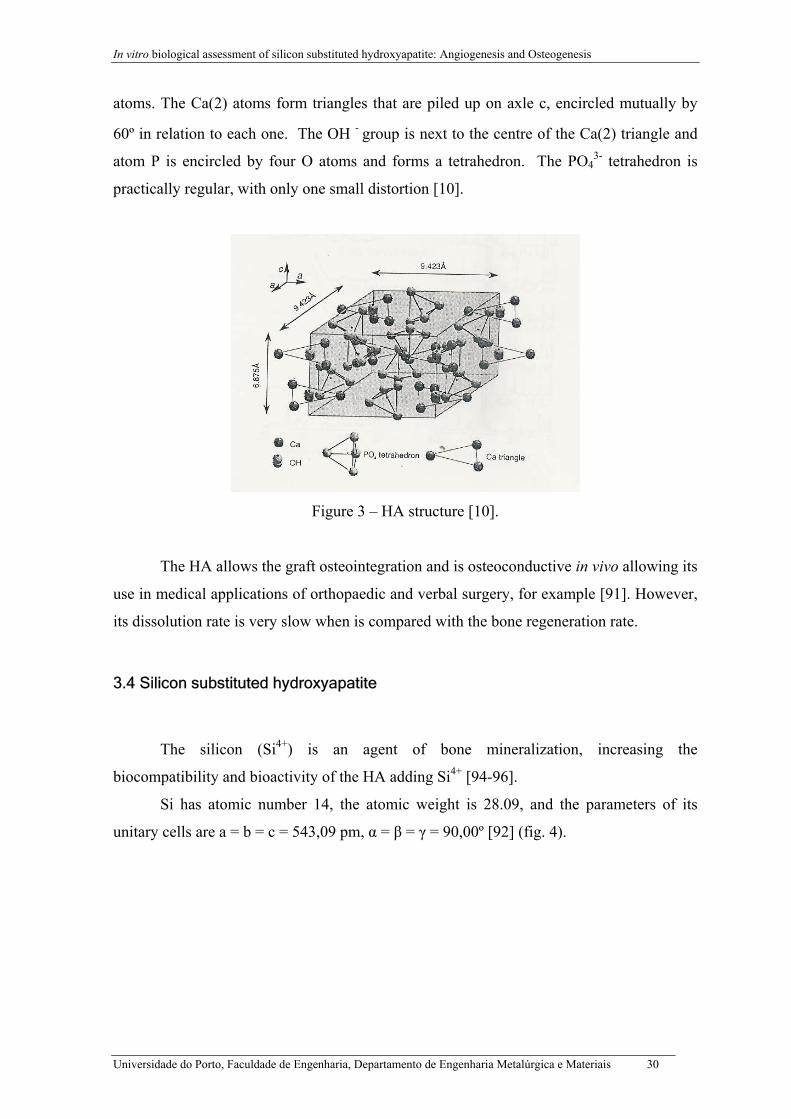
The HA lattice contains two kinds of calcium positions, assigned for Ca(1) and
Ca(2) (fig. 3). Ca(2) is surrounded by six atoms of O that belongs to the PO43- group and
one OH- group, while the atom of Ca(1) is almost octahedral and surrounded for six O
Universidade do Porto, Faculdade de Engenharia, Departamento de Engenharia Metalúrgica e Materiais 29
In vitro biological assessment of silicon substituted hydroxyapatite: Angiogenesis and Osteogenesis
atoms. The Ca(2) atoms form triangles that are piled up on axle c, encircled mutually by
60º in relation to each one. The OH - group is next to the centre of the Ca(2) triangle and
atom P is encircled by four O atoms and forms a tetrahedron. The PO43- tetrahedron is
practically regular, with only one small distortion [10].
Figure 3 – HA structure [10].
The HA allows the graft osteointegration and is osteoconductive in vivo allowing its
use in medical applications of orthopaedic and verbal surgery, for example [91]. However,
its dissolution rate is very slow when is compared with the bone regeneration rate.
3.4 Silicon substituted hydroxyapatite
The silicon (Si4+) is an agent of bone mineralization, increasing the
biocompatibility and bioactivity of the HA adding Si4+ [94-96].
Si has atomic number 14, the atomic weight is 28.09, and the parameters of its
unitary cells are a = b = c = 543,09 pm, α = β = γ = 90,00º [92] (fig. 4).
Universidade do Porto, Faculdade de Engenharia, Departamento de Engenharia Metalúrgica e Materiais 30
In vitro biological assessment of silicon substituted hydroxyapatite: Angiogenesis and Osteogenesis
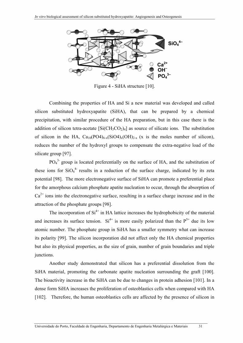
Figure 4 - SiHA structure [10].
Combining the properties of HA and Si a new material was developed and called
silicon substituted hydroxyapatite (SiHA), that can be prepared by a chemical
precipitation, with similar procedure of the HA preparation, but in this case there is the
addition of silicon tetra-acetate [Si(CH3CO2)4] as source of silicate ions. The substitution
of silicon in the HA, Ca10(PO4)6-x(SiO4)x(OH)2-x (x is the moles number of silicon),
reduces the number of the hydroxyl groups to compensate the extra-negative load of the
silicate group [97].
PO43- group is located preferentially on the surface of HA, and the substitution of
these ions for SiO44- results in a reduction of the surface charge, indicated by its zeta
potential [98]. The more electronegative surface of SiHA can promote a preferential place
for the amorphous calcium phosphate apatite nucleation to occur, through the absorption of
Ca2+ ions into the electronegative surface, resulting in a surface charge increase and in the
attraction of the phosphate groups [98].
The incorporation of Si4+ in HA lattice increases the hydrophobicity of the material
and increases its surface tension. Si4+ is more easily polarized than the P5+ due its low
atomic number. The phosphate group in SiHA has a smaller symmetry what can increase
its polarity [99]. The silicon incorporation did not affect only the HA chemical properties
but also its physical properties, as the size of grain, number of grain boundaries and triple
junctions.
Another study demonstrated that silicon has a preferential dissolution from the
SiHA material, promoting the carbonate apatite nucleation surrounding the graft [100].
The bioactivity increase in the SiHA can be due to changes in protein adhesion [101]. In a
dense form SiHA increases the proliferation of osteoblastics cells when compared with HA
[102]. Therefore, the human osteoblastics cells are affected by the presence of silicon in
Universidade do Porto, Faculdade de Engenharia, Departamento de Engenharia Metalúrgica e Materiais 31
In vitro biological assessment of silicon substituted hydroxyapatite: Angiogenesis and Osteogenesis
HA lattice and the duration of these effects can depend on the degree of substitution by
silicon [103]. SiHA also has a stimulator effect in the osteoclasts [104].
The increased structural defects in SiHA can be very important in the increase of
the material solubility and in bond between bone and ceramic. The in vivo dissolution
decreases in the following order 1.5 wt% SiHA > 0.8 wt% SiHA pure > HA and it is
particularly observed on the grain boundaries and triple junctions [105-107].
An in vivo study [108] demonstrated that the morphology of the apatite deposition
and the sequence of events in the interface between bone/HA and between bone/SiHA are
different. The organized staple fibres of collagen appear earlier in the interface bone/SiHA
then in the synthetic HA grafts. It was observed a trabecular zone in bone in contact with
the SiHA graft and the collagen staple fibres form a strong bond with SiHA ceramic graft
[108]. The beneficial effects of silicon have been reported for many years, namely by
Schwarz [109] and Carlisle [100], whom demonstrated that a deficient diet in silicon in rats
and chicks retarded the growth and disturbed the development of bone structure. Reffit et
al (2003) [111] showed that at a physiologic concentration, silicon stimulates collagen I
synthesis, differentiation and alkaline phosphatase activity in human osteoblastic like cells.
Botelho et al [103] demonstrated that dense SiHA stimulated the adhesion,
proliferation and differentiation of human osteoblasts. It was also possible to observe
calcium phosphate mineral deposits on cell layer. Additionally, Botelho et al [112]
demonstrated that this material allows the differentiation of osteoclasts precursors into
mature osteoclasts. These cells grown on the surface of the material expressed typical
phenotype characteristics, such as: actin rings, several nuclei, TRAP expression and
expression of vitronectin receptors.
It is know that titanium or titanium implants have a low bioactivity, so one way to
improve its bioactivity is by coating its surface with a ceramic biomaterials. Therefore, in
order to take advantage of the positive effect of the SiHA, a titanium alloy was coated with
SiHA and its biological behaviour assessed.
3.5 Coating techniques
Many techniques are available for the deposition of ceramic coatings, including
physical vapour deposition techniques, conversion of superficial metallic fractions to
ceramic-like oxides, immersion in a ceramic melt, direct melting or chemical reaction of
components placed directly onto the surface, electroforming, gas-pressure bonding,
Universidade do Porto, Faculdade de Engenharia, Departamento de Engenharia Metalúrgica e Materiais 32
In vitro biological assessment of silicon substituted hydroxyapatite: Angiogenesis and Osteogenesis
welding by diffusion, ultrasound or field-assisted diffusion bonding, reaction with gas
induced by laser, and covering with refractory salts or metallic oxides by plasma spraying
[91].
Plasma spraying technique is used to coat a metal substrate surface with ceramic
through a plasma flame. In the coating process an electric arc is struck between two
electrodes and stream of gases is passed through the arc. The coating powder is injected
into the plasma with a carrier gas (usually argon or nitrogen), and the heated powders melt
and reach the substrate with high velocity. There are many variables in the process
including the gases used, the electrical settings, the nozzle/substrate separation and the
morphology, particle size, and particle size distribution of the powder [91,109].
Plasma spray process parameters affect the structure of the coatings, specially the
crystallinity, porosity, density, adhesion, cohesion, and as a consequence the bone bonding
mechanism and the rate of bone formation [91, 92, 113]. The structure of the coatings will
depend on the time of performance of the particles in the flame, and on the solidification
and cooling conditions. The high temperatures of the plasma flame and the high cooling
rates promote the formation of amorphous phases. The way by which a coating adheres to
a substrate is very complex and it is not fully understood. However, many factors seem to
influence the establishment of coating-to-substrate adhesion: mechanical anchorage, Van
der Waals physical interaction forces, chemical interaction and metallurgical process [114].
3.6 In vitro biological studies
The most important characteristics that will allow the selection a material for
medical use are its biocompatibility, absence of harmful or toxic effect for the organism
and its biomechanics properties capable to answer to the dynamic and static requests that
will be subject during its useful life. Cell death, reduced cell adhesion, altered cell
morphology, reduced cell proliferation and reduced biosynthetic activity are examples of
toxicity in vitro [115].
In vitro research is used to assess the good performance of a biomaterial. With
fewer variables and perceptually amplified reactions to subtle causes, results are more
discernible. Evaluation under in vitro conditions may provide rapid and not expensive data
on biological interaction. However, these results may allow us to partially predict its
performance in vivo, because there are many variables that are not controlled. The
biomaterials must include biocompatibility studies, cell cultures, prior to any in vivo testing
Universidade do Porto, Faculdade de Engenharia, Departamento de Engenharia Metalúrgica e Materiais 33

In vitro biological assessment of silicon substituted hydroxyapatite: Angiogenesis and Osteogenesis
[116] due to the legal and ethical rules that restrict animal experimentation and because the
correlations between in vitro and in vivo tests provide a quite good results of the expected
biological performance of a biomaterial.
The studies in vitro are important because it can have an important influence for the
studies in vivo and also because the in vitro approach is the unique possibility to test
human cells reaction on the material.
Universidade do Porto, Faculdade de Engenharia, Departamento de Engenharia Metalúrgica e Materiais 34
In vitro biological assessment of silicon substituted hydroxyapatite: Angiogenesis and Osteogenesis
CHAPTER 2
EXPERIMENTAL PROCEDURES
Universidade do Porto, Faculdade de Engenharia, Departamento de Engenharia Metalúrgica e Materiais 35
In vitro biological assessment of silicon substituted hydroxyapatite: Angiogenesis and Osteogenesis
Chapter 2
Introduction
The objective of this chapter is to describe the laboratorial procedures:
1. The preparation and characterization of HA and SiHA as dense samples and as
plasma-sprayed coated disc of Ti;
2. In vitro biological studies with Human Umbilical Vein Endothelial Cells;
3. In vitro biological studies with Human Osteoblastic Cells;
The HA and SiHA powders were prepared through a precipitation method.
The physico-chemical and structural characterization of the bioceramics prepared
were performed using the X-ray diffraction (XRD), Fourier transform infrared (FTIR) and
Scanning Electron Microscopy (SEM) techniques.
In vitro biological studies with human umbilical vein endothelial cells cultured in
dense samples of HA and SiHA were evaluated through MTT assay and Confocal laser
scanning microscopy (CLSM).
In vitro biological studies of human osteoblastic cells cultured on the surface of Ti
alloy coated with HA and SiHA, were evaluated through MTT assay, CLSM and SEM.
Universidade do Porto, Faculdade de Engenharia, Departamento de Engenharia Metalúrgica e Materiais 36
In vitro biological assessment of silicon substituted hydroxyapatite: Angiogenesis and Osteogenesis
1. Preparation of HA and SiHA
1.1 HA preparation
HA was prepared through a precipitation method consisting on the reaction
between calcium hydroxide (Ca(OH)2) and orthophosphoric acid (H3PO4), following the
reaction:
10Ca(OH)2 + 6H3PO4 → Ca10(PO4)6(OH)2 + 18H20
During the synthesis process 0.500 moles of calcium hydroxide (Ca(OH)2) were
dissolved in 1 litre of deionised water, and 0.299 moles of orthophosphoric acid (H3PO4)
in 1 litre of deionised water. The solution of orthophosphoric acid was added drop to drop
to the calcium hydroxide solution, at room temperature, for a period of 3 hours, and the pH
of the suspension was kept at 10.5 by the addition of ammonia (NH4OH). The suspension
was stirred for one hour and left aging for 6 hours.
1.2 SiHA preparation
The method used in the preparation of SiHA was similar to that used in preparation
of HA, but in this case silicon tetra-acetate [Si(CH3COO4] was added to the mixture, as a
source of silicate ions.
In the preparation of SiHA the amount of reagents used to prepare was calculated
on the basis of the following equation:
10Ca2+ + (6-x)PO43- + xSiO4
4- + (2-x)OH- → Ca10(PO4)6-x(SiO4)x(OH)2-x
(x is the moles number of silicon tetra-acetate)
To prepare 0.8 wt (%) of SiHA each reagent was dissolved in 1 litre of deionised
water: 0.500 moles of (Ca(OH)2), 0.285 moles of (H3PO4) and 0.014 moles of
[Si(CH3COO4]. The solutions were mixed for 30 minutes and the orthophosphoric acid
solution was added drop to drop to the Ca(OH)2 solution, for a period of 3 hours. Such as
in the preparation of HA, during the precipitation reaction, the pH of the suspension was
kept above 10.5 by the addition of ammonia. The suspension was stirred during one hour
and left aging for 6 hours.
Universidade do Porto, Faculdade de Engenharia, Departamento de Engenharia Metalúrgica e Materiais 37
In vitro biological assessment of silicon substituted hydroxyapatite: Angiogenesis and Osteogenesis
1.3 Dense Samples
HA and SiHA dense samples of approximately 15 mm of diameter of were
prepared by filling a cylindrical die with 1g of powder of each material, and then they were
pressed using a uniaxial press until 180 Bar. The HA and SiHA dense samples were then
sintered in a furnace at 1300ºC using a heating rate of 2.5ºC/min with 120 min dwelling
time, followed by natural cooling inside the furnace.
1.4 Milling and sieving
After uniaxial pressing and sinterization, the samples were, milled and sieved until
obtain the following distribution (optimize to for plasma spray):
30% - 90 µm and 125µm
40% - 75µm-90µm
20% - 63µm-75µm
10% - 45µm-63µm.
1.5 Plasma-spray
A commercial Ti rod (Ti-6Al-4V) of 14 mm of diameter was coated with HA and
SiHA. The thickness of the coating was 120 µm.
2. Physical-chemical and structural characterization of HA and SiHA
X-ray diffraction (XRD), Fourier transform infrared (FTIR), Scanning Electron
Microscopy (SEM) and Confocal Laser Scanning Microscopy (CLSM) were used to
characterize the HA and Si-HA samples.
X-ray powder diffraction (XRD)
To determine the phase purity of the HA and SiHA, the samples were grounded to
a fine powder and analysed using a Rigaku Dmax-III-VC X-ray diffractometer, with CU-
Kα radiation (Kα = 1.54056 Aº). Data was collected from 4º to 80º (2θ), with step size of
0.02º/s.
Universidade do Porto, Faculdade de Engenharia, Departamento de Engenharia Metalúrgica e Materiais 38
In vitro biological assessment of silicon substituted hydroxyapatite: Angiogenesis and Osteogenesis
Fourier transform infrared (FTIR)
The infrared spectra analysis was performed with a System 2000 FT-IR, Perkin
Elmer, with a 4cm-1 resolution and 100 scans.
Scanning electron microscopy (SEM)
For morphological evaluation, the HA and SiHA samples were coated with gold
and were observed in a JOEL JSM-63 10F scanning electron microscope, with detection of
secondary electrons and Backscattered electrons.
3. In vitro biological studies of Human Umbilical Vein Endothelial Cells
3.1 Human Umbilical Vein Endothelial Cells
Human Umbilical Vein Endothelial Cells (HUVECs) (5º passage) were cultured in
M199 medium, supplemented with 10% of Simulated body fluid (SBF), 1% of
Penicillin/Streptomycin, 1% of heparin (10mg/ml) and 2 µl/ml of ECGS. After cells
reached confluence, they were enzymatically released by trypsin-EDTA solution and
cultured on the HA and SiHA dense surface at a density of 3x10⁴ cells/cm2. The samples
were previously coated with 0.2% gelatine (1hour immersion at 37ºC).
3.2 Cell proliferation
For cell proliferation evaluation, the colonized samples were incubated with 3-[4,5-
dimethylthiazol-2-yl]-2,5-diphenyltetrasodium bromide (MTT) (5mg/mL) at 37º C for 4
hours. After treatment with DMSO, the absorbance of the solution was evaluated at 610
nm, at day 1 and 3.
3.3 Morphologic Evaluation
Cells were fixed with 4% formaldehyde (methanol free), permeabilized with 0.1%
triton and incubated in 10 mg/ml bovine serum albumin (BSA) with 100µg/ml RNAse. F-
actin filaments were stained with Alexafluor®-conjugated phalloidin and nuclei were
counterstained with 10µg/ml propidium iodide. Samples were washed with phosphate
Universidade do Porto, Faculdade de Engenharia, Departamento de Engenharia Metalúrgica e Materiais 39
In vitro biological assessment of silicon substituted hydroxyapatite: Angiogenesis and Osteogenesis
buffered saline (PBS) and covered with Vectashield. Images were acquired on a Leica TCP
SP2 AOBS with a LCS software.
To prove that the cells in the material surface were in fact endothelial cells, they
were staining with a platelet/endothelail cell adhesion molecule-1 (PECAM-1). After cells
fixation with 4% formaldehyde, the cells were permeabilized with triton 0.1% during 5
minutes, after which they were incubated with the primary antibody, Pecam-1 anti-rabbit,
in 1:50 dilution on 1% BSA/PBS solution during 45 minutes. The samples were then
washed with PBS and incubated with secondary antibody AlexaFlour 488 anti-rabbit in 1%
BSA/PBS solution, in a 1:1000 dilution for 45 minutes. The nucleus was stained with
1.5µg/ml propidium iodide for 7 minutes.
One of the characteristics of endothelial cells is the capacity to form tubular
structures, so 1.5% of collagen gel was added to the samples. The gel was added during 24
hours. As control positive, endothelial cells were cultured in specific cell culture material.
4. In vitro biological studies of Human Osteoblastic Cells
4.1 Human osteoblastic cells
The human osteoblastic cells were isolated from bone marrow of the kneecap of a
28 years old male patient. Trabecular bone samples were mechanically disaggregated and
washed with standard culture medium, as result a single-cell suspension was obtained. The
cells were cultured in minimum essential medium Eagle, alpha modification (α-MEM)
containing 10% foetal bovine serum (FBS), 100µg/ml penicillin, 10IU/ml streptomycin
and 2.5 µg/ml fungizone. Incubation was carried out in a humidified atmosphere of 95%
air and 5% CO2 at 37ºC. The passage was made when primary culture was near
confluence. Adherent cells were enzymatically released by trypsin-EDTA solution and
counted using a hemocytometer. The human bone marrow cells (first subculture) were
seeded on surface biomaterials at a density of 2x10⁴/cm2 for 28 days, at conditions
favouring osteogenic differentiation. The cells were cultured at 37ºC in a humidified
atmosphere of a 95% air and 5% CO2 in plates containing 10% of foetal bovine serum
(protein source), 1% of fungizone and streptomycin/penicillin (antibacterial), 50µg/ml
ascorbic acid (vitamin C source for collagen formation), 10nM dexamathasone (increases
Universidade do Porto, Faculdade de Engenharia, Departamento de Engenharia Metalúrgica e Materiais 40
In vitro biological assessment of silicon substituted hydroxyapatite: Angiogenesis and Osteogenesis
the cellular proliferation and it induces osteoblasts differentiation) and 10mM β-
glycerolphosphate. The culture medium was changed every three days.
4.2 Cell proliferation
For cell proliferation evaluation, the colonized samples were incubated with MTT
(5mg/mL) at 37º C for 4 hours. After treatment with DMSO, the absorbance of the solution
was read at 610 nm, at days 7, 14, 21 and 28.
4.3 Morphologic Evaluation
For the morphologic evaluation, the cells were analyzed by Confocal Laser
Scanning Microscopy (CLSM) and Scanning Electron Microscopy (SEM).
Confocal laser scanning microscopy (CLSM)
In this case a similar method to the HUVECs was used. Cells were fixed with 4%
formaldehyde (methanol free), permeabilized with 0.1% triton and incubated in 10 mg/ml
bovine serum albumin (BSA) with 100µg/ml RNAse. F-actin filaments were stained with
Alexafluor®-conjugated phalloidin and nuclei were counterstained with 10µg/ml
propidium iodide. Samples were washed with PBS and covered with Vectashield. Images
were acquired on a Leica TCP SP2 AOBS with a LCS software.
Scanning electron microscopy (SEM)
For SEM observation the cells were fixed with 1.5% glutaraldehyde in 0.14 M
sodium cacodylate, and then dehydrated in graded series of alcohols and critical-point
dried. Specimens were mounted onto aluminium supports using araldite and then sputter-
coated with gold and observed in a Joel JSM 35C scanning electron microscope equipped
with an X-ray energy dispersive spectroscopy voyager XRMA System, Noran Instruments,
at days 3, 7, 14, 21 and 28.
Universidade do Porto, Faculdade de Engenharia, Departamento de Engenharia Metalúrgica e Materiais 41

In vitro biological assessment of silicon substituted hydroxyapatite: Angiogenesis and Osteogenesis
RESULTS
Universidade do Porto, Faculdade de Engenharia, Departamento de Engenharia Metalúrgica e Materiais 42
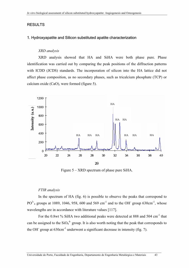
In vitro biological assessment of silicon substituted hydroxyapatite: Angiogenesis and Osteogenesis
RESULTS
1. Hydroxyapatite and Silicon substituted apatite characterization
XRD analysis
XRD analysis showed that HA and SiHA were both phase pure. Phase
identification was carried out by comparing the peak positions of the diffraction patterns
with ICDD (JCDS) standards. The incorporation of silicon into the HA lattice did not
affect phase composition, as no secondary phases, such as tricalcium phosphate (TCP) or
calcium oxide (CaO), were formed (figure 5).
HA
HA
HA
HA
HA
HA
HA
HA
2θ
Figure 5 – XRD spectrum of phase pure SiHA.
FTIR analysis
In the spectrum of HA (fig. 6) is possible to observe the peaks that co
PO3-4 groups at 1089, 1046, 958, 600 and 569 cm-1 and to the OH- group 630
wavelengths are in accordance with literature values [117].
For the 0.8wt % SiHA two additional peaks were detected at 888 and 5
can be assigned to the SiO44- group. It is also worth noting that the peak that co
the OH- group at 630cm-1 underwent a significant decrease in intensity (fig. 7).
Universidade do Porto, Faculdade de Engenharia, Departamento de Engenharia Metalúrgica e Materiais
HA
rrespond to
cm-1, whose
04 cm-1 that
rresponds to
43
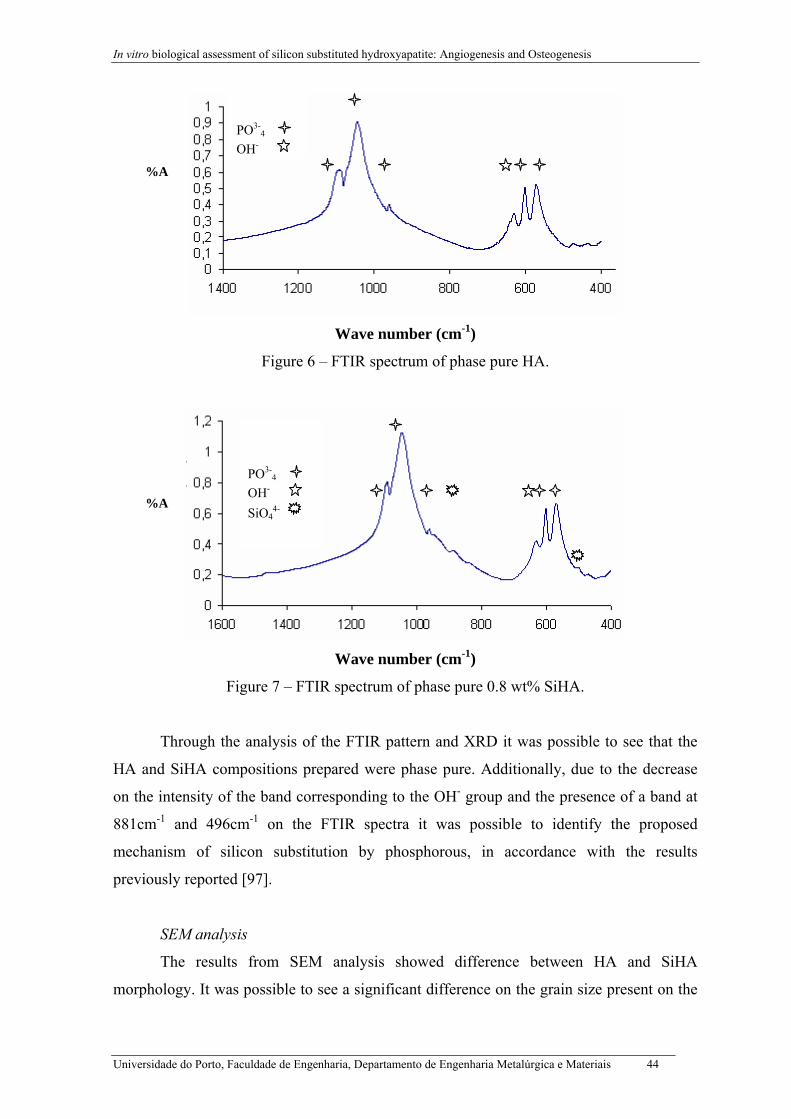
In vitro biological assessment of silicon substituted hydroxyapatite: Angiogenesis and Osteogenesis
%A
%A
F
Through th
HA and SiHA co
on the intensity of
881cm-1 and 496
mechanism of si
previously reporte
SEM analy
The result
morphology. It wa
Universidade do Porto, F
PO3-
4
OH-
Wave number (cm-1)
Figure 6 – FTIR spectrum of phase pure HA.
PO3-
4
OH- SiO4
4-
Wave number (cm-1)
igure 7 – FTIR spectrum of phase pure 0.8 wt% SiHA.
e analysis of the FTIR pattern and XRD it was possible to see that the
mpositions prepared were phase pure. Additionally, due to the decrease
the band corresponding to the OH- group and the presence of a band at
cm-1 on the FTIR spectra it was possible to identify the proposed
licon substitution by phosphorous, in accordance with the results
d [97].
sis
s from SEM analysis showed difference between HA and SiHA
s possible to see a significant difference on the grain size present on the
aculdade de Engenharia, Departamento de Engenharia Metalúrgica e Materiais 44
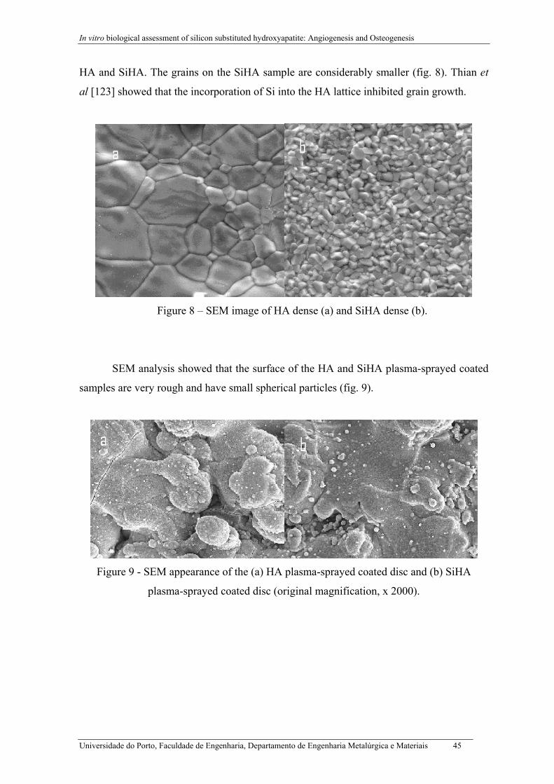
In vitro biological assessment of silicon substituted hydroxyapatite: Angiogenesis and Osteogenesis
HA and SiHA. The grains on the SiHA sample are considerably smaller (fig. 8). Thian et
al [123] showed that the incorporation of Si into the HA lattice inhibited grain growth.
Figure 8 – SEM image of HA dense (a) and SiHA dense (b).
SEM analysis showed that the surface of the HA and SiHA plasma-sprayed coated
samples are very rough and have small spherical particles (fig. 9).
Figure 9 - SEM appearance of the (a) HA plasma-sprayed coated disc and (b) SiHA
plasma-sprayed coated disc (original magnification, x 2000).
Universidade do Porto, Faculdade de Engenharia, Departamento de Engenharia Metalúrgica e Materiais 45
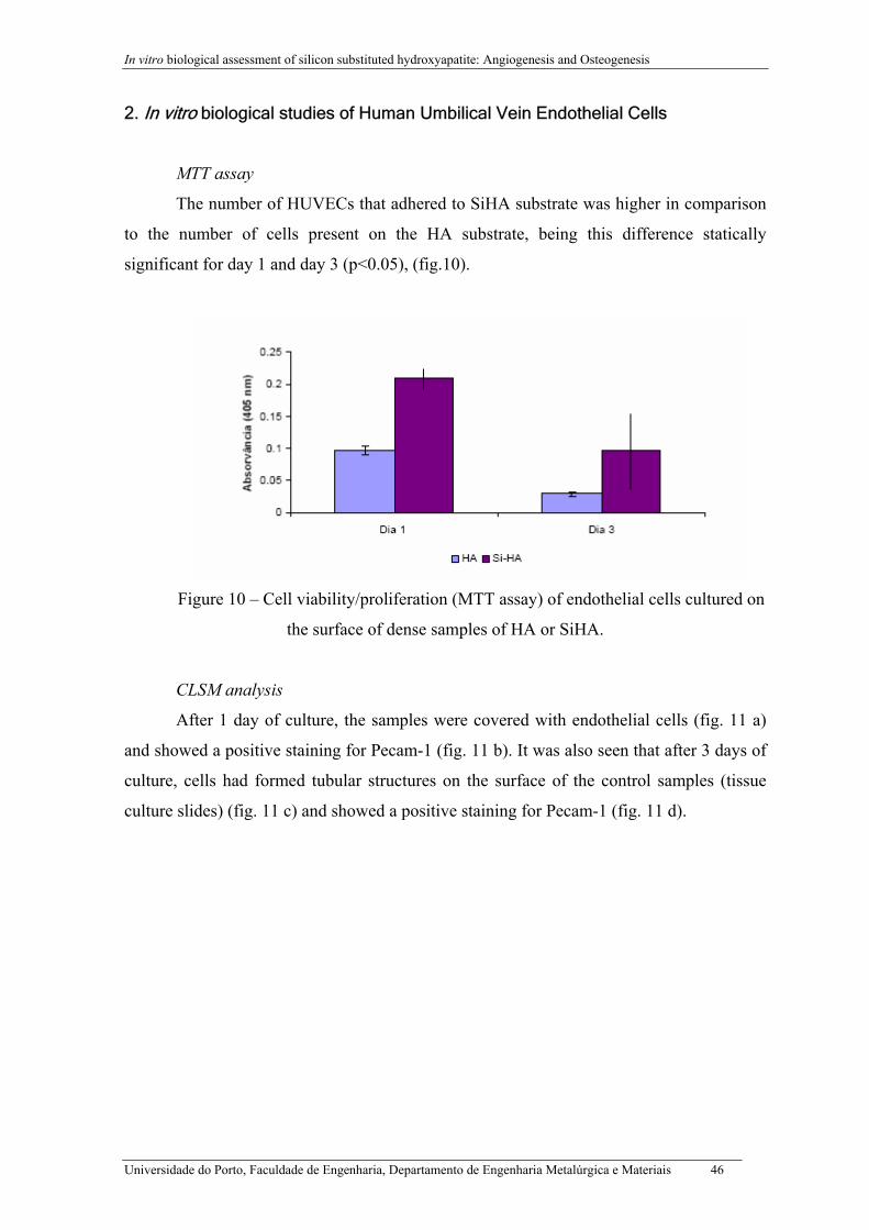
In vitro biological assessment of silicon substituted hydroxyapatite: Angiogenesis and Osteogenesis
2. In vitro biological studies of Human Umbilical Vein Endothelial Cells
MTT assay
The number of HUVECs that adhered to SiHA substrate was higher in comparison
to the number of cells present on the HA substrate, being this difference statically
significant for day 1 and day 3 (p<0.05), (fig.10).
Figure 10 – Cell viability/proliferation (MTT assay) of endothelial cells cultured on
the surface of dense samples of HA or SiHA.
CLSM analysis
After 1 day of culture, the samples were covered with endothelial cells (fig. 11 a)
and showed a positive staining for Pecam-1 (fig. 11 b). It was also seen that after 3 days of
culture, cells had formed tubular structures on the surface of the control samples (tissue
culture slides) (fig. 11 c) and showed a positive staining for Pecam-1 (fig. 11 d).
Universidade do Porto, Faculdade de Engenharia, Departamento de Engenharia Metalúrgica e Materiais 46
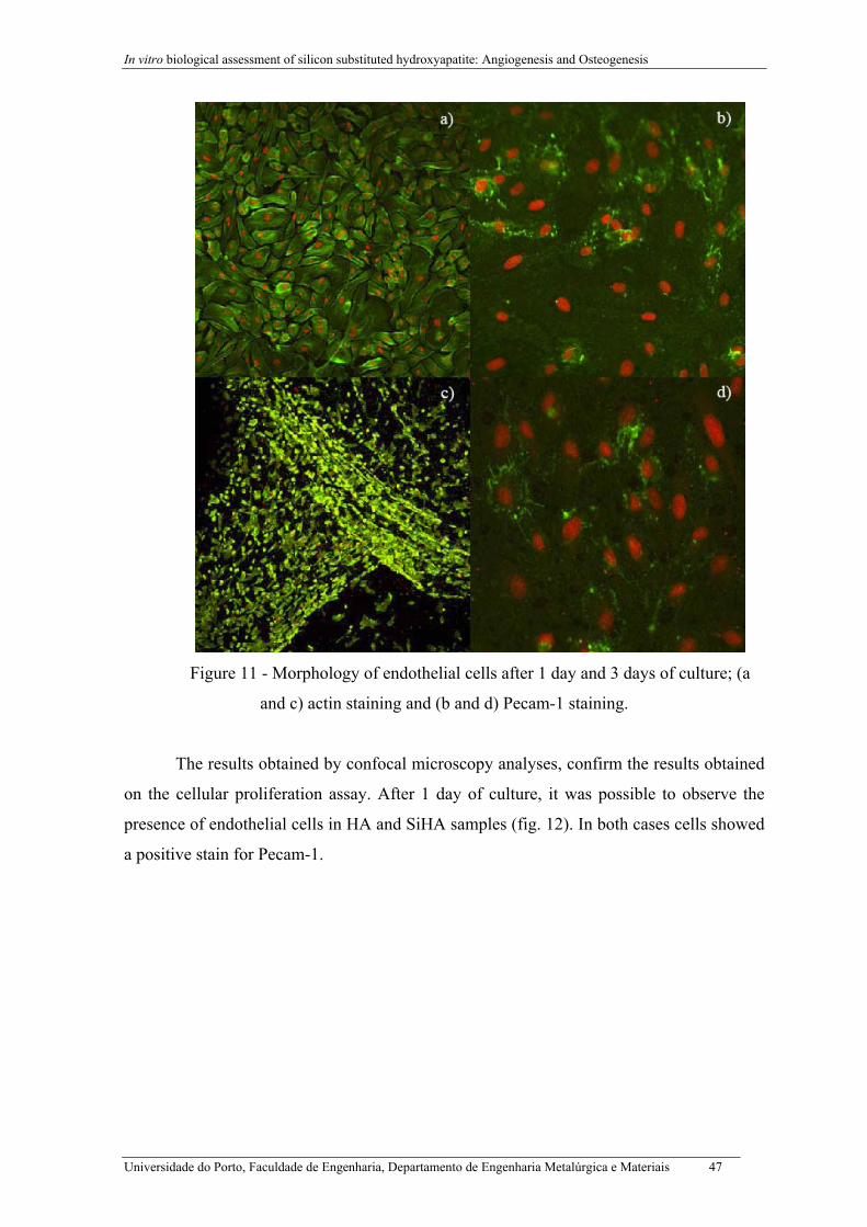
In vitro biological assessment of silicon substituted hydroxyapatite: Angiogenesis and Osteogenesis
Figure 11 - Morphology of endothelial cells after 1 day and 3 days of culture; (a
and c) actin staining and (b and d) Pecam-1 staining.
The results obtained by confocal microscopy analyses, confirm the results obtained
on the cellular proliferation assay. After 1 day of culture, it was possible to observe the
presence of endothelial cells in HA and SiHA samples (fig. 12). In both cases cells showed
a positive stain for Pecam-1.
Universidade do Porto, Faculdade de Engenharia, Departamento de Engenharia Metalúrgica e Materiais 47
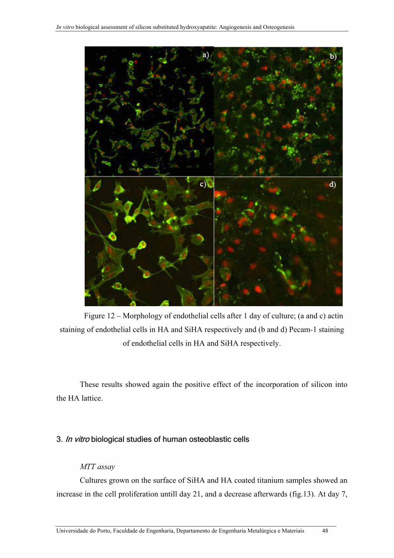
In vitro biological assessment of silicon substituted hydroxyapatite: Angiogenesis and Osteogenesis
Figure 12 – Morphology of endothelial cells after 1 day of culture; (a and c) actin
staining of endothelial cells in HA and SiHA respectively and (b and d) Pecam-1 staining
of endothelial cells in HA and SiHA respectively.
These results showed again the positive effect of the incorporation of silicon into
the HA lattice.
3. In vitro biological studies of human osteoblastic cells
MTT assay
Cultures grown on the surface of SiHA and HA coated titanium samples showed an
increase in the cell proliferation untill day 21, and a decrease afterwards (fig.13). At day 7,
Universidade do Porto, Faculdade de Engenharia, Departamento de Engenharia Metalúrgica e Materiais 48
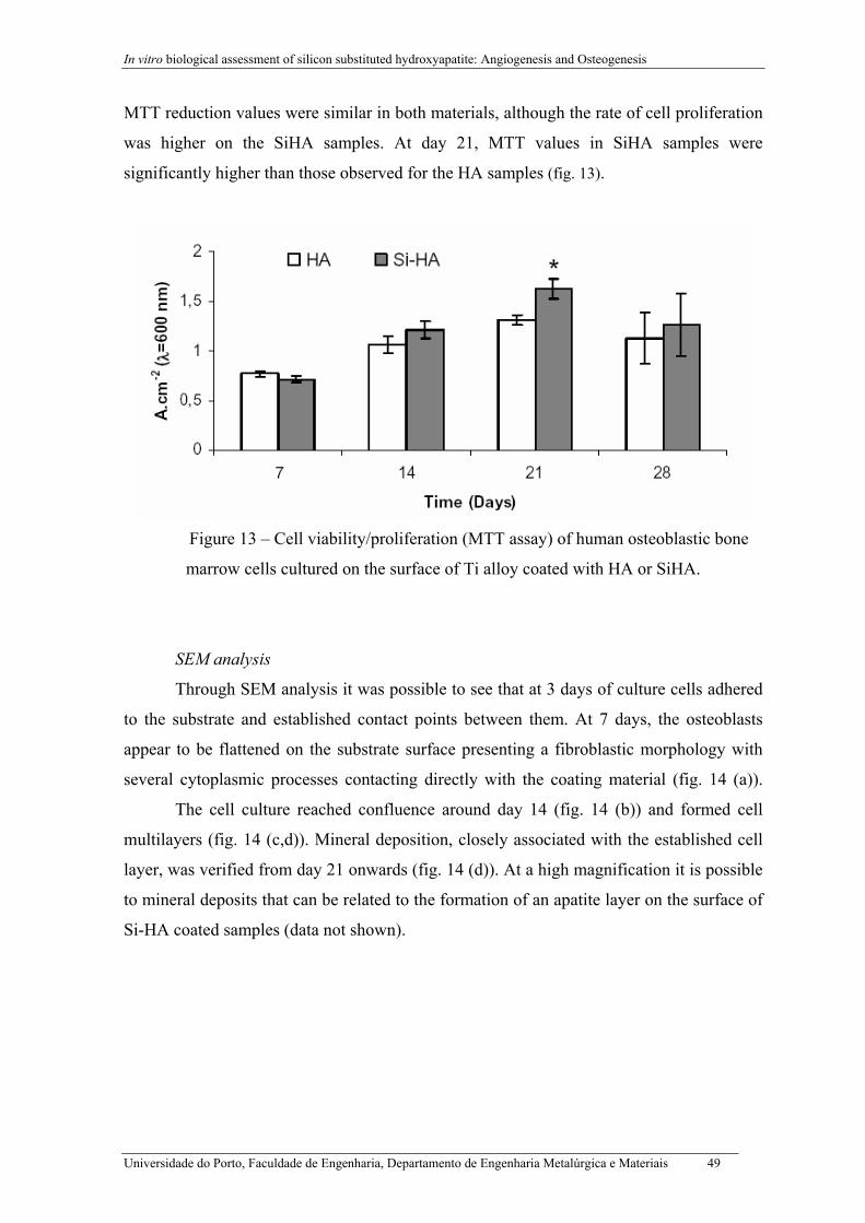
In vitro biological assessment of silicon substituted hydroxyapatite: Angiogenesis and Osteogenesis
MTT reduction values were similar in both materials, although the rate of cell proliferation
was higher on the SiHA samples. At day 21, MTT values in SiHA samples were
significantly higher than those observed for the HA samples (fig. 13).
Figure 13 – Cell viability/proliferation (MTT assay) of human osteoblastic bone
marrow cells cultured on the surface of Ti alloy coated with HA or SiHA.
SEM analysis
Through SEM analysis it was possible to see that at 3 days of culture cells adhered
to the substrate and established contact points between them. At 7 days, the osteoblasts
appear to be flattened on the substrate surface presenting a fibroblastic morphology with
several cytoplasmic processes contacting directly with the coating material (fig. 14 (a)).
The cell culture reached confluence around day 14 (fig. 14 (b)) and formed cell
multilayers (fig. 14 (c,d)). Mineral deposition, closely associated with the established cell
layer, was verified from day 21 onwards (fig. 14 (d)). At a high magnification it is possible
to mineral deposits that can be related to the formation of an apatite layer on the surface of
Si-HA coated samples (data not shown).
Universidade do Porto, Faculdade de Engenharia, Departamento de Engenharia Metalúrgica e Materiais 49
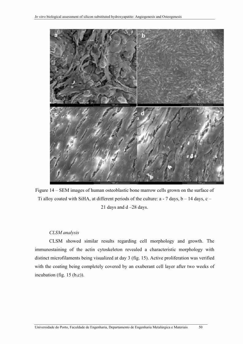
In vitro biological assessment of silicon substituted hydroxyapatite: Angiogenesis and Osteogenesis
Figure 14 – SEM images of human osteoblastic bone marrow cells grown on the surface of
Ti alloy coated with SiHA, at different periods of the culture: a - 7 days, b – 14 days, c –
21 days and d –28 days.
CLSM analysis
CLSM showed similar results regarding cell morphology and growth. The
immunostaining of the actin cytoskeleton revealed a characteristic morphology with
distinct microfilaments being visualized at day 3 (fig. 15). Active proliferation was verified
with the coating being completely covered by an exuberant cell layer after two weeks of
incubation (fig. 15 (b,c)).
Universidade do Porto, Faculdade de Engenharia, Departamento de Engenharia Metalúrgica e Materiais 50
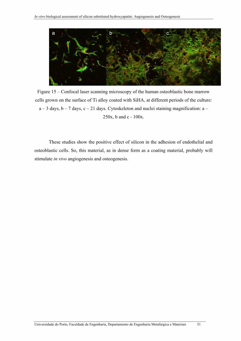
In vitro biological assessment of silicon substituted hydroxyapatite: Angiogenesis and Osteogenesis
Figure 15 – Confocal laser scanning microscopy of the human osteoblastic bone marrow
cells grown on the surface of Ti alloy coated with SiHA, at different periods of the culture:
a – 3 days, b – 7 days, c – 21 days. Cytoskeleton and nuclei staining magnification: a –
250x, b and c - 100x.
These studies show the positive effect of silicon in the adhesion of endothelial and
osteoblastic cells. So, this material, as in dense form as a coating material, probably will
stimulate in vivo angiogenesis and osteogenesis.
Universidade do Porto, Faculdade de Engenharia, Departamento de Engenharia Metalúrgica e Materiais 51

In vitro biological assessment of silicon substituted hydroxyapatite: Angiogenesis and Osteogenesis
DISCUSSION AND CONCLUSIONS
Universidade do Porto, Faculdade de Engenharia, Departamento de Engenharia Metalúrgica e Materiais 52
In vitro biological assessment of silicon substituted hydroxyapatite: Angiogenesis and Osteogenesis
DISCUSSION
In this thesis it was proposed to characterise Hydroxyapatite and Silicon Substituted
Hydroxyapatite, in a dense form, regarding its ability to stimulate angiogenesis and HA
and SiHA as a coating material regarding its ability to stimulate osteogenese.
The angiogenesis was evaluated by in vitro cultures with human umbilical vein
endothelial cells, which were seeded on the surface of HA and SiHA. Several parameters
were assessed, namely cellular growth and functional activity at time points of the culture.
The osteogenesis evaluation was performed by culturing human osteoblastic cells on the
surface of Ti alloy coated samples with HA and SiHA.
If a coating material is too soluble, there is high probability that it will dissolve
completely into the physiological environment, even before it is able to accelerate the
promotion of direct attachment at the bone/implant interface [118]. Variables like
temperature, pH, dissolution time, reagent type, must be controlled because small changes
in these variables can be responsible for disarrangement of chemical reaction. According to
the literature, [119, 120] the XRD pattern of the Si-HA powder before plasma spraying has
a similar pattern to crystalline phase pure HA and after plasma spraying the SiHA coating
becomes more amorphous and suffers some conversion to secondary phases, having the
following chemical composition: HA (34.1%), calcium oxide (CaO: 12.6%) and tri-
calcium phosphate phases (α-TCP: 27.4% and β-TCP: 25.9%).
On the FTIR spectra it was possible to see the characteristic wavelengths for the
phosphate groups present on the HA lattice. When comparing the FTIR spectra of HA and
SiHA it was possible to observe a decrease on the intensity of the band corresponding to
the OH- group. It is known that PO3-4 groups are preferentially located at HA surface and
therefore the substitution of these ions for SiO44- groups results in a decrease in surface
charge [121]. The decrease in intensity of the peak on the FTIR spectra that corresponds to
the OH- group on the SiHA was expected since according to the mechanism described by
the following equation: 10Ca2+ + (6-x)PO43- + xSiO4
4- + (2-x)OH- → Ca10(PO4)6-
x(SiO4)x(OH)2-x, the substitution of the phosphate group for the silicate group leads to the
loss of some OH- to maintain the charge balance [122]. Si4- ions are structurally
incorporated into the HA lattice in solid solution, and are not segregated as a secondary
phase.
Universidade do Porto, Faculdade de Engenharia, Departamento de Engenharia Metalúrgica e Materiais 53
In vitro biological assessment of silicon substituted hydroxyapatite: Angiogenesis and Osteogenesis
Thian et al [123] showed that with increasing Si addition, the crystalline size of
SiHA decreased and the incorporation of Si inhibited grain growth, being the effect more
significant as the level of Si increased. These results are in agreement with the results
obtained through SEM analysis, that showed a significant difference between the grain size
of HA and SiHA dense sample, being smaller in the SiHA. Additionally, it would be
expected that an increased of Si would cause HA crystals to be more soluble, releasing
more Ca2+ and P5+ ions into the culture medium. A rapid re-precipitating and formation of
a newly-formed CaP-rich layer on the coating surface, with features very similar to the
mineral phase of bone, would be an ideal site for osteoblast cells to attach, grow and form
new bone [124]. Thian et al [123] showed that although the addition of Si increased the
unit cell parameters and reduced the crystalline size, the XRD showed no changes in the
relative intensities. In this work, XRD also showed no changes in the relative intensities
what can be explained because Si4+ and P5+ are adjacent in the Periodic Table (differ by
only 1 in atomic number) and the Si level is relatively low (< 5 wt.%), although such
changes are normally expected when there is an atomic substitution [123].
Implantation involves tissue trauma, which evokes an inflammatory response,
coupled to a wound healing reaction, involving angiogenesis, fibroblast activation and
matrix remodelling. Three principal fields of research can yield useful data to better
understand these phenomena: physical-chemical studies of biomaterials, in vitro studies
and animal models. Until now the type and extent of such reactions to give optimal
integration of various biomaterials are practically unknown [125]. In vitro biological
studies with endothelial cells allows a better understanding of mechanisms involved in the
interactions between cells and a material surface, important for the rational development of
medical devices with optimal biocompatibility [126,127]. The success of biomaterial
applications is not only dependent on the growth and function of the tissue-specific cells on
the biomaterials but is also dependent in most cases on successful vascularization after
implantation [128]. So, there is a significant importance to characterise the biological
response of HA and SiHA in the presence of HUVECs.
The MTT assay measures the metabolic activity of the cells, which can be
correlated with the number of viable cells. The number of endothelial cells that adhere to
SiHA substrate was superior to the number of endothelial cells that adhere to HA substrate,
being this difference statistically significant at day 1 and day 3 (p<0.05). Similar result was
obtained at day 3 of culture. On the surface of the control used in this study (tissue culture
Universidade do Porto, Faculdade de Engenharia, Departamento de Engenharia Metalúrgica e Materiais 54
In vitro biological assessment of silicon substituted hydroxyapatite: Angiogenesis and Osteogenesis
plastic), the HUVECs formed tubular structures and also showed a positive stain for
Pecam-1, which proved that the cells culture on the surface of these materials expressed
the endothelial phenotype. Therefore, the results obtained by confocal microscopy analyses
confirm the results obtained in cellular proliferation assay. These results showed again the
positive effect of the incorporation of silicon into the HA lattice.
Bone formation is dependent on several molecules such as BMPs, molecules that
will initiate vascularization and osteogenic precursor cells. BMPs are also involved on the
molecular cascade that will lead to the formation of cartilage and bone progenitor cell
differentiation. Multipotent stem cells originating from bone marrow stromal cells are a
particularly attractive source for osteogenic precursors for bone tissue engineering, because
they can be easily harvested and expanded in vitro and can differentiate into bone-forming
cells. Human osteoblastic cells were used to evaluate osteogenesis on coated HA and
SiHA. In vitro studies with human osteoblastic cells showed that cultures grown on the
surface of SiHA coated titanium had an increase in cell proliferation untill day 21, and a
decrease afterwards. This behaviour is in accordance with the literature, where it is
described the in vitro development of osteoblastic phenotype. At day 14 and 21, cell
growth on the SiHA was higher (statistically significant) compared with the HA as
expected according to the literature [94,95,102,108].
Ferraz et al [129] showed that the resulting surface modifications improved
osteoblasts growth, probably due to the formation of a relatively stable calcium-
phosphorous rich layer on the coatings surface that is a strong indicator of bioactivity.
Furthermore, the adsorption of biological molecules from the culture medium by the
material, have an important role in the adhesion process and functional behaviour of the
osteoblasts cells [123,130]. The differences in the proliferation behaviour of human
osteoblasts seeded into the surface of HA and of SiHA, are probably related to differences
in surface characteristics, as their chemical composition and physical properties differ
[94,95,102,108, 98-101, 130].
Although, in this study, no osteoblastic differentiation markers were assessed, the
cell morphology and the formation of a mineralized matrix indicate that the cell culture
reached a differentiated stage on the SiHA coated substrate. Compared to the HA coated
titanium, the enhanced behaviour of the SiHA coating might be explained by the presence
of silicon into the culture medium. It is known that the formation of an apatite layer is
Universidade do Porto, Faculdade de Engenharia, Departamento de Engenharia Metalúrgica e Materiais 55
In vitro biological assessment of silicon substituted hydroxyapatite: Angiogenesis and Osteogenesis
preceded by a dissolution stage, which indicates that the silicon could be released into the
culture medium and incorporated by the osteoblasts. Similar results were reported by
Botelho et al [103] who showed that the SiHA, in a dense form, stimulates osteoblasts
proliferation, differentiation and formation of mineral deposits. Thian et al [123] also
reported that a SiHA coating prepared by magnetron co-sputtering can also stimulate the
proliferation of osteoblasts. This is in line with that reported by several authors suggesting
that silicon is a key element for bone regeneration [102,107].
These results clearly demonstrate the beneficial effect of silicon on the adhesion of
endothelial cells and osteoblastic cells.
Universidade do Porto, Faculdade de Engenharia, Departamento de Engenharia Metalúrgica e Materiais 56
In vitro biological assessment of silicon substituted hydroxyapatite: Angiogenesis and Osteogenesis
CONCLUSIONS
The main conclusions of this work are:
Analysis of the SiHA compositions prepared for this study using XRD and FTIR
confirmed the substitution mechanism of silicon by phosphorous, in accordance with the
results reported previously.
In vitro biological studies of human umbilical vein endothelial cells cultured in
dense samples of HA and SiHA showed the positive effect the incorporation of Si into the
HA lattice.
The SiHA coating allowed the adhesion, proliferation and differentiation of human
bone marrow osteoblastic cells, which suggest that this coating is suitable to be used in
several biomedical applications. Although, this study revealed an enhanced activity of the
osteoblasts cultured on the SiHA coating, the mechanism behind this behaviour is still
unknown and further evaluation is required.
The results achieved in this work are in agreement with previous in vitro studies
suggesting that SiHA has better characteristics for bone cell growth and function than HA,
using human osteoblasts to evaluate the biological response of the materials.
SiHA is suitable material to be used as bone graft, as in dense form or as a coating
material. This material elicits a positive response in different cell types, such as: human
umbilical vein endothelial cells and human osteoblastic cells.
As future work it is proposed to optimize the ideal amount of Si to incorporate into
the HA lattice in order to improve the response of human umbilical vein endothelial cells
to the material in a dense form and as a coating.
Universidade do Porto, Faculdade de Engenharia, Departamento de Engenharia Metalúrgica e Materiais 57
In vitro biological assessment of silicon substituted hydroxyapatite: Angiogenesis and Osteogenesis
REFERENCES
[1] Wynsberghe, D. V., Noback, C. R., Carola, R.; “Human Anatomy & Physiology”,
McGraw-Hill, 1995, Third Edition.
[2] Hench LL, Wilson J. In: An Introduction to Bioceramics. Singapore: World Sientific,
1993.
[3] Bronzino, J.D., “The Biomedical Engeneering Handbook”, IEEEPress, 1995.
[4] Aoki, Hideki: “Medical Applications of Hydroxyapatite”, Ishiyaku EuroAmerica Inc,
Tokyo, 1994.
[5] Seeley, R., Stephens, T., Tate, P., “Anatomia e Fisiologia”, Lusodidacta, 1997.
[6] Guyton & Hall, “Tratado de Fisiologia Médica”, 9ª edição.
[7] An, Y. H.; “Orthopaedic Issues in Osteoporosis”, CRC Press, 2003, U.S.
[8] Rosen, C.J., Glowacki, J, Bilezikian, J.P., “The Aging Skeleton”, Academic Press,
1999, USA.
[9] Marcus, R., Feldman, D., Kelsey, J., “Osteoporosis”, Academic Press, 1996, USA.
[10] Bilezikian, J. P., Raisz, L.G., Rodan, G.A.; “Principles of Bone Biology”, Academic
Press, 1996, USA.
[11] Seibel, M. J., Robins, S., Bilezikian, J. P.; “Dynamics of Bone and Cartilage
Metabolism”, Academic Press, 1999, U.K.
[12] Carmeliet, P., Mechanisms of angiogenesis and arteriogenesis. Nature Medicine 6:
389-395 (2000).
Universidade do Porto, Faculdade de Engenharia, Departamento de Engenharia Metalúrgica e Materiais 58
In vitro biological assessment of silicon substituted hydroxyapatite: Angiogenesis and Osteogenesis
[13] Carmeliet, P., Jain, R.K., Angiogenesis in cancer and other diseases. Nature 407: 249-
257 (2000).
[14] Carmeliet, P., Manipulating angiogenesis in medicine. Journal of Internal Medicine
255(5): 538-561 (2004).
[15]Carmeliet, P., Angiogenesis in health and disease, Nature Medicine 9, 653-660 (2003).
[16] Patan, S., Vasculogenesis and angiogenesis as mechanism of vascular network
formation, growth and remodelling, Neurooncol. 2000 Oct-Nov; 50(1-2):1-15.
[17] Luttun, A., Carmeliet, G. & Carmeliet, P. Vascular progenitors: from biology to
treatment. Trends Cardiovasc. Med. 12, 88-96 (2002).
[18] Asahara, T. & Isner, J.M. Endothelial progenitor cells for vascular regeneration. J.
Hematother. Stem Cell Res. 11, 171-178 (2002).
[19] Reyes, M. et al. Origin of endothelial progenitors in human postnatal bone marrow. J.
Clin. Invest. 109, 337-346 (2002).
[20] Rehman, J., Li, J., Orschell, C.M. & March, K.L. Peripheral blood "endothelial
progenitor cells" are derived from monocyte/macrophages and secrete angiogenic growth
factors. Circulation 107, 1164-1169 (2003).
[21] Takakura, N. et al. A role for hematopoietic stem cells in promoting angiogenesis.
Cell 102, 199-209 (2000).
[22] Gerber, H.P. et al. VEGF regulates haematopoietic stem cell survival by an internal
autocrine loop mechanism. Nature 417, 954-958 (2002).
[23] Hattori, K. et al. Placental growth factor reconstitutes hematopoiesis by recruiting
VEGFR1+ stem cells from bone-marrow microenvironment. Nat. Med. 8, 841-849 (2002).
Universidade do Porto, Faculdade de Engenharia, Departamento de Engenharia Metalúrgica e Materiais 59
In vitro biological assessment of silicon substituted hydroxyapatite: Angiogenesis and Osteogenesis
[24] Luttun, A. et al. Revascularization of ischemic tissues by PlGF treatment, and
inhibition of tumor angiogenesis, arthritis and atherosclerosis by anti-Flt1. Nat. Med. 8,
831-840 (2002).
[25] Rafii, S. & Lyden, D. Therapeutic stem and progenitor cell transplantation for organ
vascularization and regeneration. Nat. Med. 9, 702-712 (2003).
[26] Lawson, N.D. et al. Notch signaling is required for arterial-venous differentiation
during embryonic vascular development. Development 128, 3675-3683 (2001).
[27] Zhong, T.P., Childs, S., Leu, J.P. & Fishman, M.C. Gridlock signalling pathway
fashions the first embryonic artery. Nature 414, 216-220 (2001).
[28] Lawson, N.D., Vogel, A.M. & Weinstein, B.M. Sonic hedgehog and vascular
endothelial growth factor act upstream of the Notch pathway during arterial endothelial
differentiation. Dev. Cell 3, 127-136 (2002).
[29] Visconti, R.P., Richardson, C.D. & Sato, T.N. Orchestration of angiogenesis and
arteriovenous contribution by angiopoietins and vascular endothelial growth factor
(VEGF). Proc. Natl. Acad. Sci. USA 99, 8219-8224 (2002).
[30] Suri, C. et al. Increased vascularization in mice overexpressing angiopoietin-1.
Science 282, 468-471 (1998).
[31] LeCouter, J. et al. Identification of an angiogenic mitogen selective for endocrine
gland endothelium. Nature 412, 877-884 (2001).
[32] Wang, H.U., Chen, Z.F. & Anderson, D.J. Molecular distinction and angiogenic
interaction between embryonic arteries and veins revealed by ephrin-B2 and its receptor
Eph-B4. Cell 93, 741-753 (1998).
[33] Gerety, S.S., Wang, H.U., Chen, Z.F. & Anderson, D.J. Symmetrical mutant
phenotypes of the receptor EphB4 and its specific transmembrane ligand ephrin-B2 in
cardiovascular development. Mol. Cell 4, 403-414 (1999).
Universidade do Porto, Faculdade de Engenharia, Departamento de Engenharia Metalúrgica e Materiais 60
In vitro biological assessment of silicon substituted hydroxyapatite: Angiogenesis and Osteogenesis
[34] Zhang, X.Q. et al. Stromal cells expressing ephrin-B2 promote the growth and
sprouting of ephrin-B2(+) endothelial cells. Blood 98, 1028-1037 (2001).
[35] Pugh, C.W. & Ratcliffe, P.J. Regulation of angiogenesis by hypoxia: role of the HIF
system. Nat. Med. 9, 677-684 (2003).
[36] Hangai, M. et al. Matrix metalloproteinase-9-dependent exposure of a cryptic
migratory control site in collagen is required before retinal angiogenesis. Am. J. Pathol.
161, 1429-1437 (2002).
[37] Hood, J.D. & Cheresh, D.A. Role of integrins in cell invasion and migration. Nat.
Rev. Cancer 2, 91-100 (2002).
[38] Hynes, R.O. A reevaluation of integrins as regulators of angiogenesis. Nat. Med. 8,
918-921 (2002).
[39] Pepper, M.S. Extracellular proteolysis and angiogenesis. Thromb. Haemost. 86, 346-
355 (2001).
[40] Jackson, C. Matrix metalloproteinases and angiogenesis. Curr. Opin. Nephrol.
Hypertens. 11, 295-299 (2002).
[41] Luttun, A., Dewerchin, M., Collen, D. & Carmeliet, P. The role of proteinases in
angiogenesis, heart development, restenosis, atherosclerosis, myocardial ischemia, and
stroke: insights from genetic studies. Curr. Atheroscler. Rep. 2, 407-416 (2000).
[42] Bajou, K. et al. Absence of host plasminogen activator inhibitor 1 prevents cancer
invasion and vascularization. Nat. Med. 4, 923-928 (1998).
[43] Hellstrom, M. et al. Lack of pericytes leads to endothelial hyperplasia and abnormal
vascular morphogenesis. J. Cell Biol. 153, 543-553 (2001).
Universidade do Porto, Faculdade de Engenharia, Departamento de Engenharia Metalúrgica e Materiais 61
In vitro biological assessment of silicon substituted hydroxyapatite: Angiogenesis and Osteogenesis
[44] Abramsson, A. et al. Analysis of mural cell recruitment to tumor vessels. Circulation
105, 112-117 (2002).
[45] Takagi, H. et al. Potential role of the angiopoietin/tie2 system in ischemia-induced
retinal neovascularization. Invest. Ophthal. Mol. Vis. Sci. 44, 393-402 (2003).
[46] Shim, W.S. et al. Angiopoietin 1 promotes tumor angiogenesis and tumor vessel
plasticity of human cervical cancer in mice. Exp. Cell Res. 279, 299-309 (2002).
[47] van den Driesche, S., Mummery, C.L. & Westermann, C.J. Hereditary hemorrhagic
telangiectasia: an update on transforming growth factor signaling in vasculogenesis and
angiogenesis. Cardiovasc. Res. 58, 20-31 (2003).
[48] Helisch, A. & Schaper, W. Arteriogenesis: the development and growth of collateral
arteries. Microcirculation 10, 83-97 (2003).
[49] Kamihata, H. et al. Improvement of collateral perfusion and regional function by
implantation of peripheral blood mononuclear cells into ischemic hibernating myocardium.
Arterioscler. Thromb. Vasc. Biol. 22, 1804-1810 (2002).
[50] Heil, M. et al. Blood monocyte concentration is critical for enhancement of collateral
artery growth. Am. J. Physiol. Heart Circ. Physiol. 283, H2411-H2419 (2002).
[51] Buschmann, I.R. et al. GM-CSF: a strong arteriogenic factor acting by amplification
of monocyte function. Atherosclerosis 159, 343-356 (2001).
[52] Pipp, F. et al. VEGFR-1-selective VEGF homologue PlGF is arteriogenic: evidence
for a monocyte-mediated mechanism. Circ. Res. 92, 378-385 (2003).
[53] Cao, R. et al. Angiogenic synergism, vascular stability and improvement of hind-limb
ischemia by a combination of PDGF-BB and FGF-2. Nat. Med. (2003).
Universidade do Porto, Faculdade de Engenharia, Departamento de Engenharia Metalúrgica e Materiais 62
In vitro biological assessment of silicon substituted hydroxyapatite: Angiogenesis and Osteogenesis
[54] Vacca, A. et al. Human lymphoblastoid cells produce extracellular matrix-degrading
enzymes and induce endothelial cell proliferation, migration, morphogenesis, and
angiogenesis. Int. J. Clin. Lab. Res. 28, 55-68 (1998).
[55] Norrby, K. Mast cells and angiogenesis. APMIS 110, 355-371 (2002).
[56] Schmeisser, A. & Strasser, R.H. Phenotypic overlap between hematopoietic cells with
suggested angioblastic potential and vascular endothelial cells. J. Hematother. Stem Cell
Res. 11, 69-79 (2002).
[57] Carmeliet, P. Biomedicine. Clotting factors build blood vessels. Science 293, 1602-
1604 (2001).
[58] English, D., Brindley, D.N., Spiegel, S. & Garcia, J.G. Lipid mediators of
angiogenesis and the signalling pathways they initiate. Biochim. Biophys. Acta 1582, 228-
239 (2002).
[59] Burri, PH (2004). "Intussusceptive angiogenesis: its emergence, its characteristics,
and its significance.". Dev Dyn. 231(3): 474-88.
[60] Prior, B. M., Yang, H. T., & Terjung, R. L. What makes vessels grow with exercise
training? J App Physiol 97: 1119-28, 2004.
[61] Ding, Y. H., Luan, X. D., Li, J., Rafols, J. A., Guthinkonda, M., & Diaz, F. G. et al.
Exercise-induced overexpression of angiogenic factors and reduction of
ischemia/reperfusion injury in stroke. Curr Neurovasc Res 1: 411-20, 2004.
[62] Gavin, T. P., Robinson, C. B., Yeager, R. C., England, J. A., Nifong, L. W., &
Hickner, R. C. Angiogenic growth factor response to acute systemic exercise in human
skeletal muscle. J App Physiol 96: 19-24, 2004.
[63] Kraus, R. M., Stallings, H. W., Yeager, R. C., & Gavin, T. P. Circulating plasma
VEGF response to exercise in sedentary and endurance-trained men. J App Physiol 96:
1445-50, 2004.
Universidade do Porto, Faculdade de Engenharia, Departamento de Engenharia Metalúrgica e Materiais 63
In vitro biological assessment of silicon substituted hydroxyapatite: Angiogenesis and Osteogenesis
[64] Lloyd, P. G., Prior, B. M., Yang, H. T., & Terjung, R. L. Angiogenic growth factor
expression in rat skeletal muscle in response to exercise training. Am J Physiol Heart Circ
Physiol 284: 1668-78, 2003.
[65] Karkkainen, M.J., Makinen, T. & Alitalo, K. Lymphatic endothelium: a new frontier
of metastasis research. Nat. Cell Biol. 4, E2-E5 (2002).
[66] Ferrara, N. & Davis-Smyth, T. The biology of vascular endothelial growth factor.
Endocr. Rev. 18, 4-25 (1997).
[67] Neufeld, G., Cohen, T., Gengrinovitch, S. & Poltorak, Z. Vascular endothelial growth
factor (VEGF) and its receptors. FASEB J. 13, 9-22 (1999).
[68] Leung, D.W., Cachianes, G., Kuang, W.J., Goeddel, D.V. & Ferrara, N. Vascular
endothelial growth factor is a secreted angiogenic mitogen. Science 246, 1306-1309
(1989).
[69] Plouet, J., Schilling, J. & Gospodarowicz, D. Isolation and characterization of a newly
identified endothelial cell mitogen produced by AtT20 cells. EMBO J. 8, 3801-3808
(1989).
[70] Giavazzi, R., Albini, A., Bussolino, F., DeBraud, F., Presta, M., Ziche, M., Costa, A.,
“The biological basi for antiangiogenic therapy”. European Journal of Cancer 36 (2000)
1913-1918.
[71] Gerber, H.P., Condorelli, F., Park, J. & Ferrara, N. Differential transcriptional
regulation of the two VEGF receptor genes. Flt-1, but not Flk-1/KDR, is up-regulated by
hypoxia. J. Biol. Chem. 272, 23659-23667 (1997).
[72] Clauss, M. et al. Vascular permeability factor: a tumor-derived polypeptide that
induces endothelial cell and monocyte procoagulant activity, and promotes monocyte
migration. J. Exp. Med. 172, 1535-1545 (1990).
Universidade do Porto, Faculdade de Engenharia, Departamento de Engenharia Metalúrgica e Materiais 64
In vitro biological assessment of silicon substituted hydroxyapatite: Angiogenesis and Osteogenesis
[73] Broxmeyer, H.E. et al. Myeloid progenitor cell regulatory effects of vascular
endothelial cell growth factor. Int. J. Hematol. 62, 203-215 (1995).
[74] Gabrilovich, D.I. et al. Production of vascular endothelial growth factor by human
tumors inhibits the functional maturation of dendritic cells. Nat. Med. 2, 1096-1103
(1996).
[75] Hattori, K. et al. Vascular endothelial growth factor and angiopoietin-1 stimulate
postnatal hematopoiesis by recruitment of vasculogenic and hematopoietic stem cells. J.
Exp Med. 193, 1005-1014 (2001).
[76] Senger, D.R. et al. Tumor cells secrete a vascular permeability factor that promotes
accumulation of ascites fluid. Science 219, 983-985 (1983).
[77] Dvorak, H.F., Brown, L.F., Detmar, M. & Dvorak, A.M. Vascular permeability
factor/vascular endothelial growth factor, microvascular hyperpermeability, and
angiogenesis. Am. J. Pathol. 146, 1029-1039 (1995).
[78] Bates, D.O. & Curry, F.E. Vascular endothelial growth factor increases microvascular
permeability via a Ca(2+)-dependent pathway. Am. J. Physiol. 273, H687-H694 (1997).
[79] Haas, T. L., Milkiewicz, M., Davis, S. J., Zhou, A. L., Egginton, S., Brown, M. D.,
Madri, J. A., Hudlicka, O. Matrix metalloproteinase activity is required for activity-
induced angiogenesis in rat skeletal muscle. Am J Physiol Heart Circ Physiol 279: H1540-
H1547, 2000.
[80] Street, J., Bao, M., deGuzman, L., Bunting, S., Peale F. V., Ferrara, N, Vascular
endothelial growth factor stimulates bone repair by promoting angiogenesis and bone
turnover, PNAS, July 23, 2002, vol. 99, no. 15, 9656-9661.
[81] Mandracchia, V. J. , Nelson, S. C. & Barp, E. A. (2001) Clin. Pod. Med. Surg. 18, 55-
77.
[82] Gittens, S. A. & Uludag, H. (2001) J. Drug Targeting 9, 407-429.
Universidade do Porto, Faculdade de Engenharia, Departamento de Engenharia Metalúrgica e Materiais 65
In vitro biological assessment of silicon substituted hydroxyapatite: Angiogenesis and Osteogenesis
[83] Gerber, H. P. , Vu, T. H. , Ryan, A. M. , Kowalski, J. , Werb, Z. & Ferrara, N. (1999)
Nat. Med. 5, 623-628.
[84] Ferguson, C. , Alpern, E. , Miclau, T. & Helms, J. A. (1999) Mech. Dev. 87, 57-66.
[85] Tatsuyama, K. , Maezawa, Y. , Bab, H. , Imamura, Y. & Fukuda, M. (2000) Eur.
J. Histochem. 44, 269-278.
[86] Glowacki, J. (1998) Clin. Orthop. 355, S82-S89.
[87] Huang YC, Kaigler D, Rice KG, Krebsbach PH, Mooney DJ, Combined Angiogenic
and Osteogenic Factor Delivery Enhances Bone Marrow Stromal Cell-Driven Bone
Regeneration, Journal of Bone and Mineral Research, May 2005:20:848-857.
[88] Hanada K, Solchaga LA, Caplan AI, Hering TM, Goldberg VM, Yoo JU, Johnstone B
2001 BMP-2 induction and TGF-beta 1 modulation of rat periosteal cell chondrogenesis. J
Cell Biochem 81:284294.
[89] Bonadio J, Goldstein SA, Levy RJ 1998 Gene therapy for tissue repair and
regeneration. Adv Drug Deliv Rev 33:5369.
[90] Krebsbach PH, Kuznetsov SA, Bianco P, Robey PG 1999 Bone marrow stromal cells:
Characterization and clinical application. Crit Rev Oral Biol Med 10:165181.
[91] RATNER, B. D., HOFFMAN, A. S., SCHOEN, F. J., and LEMONS, J. E.,
“Biomaterials Science, An Introduction to Materials in Medicine”, 2nd edition, 2004,
Elsevier Inc.
[92] SMITH, WILLIAM F., “Princípios de Ciência e Engenharia dos Materiais”,
McGRAW-HILL, 1998.
[93] Thian ES, Huang J, Best SM, Barber ZH, Bonfield W. Novel silicon-doped
hydroxyapatite (Si-HA) for biomedical coatings: an in vitro study using acellular simulated
Universidade do Porto, Faculdade de Engenharia, Departamento de Engenharia Metalúrgica e Materiais 66
In vitro biological assessment of silicon substituted hydroxyapatite: Angiogenesis and Osteogenesis
body fluid. Journal of Biomedical Materials Research Part B: Applied Biomaterial 2006;
76B:2: 326-333.
[94] BOTELHO, C. M., BROOKS, R., BEST, S. M., LOPES, M. A., SANTOS, J. D.,
RUSHTON, N., and BONFIELD, W.: “Biological and Physical-Chemical Characterization
of Phase Pure HA and Si-HA Apatite by Different Microscopy Techniques”, Key
Engineering Materials, 254-256, 845-848, 2004.
[95] GIBSON I. R., HING K. A., REVELL P. A., SANTOS J. D., BEST S. M., and
BONFIELD W.: “Enhanced in vivo response to silicate-substituted Hydroxyapatite”, Key
Engineering Materials, vols. 218-220 (2002), pp. 203-206.
[96] PATEL N., BEST S. M., BONFIELD W., GIBSON I. R., HING K. A. and DAMIEN,
E.: “A Comparative Study on the in vivo Behaviour of Hydroxyapatite and Silicon
Substituted Hydroxyapatite Granules”, Journal of Materials Science: Materials in
Medicine, vol. 13, pp. 1199-1206.
[97] GIBSON, I. R., BEST, S. M., and BONFIELD, W.: “Chemical Characterization of
Silicon-Substituted Hydroxyapatite”, Journal of Biomedical Materials Research 44 (4):
422-428, Mar 1999.
[98] BOTELHO, C. M., LOPES, M. A., GIBSON, S. M., BEST, S. M., SANTOS, J. D.:
“Structural Analysis of Si-Substituted Hydroxyapatite: Zeta Potential and X-ray
Photoelectron Spectroscopy (XPS)”, Journal of Materials in Medicine, 13:1123-1127,
2002.
[99] BOTELHO, C. M., LOPES, M. A., GIBSON, S. M., BEST, S. M., SANTOS, J. D.:
“Structural Analysis of Si-Substituted Hydroxyapatite: Part II.”
[100] PORTER, A. E., BOTELHO, C. M., LOPES, M. A., SANTOS, J. D., BEST, S. M.,
BONFIELD, W.: “Ultrastructural Evaluation of Dissolution and Apatite Precipitation on
Hydroxyapatite and Silicon-Substituted Hydroxyapatite in vitro”, Journal of Biomedical
Materials Research, 69a: 670-679, 2004.
Universidade do Porto, Faculdade de Engenharia, Departamento de Engenharia Metalúrgica e Materiais 67
In vitro biological assessment of silicon substituted hydroxyapatite: Angiogenesis and Osteogenesis
[101] BOTELHO, C. M., BROOKS, R. A., KAWAI, T., OGATA, S., OHTSUKI, C.,
BEST, S. M., LOPES, M. A., SANTOS, J. D., RUSHTON, N., BONFIELD, W.: “In Vitro
analysis of protein adhesion to phase pure hydroxyaptite and silicon substituted
Hydroxyapatite”, Key Engineering Materials, vols. 284-286, 461-464, 2005.
[102] BOTELHO, C. M., BROOKS, R. A., BEST, S. M., LOPES, M. A., SANTOS, J. D.,
RUSHTON, N., BONFIELD, W.: “Biological and physical characterization of phase pure
HA and Si-substituted Hydroxyapatite by different microscopy techniques”, Key
Engineering Materials, 254-256, 2004, 845-848.
[103] BOTELHO, C. M., BROOKS, R. A., BEST, S. M., LOPES, M. A., SANTOS, J. D.,
RUSHTON, N., BONFIELD, W.: “Human Osteoblast response to Silicon-Substituted
Hydroxyapatite”, Journal of Biomaterial Materials, 2006;79A:723-730.
[104] BOTELHO, C. M., BROOKS, SPENCE, G., MCFARLANE, I., LOPES, M. A.,
BEST, S. M., SANTOS, J. D., RUSHTON, N., BONFIELD,W.: “Osteoclast formation on
Si-Substituted Hydroxyapatite”, Journal of Biomaterial Materials.
[105] PORTER, A. E., BEST, S., and BONFIELD, W.: “Ultrastructural Characterisation of
Hydroxyapatite and Silicon-Substituted Hydroxyapatite”, Key Engineering Materials, vol.
240-242, pp. 505-508, 2003.
[106] PORTER, A. E., PATEL, N., SKEPPER, J. N., BEST, S. M., and BONFIELD, W.:
“Comparison of in Vivo Dissolution Processes in Hydroxyapatite and Silicon-Substituted
Hydroxyapatite Bioceramics”, Biomaterials, vol. 24, pp. 4609-4620, Nov 2003.
[107] PORTER, A. E., BEST, S. M. and BONFIELD, W.: “Ultrastructural Comparison of
Hydroxyapatite and Silicon-Substituted Hydroxyapatite for Biomedical Applications”,
Journal of Biomedical Materials Research Part A ,vol. 68A, pp. 133-141, Jan 2004.
[108] PORTER, A. E., PATEL, N., SKEPPER, J. N., BEST, S. M., and BONFIELD, W.:
“Effect of Sintered Silicate-Substituted Hydroxyapatite on Remodelling Processes at the
Bone-Interface”, Biomaterials, 25 (16), pp. 3303-3314, Jul 2004.
Universidade do Porto, Faculdade de Engenharia, Departamento de Engenharia Metalúrgica e Materiais 68
In vitro biological assessment of silicon substituted hydroxyapatite: Angiogenesis and Osteogenesis
[109] SCHWARZ K, MILNE D.,Growth-promoting effects of silicon in rats. Nature.
1972;239:333.
[110] CARLISLE E.; Silicon: a possible factor in bone calcification. Science 1970; 167:
179-280.
[111] REFFIT D., OGSTON N., Orthosilicic acid stimulates collagen type I synthesis and
osteoblastic differentiation in human osteoblastic-like cells in vitro. Bone 2003;32:127.
[112] BOTELHO CM, BROOKS RA, BEST S, LOPES MA, SANTOS JD, RUSHTON N,
BONFIELD W; Differentiation of mononuclear percursors into osteoclasts on the surface
of Si-substituted hydroxyapatite. Journal of Biomedical materails research 2006; 78A:709-
720.
[113] Liang H, Shi B, Fairchild A, Cale T. Applications of plasma coatings in artificial
joints: an overview. Vacuum 2004; 73: 317-326.
[114] Silva PL, Santos JD, Monteiro FJ, Knowles JC. Adhesion and microstructural
characterization of plasma-sprayed hydroxyapatite/glass ceramic coatings onto Ti-6Al-4V
substrates. Surface and Coatings Technology 1998; 102: 191-196.
[115] KIRKPATRICK,CJ, MITTERMAYER,C; “Theoretical and pratical aspects of
testing potential biomaterials in vitro.” J Mater Sci Mater Med 1990; 1:9-13.
[116] FERRAZ, M. P., “Bioactive ceramic coatings: studies in physiological conditions”,
tese submetida à Faculdade de Engenharia da Universidade do Porto, 2000.
[117] NYQUIST, R. A., KAGEL, R. O.: “Infrared and Raman Spectra of Inorganic
Compounds and Organic Salts”, 4º vol.
[118] THIAN E. S., HUANG J., BEST S. M., BARBER Z. H., BONFIELD W., Silicon-
substituted Hydroxyapatite: The next generation of bioactive coatings, Materials Science
and Engineering C 27 (2007) 251-256.
Universidade do Porto, Faculdade de Engenharia, Departamento de Engenharia Metalúrgica e Materiais 69
In vitro biological assessment of silicon substituted hydroxyapatite: Angiogenesis and Osteogenesis
[119] E. Frias, M. A. Lopes, I. R. Gibson, E. Ascensão, J. D. Santos. Si-HA plasma
sprayed coatings: structural characterization and degradation studies. Surface & Coatings
Technology. Submitted.
[120] E. Frias, M. A. Lopes, P. L. Silva, I. R. Gibson, E. Ascensão, J. D. Santos. Silicon-
substituted hydroxyapatite plasma sprayed coating on Ti-6Al-4V alloy: mechanical
characterization and corrosion behaviour. Submitted.
[121] RAEMDONCK W. V., DUCHEYNE P., MEESTER P., Metal and Ceramic
Biomaterials (CRC Press, 1984) 143-146.
[122] GIBSON, I. R., J. JHA, J. D. SANTOS, BEST, S. M., and BONFIELD, W., in
Bioceramics, vol 11, Proceedings of the 11th International Symposium on Ceramics in
Medicine (New York, USA, November 1998).
[123] THIAN E. S., HUANG J., VICKERS M. E., BEST S. M., BARBER Z. H.,
BONFIELD W., Silicon-substituted Hydroxyapatite (SiHA): A novel calcium phosphate
coating for biomedical applications, J Mater SCI 41 (2006) 709-717.
[124] NEO M., NAKAMURA T., OHTSUKI C., KOKUBO, YAMAMURO T., J.
Biomed. Mater. Res. 27 (1993) 999.
[125] Kirkpatrick CJ, Krump-Konvalinkova V, Unger RE, Bittinger F, Otto M, Peters K.
Tissue response and biomaterial integration: the efficacy of in vitro methods. Biomol Eng.
2002 Aug, 19 (2-6) 211-7.
[126] Kirkpatrick CJ, Wagner M, Köhler H, Bittinger F, Otto M, Klein CL. The cell and
molecular biological approach to biomaterial research: a perspective. J Mater Sci Mater
Med, 1997 Mar, 8(3): 131-41.
[127] Kirkpatrick CJ, Barth S, Gerdes T, Krump-Konvalinkova V, Peters K.
Pathomechanisms of impaired wound healing by metallic corrosion products. Mund Kiefer
Gesichtschir 2002 May; 6(3) 183-90.
Universidade do Porto, Faculdade de Engenharia, Departamento de Engenharia Metalúrgica e Materiais 70
In vitro biological assessment of silicon substituted hydroxyapatite: Angiogenesis and Osteogenesis
[128] Kirkpatrick CJ, Unger RE, Krump-Konvalinkova V, Peters K, Schmidt H, Kamp G.
Experimental approaches to study vascularization in tissue engineering and biomaterial
applications. J Mater Sci Mater Med 2003 Aug; 14(8):677-81.
[129] FERRAZ, M. P., FERNANDES, M. H., CABRAL, A. T., SANTOS, J. D.,
MONTEIRO, F. J., “In vitro growth and differentiation of osteoblast-like human bone
marrow cells on glass reinforced hydroxyapatite plasma-spray coatings”, J. Biomed. Mater,
10(9), 567-576, 1999.
[130] GHANNAM, E.L., DUCHEYNE, P., SPAPIRO, I, “J. Biomed. Mater. Res. 28
(1994) 723.
Universidade do Porto, Faculdade de Engenharia, Departamento de Engenharia Metalúrgica e Materiais 71