reproductive biology of the angular angel shark squatina ... · en la costa de patagonia...
TRANSCRIPT

Ciencias Marinas (2008), 34(1): 17–28
17
Introduction
Elasmobranch populations around the world are harvestedby commercial and recreational fisheries, and are mostlycaught as bycatch of fisheries targeting other marine species(Walker 1998, Stevens et al. 2000). Several species of sharks,rays and skates, either targeted or caught as bycatch, haveshown substantial population declines over the past 20 years(Pauly et al. 1998, Stevens et al. 2000, Graham et al. 2001,
Introducción
Los elasmobranquios son capturados alrededor del mundopor pesquerías comerciales y deportivas, principalmente comofauna de acompañamiento en pesquerías dirigidas a otras espe-cies marinas (Walker 1998, Stevens et al. 2000). En los últimos20 años se ha observado una marcada reducción de las pobla-ciones de varias especies objetivo o no objetivo de tiburones,rayas y mantas (Pauly et al. 1998, Stevens et al. 2000, Graham
Reproductive biology of the angular angel shark Squatina guggenheim (Chondrichthyes: Squatinidae) off Patagonia (Argentina, southwestern Atlantic)
Biología reproductiva del tiburón ángel Squatina guggenheim (Chondrichthyes: Squatinidae)en la costa de Patagonia (Argentina, Atlántico suroeste)
CA Awruch1*, FL Lo Nostro2, GM Somoza3, E Di Giácomo1
1 Instituto de Biología Marina y Pesquera “Almirante Storni”, San Antonio Oeste (8520), Provincia de Río Negro, Argentina.* E-mail: [email protected]
2 Laboratorio de Embriología Animal, Departamento de Biodiversidad y Biología Experimental, Facultad de Ciencias Exactas y Naturales, Universidad de Buenos Aires, Buenos Aires (C1428EHA), Argentina.
3 IIB-INTECH (CONICET-Universidad de San Martín), Chascomús (B7130IWA), Provincia de Buenos Aires, Argentina.
Abstract
The reproductive biology of the angel shark Squatina guggenheim was described based on 584 animals sampled in the SanMatías Gulf, Argentina, between January and December 1996. Both sexes of S. guggenheim reached similar total length andmatured at similar sizes (between 73 and 76 cm total length). In males, testes were paired and showed diametric development.Adult males were predominant in the austral autumn and winter, and were capable of mating all year round. In females, only theleft ovary was functional. The maximum follicular diameter recorded (6 cm) was observed during the austral autumn and winter,with ovulation occurring during spring and summer. The size distribution of the follicles indicated that adult females presenteddifferent maturational stages in all the seasons. No post-ovulatory follicles were distinguished in any of the females examined.The seasonal analysis showed significant differences in sex abundance. Adult males were predominant in autumn and winter,juvenile males in spring, and adult females in summer. Only a few pregnant animals were caught in January, May andSeptember. The results suggest that S. guggenheim females show a biannual reproductive cycle with gestation taking at least oneyear, and that the San Matías Gulf is not one of their main breeding areas.
Key words: Chondrichthyes, Squatinidae, Squatina guggenheim, reproductive biology, Patagonia, southwestern Atlantic.
Resumen
Se describe la biología reproductiva del tiburón ángel Squatina guggenheim con base en 584 organismos muestreados en elGolfo de San Matías, Argentina. La longitud total y el tamaño de madurez sexual fueron similares para ambos sexos (entre 73 y76 cm de longitud total). Los machos exhibieron testículos pares con desarrollo diametral. Se encontraron machos adultospredominantemente en otoño e invierno (hemisferio sur), pero éstos fueron capaces de producir esperma durante todo el año. Lashembras exhibieron sólo un ovario funcional, el izquierdo. El diámetro máximo de los folículos alcanzó los 6 cm y se observó enotoño e invierno, ocurriendo la ovulación en primavera y verano. La distribución del tamaño de los folículos indicó que lashembras adultas presentaron diferentes estadios de madurez en todas las estaciones. No se observaron folículos postovulatoriosen ninguna de las hembras examinadas. La abundancia estacional de sexos reveló diferencias significativas. Los machos adultospredominaron en otoño e invierno, los machos juveniles en primavera y las hembras adultas en verano. Se capturaron pocashembras grávidas en enero, mayo y septiembre. Los resultados sugieren que las hembras poseen un ciclo reproductivo bianual,con un periodo de gestación de por lo menos un año, y que el Golfo de San Matías no es una de las principales áreas de cría deesta especie.
Palabras clave: Chondrichthyes, Squatinidae, Squatina guggenheim, biología reproductiva, Patagonia, Atlántico suroeste.

Ciencias Marinas, Vol. 34, No. 1, 2008
18
Baum et al. 2003). Consequently, and fundamental to theconservation and management of any species, reproductivedata are needed to ensure that populations contribute to futuregenerations (Walker 2005, Tavares et al. 2006).
All squatinids are bottom dwellers and are widely distrib-uted in tropical and temperate waters, ranging from inshoredown to 1000 m (Compagno 1984). Several aspects of thereproductive biology have been reported for angel sharks,including the Pacific angel shark Squatina californica(Natanson and Cailliet 1986), the ornate angel shark S.tergocellata (Bridge et al. 1998), S. occulta (Sunye and Vooren1997), the sawback angel shark S. aculeata (Capapé et al.2005), the common angel shark S. squatina, and the smooth-back angel shark S. oculata (Capapé et al. 1990, 2002). Studieson S. guggenheim (Marini 1936) from the continental shelf offnorthern Argentina (35–41º S) and southern Brazil (30–33º S),suggested that both sexes reach sexual maturity at about 75 cmtotal length, that the reproductive mode is lecithotrophic, thatthe onset of gestation occurs in summer, and that parturitionoccurs in spring (Cousseau 1973, Sunye and Vooren 1997,Cousseau and Figueroa 2001, Colonello et al. 2006). Cousseau(1973) also reported a resting period in females duringwhich the follicular cycle is temporally separated from thepregnancy.
This study has focused on the angel shark S. guggenheim.This species is distributed from Rio Grande do Sul, Brazil(30º S) to Rawson, Argentina (43º S) (Cousseau 1973, Milessi2001). Locally known as escuadro or angelote, this species iscommonly caught in Patagonian waters as a bycatch of thehake (Merluccius hubbsi) fishery (Di Giácomo and Perier1991). Since reproductive data to ensure population sustain-ability is essential for species management, the aim of thepresent work was to provide a detailed account of the repro-ductive biology of S. guggenheim off Patagonia, based onspecimens caught in the San Matías Gulf (Argentina, south-western Atlantic).
Material and methods
Field sampling
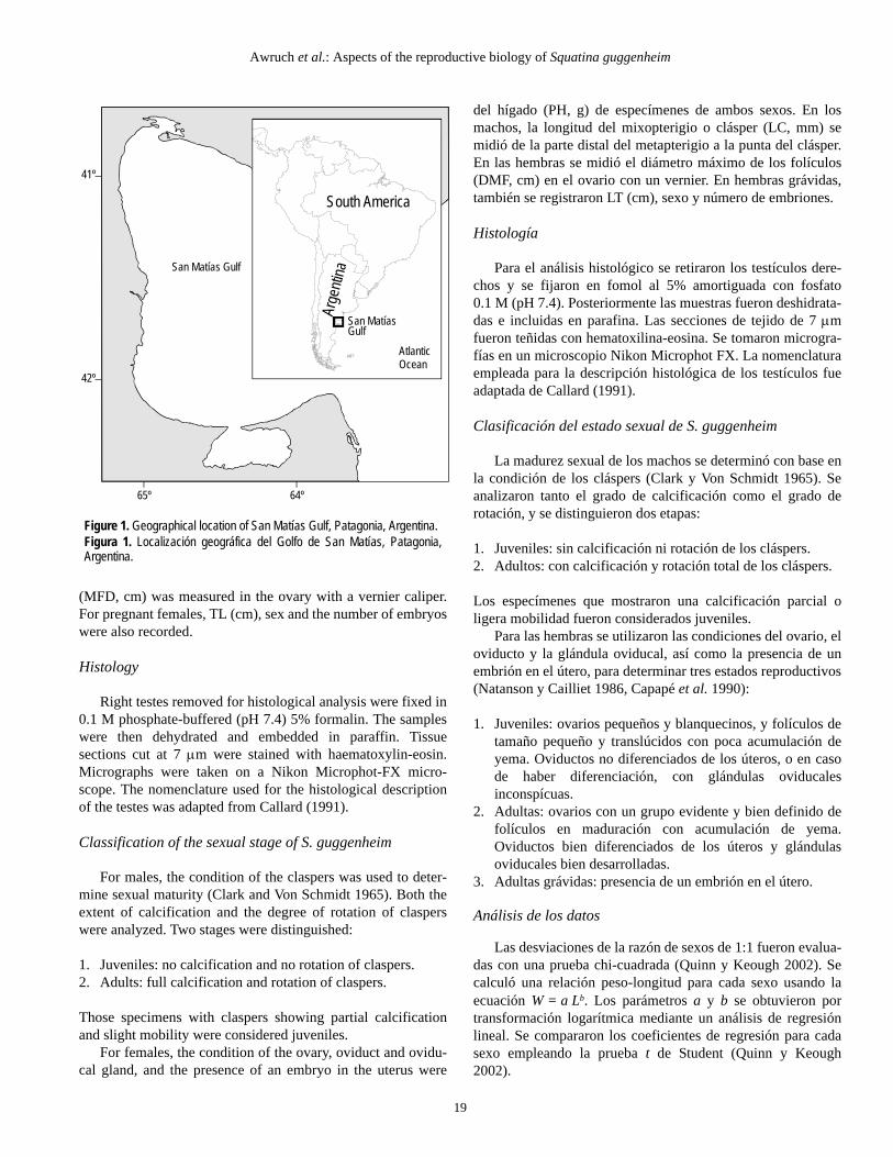
Angel sharks were obtained between January and Decem-ber 1996 from samples caught in bottom-trawl nets off thecoast of the San Matías Gulf (fig. 1). Three-hour commercialand 30-min research hauls were made at depths ranging from20 to 130 m, using a 29-m commercial otter trawl with rectan-gular doors.
Biological measurements
Measurements of total body length (TL, cm), body weight(BW, g), gonadal weight (GW, g), and liver weight (LW, g)were obtained for both sexes. In males, clasper length (CL,mm) was measured from the distal end of the metapterygium tothe tip of the clasper. In females, maximum follicular diameter
et al. 2001, Baum et al. 2003). Consecuentemente, es funda-mental para su conservación y manejo, como para el decualquier especie, contar con datos reproductivos que permitanasegurar que las poblaciones contribuyan a generacionesfuturas (Walker 2005, Tavares et al. 2006).
Todos los escuatínidos son especies demersales y seencuentran ampliamente distribuidos en aguas tropicales ytempladas desde la costa hasta 1000 m de profundidad(Compagno 1984). Se han reportado varios aspectos de labiología reproductiva de los tiburones ángeles o angelotes,incluyendo el angelote del Pacífico Squatina californica(Natanson y Cailliet 1986), el angelote adornado S.tergocellata (Bridge et al. 1998), el angelote S. occulta (Sunyey Vooren 1997), el angelote espinoso S. aculeata (Capapé et al.2005), el angelote común S. squatina, y el angelote manchadoS. oculata (Capapé et al. 1990, 2002). Los estudios sobre elangelote argentino S. guggenheim (Marini 1936) de la plata-forma continental frente al norte de Argentina (35–41º S) y elsur de Brasil (30–33º S), indican que ambos sexos alcanzan lamadurez sexual alrededor de los 75 cm de longitud total, que sereproducen lecitotróficamente, que su gestación inicia enverano y que el alumbramiento ocurre en primavera (Cousseau1973, Sunye y Vooren 1997, Cousseau y Figueroa 2001,Colonello et al. 2006). Cousseau (1973) también reportó unperiodo de descanso para las hembras durante el cual el ciclofolicular se separa temporalmente de la gravidez.
Este trabajo se ha enfocado en el tiburón ángel S.guggenheim, especie que se distribuye desde Rio Grande doSul, Brasil (30º S) hasta Rawson, Argentina (43º S) (Cousseau1973, Milessi 2001). Conocido localmente como escuadro oangelote, esta especie comúnmente se captura en aguas patagó-nicas como fauna de acompañamiento en la pesquería de lamerluza (Merluccius hubbsi) (Di Giácomo y Perier 1991).Dada la necesidad de contar con datos reproductivos para ase-gurar la sustentabilidad de la población, el objetivo delpresente estudio es proporcionar información detallada sobrela biología reproductiva de S. guggenheim en la costa dePatagonia, con base en especímenes capturados en el Golfo deSan Matías (Argentina, Atlántico suroeste).
Material y métodos
Muestreo de campo
Se capturaron especímenes de S. guggenheim entre enero ydiciembre de 1996 mediante una red de arrastre de fondo en elGolfo de San Matías (fig. 1). Se realizaron lances comercialesde 3 h, y de investigación de 30 min, a profundidades de 20 a130 m, utilizando una red de arrastre comercial de 29 m conpuertas rectángulares.
Mediciones biológicas
Se obtuvieron mediciones de longitud total del cuerpo(LT, cm), peso corporal (PC, g), peso gonadal (PG, g) y peso
Awruch et al.: Aspects of the reproductive biology of Squatina guggenheim
19
(MFD, cm) was measured in the ovary with a vernier caliper.For pregnant females, TL (cm), sex and the number of embryoswere also recorded.
Histology
Right testes removed for histological analysis were fixed in0.1 M phosphate-buffered (pH 7.4) 5% formalin. The sampleswere then dehydrated and embedded in paraffin. Tissuesections cut at 7 μm were stained with haematoxylin-eosin.Micrographs were taken on a Nikon Microphot-FX micro-scope. The nomenclature used for the histological descriptionof the testes was adapted from Callard (1991).
Classification of the sexual stage of S. guggenheim
For males, the condition of the claspers was used to deter-mine sexual maturity (Clark and Von Schmidt 1965). Both theextent of calcification and the degree of rotation of clasperswere analyzed. Two stages were distinguished:
1. Juveniles: no calcification and no rotation of claspers.2. Adults: full calcification and rotation of claspers.
Those specimens with claspers showing partial calcificationand slight mobility were considered juveniles.
For females, the condition of the ovary, oviduct and ovidu-cal gland, and the presence of an embryo in the uterus were
del hígado (PH, g) de especímenes de ambos sexos. En losmachos, la longitud del mixopterigio o clásper (LC, mm) semidió de la parte distal del metapterigio a la punta del clásper.En las hembras se midió el diámetro máximo de los folículos(DMF, cm) en el ovario con un vernier. En hembras grávidas,también se registraron LT (cm), sexo y número de embriones.
Histología
Para el análisis histológico se retiraron los testículos dere-chos y se fijaron en fomol al 5% amortiguada con fosfato0.1 M (pH 7.4). Posteriormente las muestras fueron deshidrata-das e incluidas en parafina. Las secciones de tejido de 7 μmfueron teñidas con hematoxilina-eosina. Se tomaron microgra-fías en un microscopio Nikon Microphot FX. La nomenclaturaempleada para la descripción histológica de los testículos fueadaptada de Callard (1991).
Clasificación del estado sexual de S. guggenheim
La madurez sexual de los machos se determinó con base enla condición de los cláspers (Clark y Von Schmidt 1965). Seanalizaron tanto el grado de calcificación como el grado derotación, y se distinguieron dos etapas:
1. Juveniles: sin calcificación ni rotación de los cláspers.2. Adultos: con calcificación y rotación total de los cláspers.
Los especímenes que mostraron una calcificación parcial oligera mobilidad fueron considerados juveniles.
Para las hembras se utilizaron las condiciones del ovario, eloviducto y la glándula oviducal, así como la presencia de unembrión en el útero, para determinar tres estados reproductivos(Natanson y Cailliet 1986, Capapé et al. 1990):
1. Juveniles: ovarios pequeños y blanquecinos, y folículos detamaño pequeño y translúcidos con poca acumulación deyema. Oviductos no diferenciados de los úteros, o en casode haber diferenciación, con glándulas oviducalesinconspícuas.
2. Adultas: ovarios con un grupo evidente y bien definido defolículos en maduración con acumulación de yema.Oviductos bien diferenciados de los úteros y glándulasoviducales bien desarrolladas.
3. Adultas grávidas: presencia de un embrión en el útero.
Análisis de los datos
Las desviaciones de la razón de sexos de 1:1 fueron evalua-das con una prueba chi-cuadrada (Quinn y Keough 2002). Secalculó una relación peso-longitud para cada sexo usando laecuación W = a Lb. Los parámetros a y b se obtuvieron portransformación logarítmica mediante un análisis de regresiónlineal. Se compararon los coeficientes de regresión para cadasexo empleando la prueba t de Student (Quinn y Keough2002).
Figure 1. Geographical location of San Matías Gulf, Patagonia, Argentina.Figura 1. Localización geográfica del Golfo de San Matías, Patagonia,Argentina.
65º
41º
San Matías Gulf
64º
42º
South America
Arge
ntina
AtlanticOcean
San MatíasGulf

Ciencias Marinas, Vol. 34, No. 1, 2008
20
used to classify females in three reproductive stages (Natansonand Cailliet 1986, Capapé et al. 1990):
1. Juveniles: small and whitish ovaries, follicles of small sizeand translucent with little accumulation of yolk. Oviductsnot differentiated from the uteri or, if differentiated, withinconspicuous oviducal glands.
2. Adults: ovaries containing a well-defined group of matur-ing follicles with accumulation of yolk. Oviducts welldifferentiated from the uteri and oviducal glands welldeveloped.
3. Pregnant adults: presence of embryo in uterus.
Data analysis
Sex ratio deviations from 1:1 were evaluated using the chi-square test (Quinn and Keough 2002). A length-weightrelationship was calculated for each sex using the equationW = a Lb. The parameters a and b were obtained from alogarithmic transformation using linear regression analysis.Regression coefficients for each sex were compared usingStudent’s t-test (Quinn and Keough 2002).
The gonadosomatic index (GSI) and hepatosomatic index(HSI) were calculated as follows:
GSI = (GW/BW) × 100HSI = (LW/BW) × 100
Differences in both indices between seasons were tested byone-way ANOVA (Quinn and Keough 2002), and ANOVAresidual plots were used to assess equality of variances. Datawere square-root- or log-transformed when necessary.
Size at 50% maturity was determined by logistic regressionanalysis. The logistic plot of the proportion of adult animals(P) resulted in the following equation (Walker 2005):
where Pmax is the maximum proportion (%) of animals in adultstage, l50 is the length at which 50% of the maximum propor-tion of animals is in adult condition, and l95 is the length atwhich 95% of the maximum proportion of animals is in adultcondition. The parameters for the equation were estimated byusing the Solver function in the Excel statistical package(Microsoft® Excel 2000).
The significance level was set at P = 0.05 for all analyses.
Results
A total of 254 females and 330 males were examined forthis study.
P Pmax 1 e 19( )ln– l l50–l50 l95–-------------------+
1–=
El índice gonadosomático (IGS) y el índice hepatosomático(IHS) fueron calculados de la siguiente manera:
IGS = (PG/PC) × 100IHS = (PH/PC) × 100
Se aplicó un análisis de varianza de una vía para determinarlas diferencias en ambos índices entre estaciones del año(Quinn y Keough 2002), y se utilizaron gráficas residuales detal análisis para evaluar la igualdad de varianzas. Los datosfueron transformados por raíz cuadrada o logaritmo en loscasos necesarios.
La talla de madurez de 50% se determinó mediante un aná-lisis de regresión logística. La gráfica logística de la propor-ción de animales adultos (P) dio por resultado la siguienteecuación (Walker 2005):
donde Pmax es la proporción máxima (%) de animales en estadoadulto, l50 es la longitud a la que 50% de la proporción máximade animales se encuentra en estado adulto, y l95 es la longitud ala que 95% de la proporción máxima de animales se consideraen estado adulto. Los parámetros de la ecuación fueronestimados con la función Solver del paquete estadístico Excel(Microsoft® Excel 2000).
Para todos los análisis el nivel de significancia se establecióen P = 0.05.
Resultados
En el presente estudio se examinaron un total de 254 hem-bras y 330 machos.
Sistema reproductor
El examen macroscópico del sistema reproductor de losmachos reveló testículos pares, de color rosado y formalobular, suspendidos por un mesorquio dentro de la cavidadabdominal. Se observó un órgano epigonal que cubre toda lasuperficie distal del testículo. El peso de los testículos varió de8 g (±2.33 EE) en juveniles a 36 g (±2.25 EE) en adultos. Almicroscopio óptico los testículos aparecían organizados enespermatocistos que maduraban e incrementaban de talla de lazona germinal a la zona degenerativa donde se localizan losconductos deferentes para la liberación de esperma (fig. 2).
En las hembras se encontró que sólo el ovario izquierdo erafuncional en los especímenes adultos. El ovario derecho estabareducido y aunque ocasionalmente se observaron algunosfolículos, éstos nunca se desarrollaron. El peso de los ovariosvarió de 5 g (±0.48 EE) en juveniles a 25 g (±4.88 EE) enadultas grávidas y 164 g (±13.8 EE) en adultas. El DMF fue de6 cm (media 3.2 ± 0.16 cm EE). Ambos úteros se encontraronfuncionales.
P Pmax 1 e 19( )ln– l l50–l50 l95–-------------------+
1–=

Awruch et al.: Aspects of the reproductive biology of Squatina guggenheim
21
Reproductive system
Macroscopic examination of the reproductive system inmales showed paired testes, pinkish in colour and lobular inshape, suspended by a mesorchium within the abdominalcavity. An epigonal organ was observed surrounding the entiretestis distal surface. Testis weight ranged from 8 g (±2.33 SE)in juveniles to 36 g (±2.25 SE) in adults. Under lightmicroscopy the testes were organized into spermatocysts.Spermatocysts matured and increased in size from the germinalzone to the degenerative zone where the efferent ducts arelocated for sperm release (fig. 2).
In females, only the left ovary was found to be functional inadults. The right ovary was reduced and even though somefollicles were occasionally distinguished, they were neverobserved to develop. Ovary weight ranged from 5 g (±0.48 SE)in juveniles to 25 g (±4.88 SE) in pregnant adults and 164 g(±13.8 SE) in adults. The MFD was 6 cm (mean 3.2 ± 0.16 cmSE). Both uteri were functional.
Size distribution
The female:male sex ratio was 1:1.1 for juveniles and 1:1.3for adults, not significantly different from the expected 1:1ratio (juveniles: χ2 = 2.668, d.f.1, P > 0.05; adults: χ2 = 3.80,d.f.1, P > 0.05). The seasonal analysis, however, showedsignificant differences in sex abundance. Adult males werepredominant in the austral autumn and winter, and adultfemales in summer (χ2 = 4.319, d.f.1, P < 0.05), while juvenilemales dominated in spring (χ2 = 5.523, d.f.1, P < 0.05).
The maximum TL recorded was 95 cm for both sexes.Length-frequency distribution showed a single peak withmodal frequencies of 75–90 and 80–95 cm for males andfemales, respectively (fig. 3a). Both sexes had similar length-weight relationships. The regression coefficients did not differsignificantly between sexes (males: W = 2.89 × LT
1.905, r2 =0.97; females: W = 3.13 × LT
2.308, r2 = 0.96; Student’s t-test: t =1.994, d.f.570, P > 0.05) (fig. 3b).
Length at sexual maturity
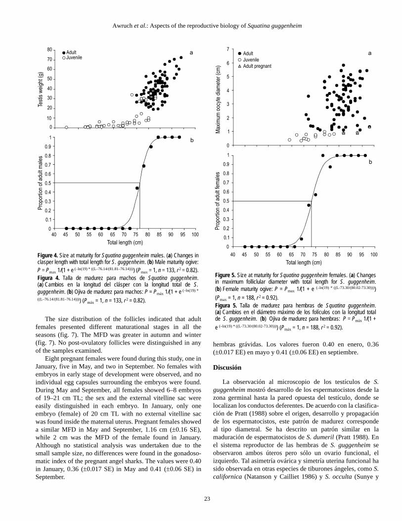
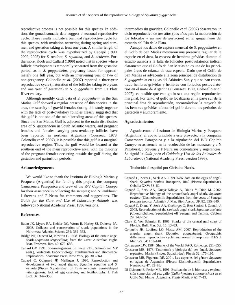
In males, CL increased abruptly at 75–80 cm TL (fig. 4a).The largest juvenile male was 84 cm TL, while the smallestadult male was 75 cm TL. Males reached 50% sexual maturityat 76 cm TL (fig. 4b). In females, MFD increased noticeably inthose specimens larger than 79 cm TL (fig. 5a). All femalessmaller than 71 cm TL were juveniles, whereas females largerthan 83 cm TL were adults. Females reached 50% sexualmaturity at 73 cm TL (fig. 5b).
Reproductive cycle
For adult males, no significant differences were found inthe values of the gonadosomatic index (F = 2.55; d.f.3, 196; P >0.05) between seasons (fig. 6a). Similarly, both macroscopicand microscopic examinations of the testes showed no change
Distribución de talla
La proporción de sexos (hembras:machos) fue 1:1.1 parajuveniles y 1:1.3 para adultos, sin diferir significativamente dela razón esperada de 1:1 (juveniles: χ2 = 2.668, g.l.1, P > 0.05;adultos: χ2 = 3.80, g.l.1, P > 0.05). Sin embargo, el análisisestacional mostró diferencias significativas en la abundanciade sexos. Los machos adultos predominaron en otoño einvierno (hemisferio sur) y las hembras adultas en verano(χ2 = 4.319, g.l.1, P < 0.05), mientras que los machos juvenilespredominaron en primavera (χ2 = 5.523, g.l.1, P < 0.05).
Se registró una LT máxima de 95 cm para ambos sexos. Ladistribución de frecuencia de longitud mostró un solo pico confrecuencias modales de 75–90 y 80–95 cm para machos yhembras, respectivamente (fig. 3a). Ambos sexos presentaronrelaciones peso-longitud similares. Los coeficientes de regre-sión no difirieron significativamente entre sexos (machos: W =2.89 × LT
1.905, r2 = 0.97; hembras: W = 3.13 × LT2.308, r2 = 0.96;
prueba t de Student: t = 1.994, g.l.570, P > 0.05) (fig. 3b).
Longitud de madurez sexual
En los machos la LC incrementó abruptamente a los 75–80 cm LT (fig. 4a). El juvenil más grande midió 84 cm LT y elmacho adulto más pequeño, 75 cm LT. Los machos alcanzaronla talla de madurez sexual de 50% a los 76 cm LT (fig. 4b).En las hembras el DMF incrementó notablemente en los espe-címenes mayores a 79 cm LT (fig. 5a). Todas las hembrasmenores de 71 cm LT fueron juveniles y las mayores de 83 cmLT fueron adultas. Las hembras alcanzaron la talla de madurezsexual de 50% a los 73 cm LT (fig. 5b).
Ciclo reproductivo
En los machos adultos no se encontraron diferencias signi-ficativas en los valores del índice gonadosomático (F = 2.55;g.l.3, 196; P > 0.05) entre las estaciones del año (fig. 6a). Lasevaluaciones macro y microscópica de los testículos tampocomostraron cambios temporales. La forma, el color y la lobula-ción de los testículos, así como la proporción de los diferentesestadios de madurez de los espermatocistos fueron similaresdurante el periodo de estudio.
En las hembras adultas los niveles del índice gonadoso-mático fueron similares durante verano y otoño, aumentandosignificativamente en invierno para luego disminuir en prima-vera (F = 6.10; g.l.3, 134; P < 0.05) (fig. 6b). En contraste, larelación entre el índice hepatosomático y la temporada nomostró ningún cambio estacional evidente (F = 2.85; g.l.3, 134;P > 0.05) (fig. 6c).
La distribución del tamaño de los folículos mostró que lashembras adultas presentaron diferentes estadios de madurez entodas las estaciones (fig. 7). El DMF fue mayor en otoño einverno (fig. 7). No se distinguieron folículos postovulatoriosen ninguna de las hembras examinadas.

Ciencias Marinas, Vol. 34, No. 1, 2008
22
over time. Similar shape, colour and lobulation of the testes,and similar proportion of the different spermatocyst matura-tional stages were found during the study period.
For adult females, similar levels in the gonadosomaticindex were found during summer and autumn, followed by asignificant increase in winter and subsequent decline in spring(F = 6.10; d.f.3, 134; P < 0.05) (fig. 6b). In contrast, the relation-ship between hepatosomatic index and season showed no clearseasonal change (F = 2.85; d.f.3, 134; P > 0.05) (fig. 6c).
Se encontraron ocho hembras grávidas durante el estudio,una en enero, cinco en mayo y dos en septiembre. No seobservaron hembras con embriones en estado temprano dedesarrollo, ni cápsulas de huevo individuales rodeando losembriones. En mayo y septiembre todas las hembras mostraronde seis a ocho embriones de 19 a 21 cm LT, siendo fácilmentedistinguibles sexo y saco vitelino exterior en cada embrión. Enenero, dentro del útero maternal sólo se encontró un embrión(hembra) de 20 cm LT sin evidencia de saco vitelino exterior.El DMF de las hembras grávidas fue similar en mayo y sep-tiembre, de 1.16 cm (±0.16 EE), mientras que el de la hembraencontrada en enero fue de 2 cm. Aunque no se realizó un aná-lisis estadístico debido al reducido tamaño de muestra, no seencontraron diferencias en el índice gonadosomático de las
Figure 2. Cross-section of Squatina guggenheim testis: DZ, degenerativezone; GZ, germinal zone. Light microscope photograph ×40. Stain:haematoxylin-eosin.Figura 2. Sección transversal de testículo de Squatina guggenheim: DZ,zona degenerativa; GZ, zona germinal. Fotografía con microscopio óptico×40. Tinción: hematoxilina-eosina.
Figure 3. Squatina guggenheim size distribution. (a) Length-frequencydistribution for males (n = 330) and females (n = 254). (b) Length-weightrelationship for males and females. Data were logarithmically transformed;the linear regression slopes of the logarithmic transformations did not differsignificantly. Males (—), females (—).Figura 3. Distribución de tallas de Squatina guggenheim. (a) Distribuciónde frecuencias de longitud para machos (n = 330) y hembras (n = 254).(b) Relación peso-longitud para machos y hembras. Los datos fuerontransformados logarítmicamente; las pendientes de la regresión lineal delas transformaciones logarítmicas no difirieron significativamente. Machos(—), hembras (—).
Awruch et al.: Aspects of the reproductive biology of Squatina guggenheim
23
The size distribution of the follicles indicated that adultfemales presented different maturational stages in all theseasons (fig. 7). The MFD was greater in autumn and winter(fig. 7). No post-ovulatory follicles were distinguished in anyof the samples examined.
Eight pregnant females were found during this study, one inJanuary, five in May, and two in September. No females withembryos in early stage of development were observed, and noindividual egg capsules surrounding the embryos were found.During May and September, all females showed 6–8 embryosof 19–21 cm TL; the sex and the external vitelline sac wereeasily distinguished in each embryo. In January, only oneembryo (female) of 20 cm TL with no external vitelline sacwas found inside the maternal uterus. Pregnant females showeda similar MFD in May and September, 1.16 cm (±0.16 SE),while 2 cm was the MFD of the female found in January.Although no statistical analysis was undertaken due to thesmall sample size, no differences were found in the gonadoso-matic index of the pregnant angel sharks. The values were 0.40in January, 0.36 (±0.017 SE) in May and 0.41 (±0.06 SE) inSeptember.
hembras grávidas. Los valores fueron 0.40 en enero, 0.36(±0.017 EE) en mayo y 0.41 (±0.06 EE) en septiembre.
Discusión
La observación al microscopio de los testículos de S.guggenheim mostró desarrollo de los espermatocistos desde lazona germinal hasta la pared opuesta del testículo, donde selocalizan los conductos deferentes. De acuerdo con la clasifica-ción de Pratt (1988) sobre el origen, desarrollo y propagaciónde los espermatocistos, este patrón de madurez correspondeal tipo diametral. Se ha descrito un patrón similar en lamaduración de espermatocistos de S. dumeril (Pratt 1988). Enel sistema reproductor de las hembras de S. guggenheim seobservaron ambos úteros pero sólo un ovario funcional, elizquierdo. Tal asimetría ovárica y simetría uterina funcional hasido observada en otras especies de tiburones ángeles, como S.californica (Natanson y Cailliet 1986) y S. occulta (Sunye y
Figure 5. Size at maturity for Squatina guggenheim females. (a) Changesin maximum folliclular diameter with total length for S. guggenheim.(b) Female maturity ogive: P = Pmax 1/(1 + e (–ln(19) * ((L-73.30/(80.02-73.30))))(Pmax = 1, n = 188, r 2 = 0.92).Figura 5. Talla de madurez para hembras de Squatina guggenheim.(a) Cambios en el diámetro máximo de los folículos con la longitud totalde S. guggenheim. (b) Ojiva de madurez para hembras: P = Pmáx 1/(1 +e (–ln(19) * ((L-73.30/(80.02-73.30)))) (Pmáx = 1, n = 188, r 2 = 0.92).
Figure 4. Size at maturity for Squatina guggenheim males. (a) Changes inclasper length with total length for S. guggenheim. (b) Male maturity ogive:P = Pmax 1/(1 + e (–ln(19) * ((L–76.14/(81.81–76.14)))) (Pmax = 1, n = 133, r 2 = 0.82).Figura 4. Talla de madurez para machos de Squatina guggenheim.(a) Cambios en la longitud del clásper con la longitud total de S.guggenheim. (b) Ojiva de madurez para machos: P = Pmáx 1/(1 + e (–ln(19) *
((L–76.14/(81.81–76.14)))) (Pmáx = 1, n = 133, r 2 = 0.82).

Ciencias Marinas, Vol. 34, No. 1, 2008
24
Discussion
The microscopic observation of S. guggenheim testesshowed spermatocyst development spreading from thegerminal zone to the opposite wall of the testis, where theefferent duct system is located. According to Pratt’s (1988)classification of the origin, development, and propagation ofspermatocysts, the present maturity pattern corresponds to thediametric type. A similar pattern of spermatocyst maturationwas described for S. dumeril (Pratt 1988). The reproductivesystem of S. guggenheim females showed both uteri and onlythe left ovary functional. In concordance, such functional uter-ine symmetry and ovarian asymmetry has been reported inother angel shark species, such as S. californica (Natanson andCailliet 1986) and S. occulta (Sunye and Vooren 1997). Incontrast, symmetrically functional uteri and ovaries weredescribed for S. oculata (Capapé et al. 1990, 2002), S.tergocellata (Bridge et al. 1998), S. squatina (Capapé et al.1990), and S. aculeata (Capapé et al. 2005).
Changes in the S. guggenheim sex ratio were not apparentthroughout the year; however, grouping the data by seasonindicated that adult males were predominant in autumn andwinter, whereas adult females dominated in summer. Changesin sex ratio (with higher proportion of adult females over malesthroughout the year) have also been reported for S. tergocellata(Bridge et al. 1998), S. oculata (Capapé et al. 2002), and S.aculeata (Capapé et al. 2005). These differences in sex ratio inSquatina species could be the consequence of sexual segrega-tion. Segregation of the sexes within a species has beenreported among elasmobranch populations as a result of acombination of social, reproductive, and sex-specific factors(Klimley 1987, Sims et al. 2001, Sims 2005).
No sexual size dimorphism was found in S. guggenheim.Both sexes reached similar TLs and showed similar length-weight relationships. In contrast, sexual dimorphism by sizehas been reported for other squatinids, such as S. tergocellata(Bridge et al. 1998), S. squatina, and S. oculata (Capapé et al.1990, 2002), with females attaining larger sizes than males. Inaddition, Bridge et al. (1998) reported that the differences insize for S. tergocellata arise at the onset of sexual maturitywhen females begin to grow larger than males. It is interestingto note that both ovaries are functional in those Squatina spe-cies showing sexual dimorphism. This fact suggests that thelarger female sizes may be associated with a greater body cav-ity in order to accommodate large quantities of reproductivematerial.
In the present study, both sexes of S. guggenheim reachedsexual maturity at similar sizes (between 73 and 76 cm).Similar size at maturity was reported for S. guggenheim fromnorthern Argentinean (Cousseau 1973, Colonello et al. 2007)and southern Brazilian waters (Sunye and Vooren 1997). Incontrast, the size at maturity of S. guggenheim females fromthe La Plata River estuary (northern Argentina and Uruguay)was slightly smaller (71 mm TL) (Colonello et al. 2007).
Vooren 1997). En contraste, se han descrito úteros y ovariossimétricamente funcionales en S. oculata (Capapé et al. 1990,2002), S. tergocellata (Bridge et al. 1998), S. squatina (Capapéet al. 1990) y S. aculeata (Capapé et al. 2005).
No se observaron cambios aparentes en la proporción desexos de S. guggenheim durante el año; sin embargo, la agrupa-ción de los datos por estación indicó que predominaron losmachos adultos en otoño e invierno y las hembras adultas enverano. Se han encontrado cambios en la razón de sexos (conmayor proporción de hembras adultas que machos a lo largodel año) para S. tergocellata (Bridge et al. 1998), S. oculata(Capapé et al. 2002) y S. aculeata (Capapé et al. 2005). Estasdiferencias en la razón de sexos de las especies de Squatinapuede ser consecuencia de una segregación sexual. Se haobservado tal segregación intraespecífica de sexos en poblacio-nes de elasmobranquios como resultado de una combinaciónde factores sociales, reproductivos y sexuales (Klimley 1987,Sims et al. 2001, Sims 2005).
No se observó dimorfismo sexual en cuanto a la talla de S.guggenheim. Ambos sexos alcanzaron LTs similares y mostra-ron relaciones peso-longitud similares. En contraste, se hainformado sobre tal dimorfismo sexual en otros escuatínidos,como S. tergocellata (Bridge et al. 1998), S. squatina y S.oculata (Capapé et al. 1990, 2002), con las hembras alcan-zando mayores tallas que los machos. Asimismo, Bridge et al.(1998) encontraron que las diferencias en las tallas de S.tergocellata comienzan al inicio de la madurez sexual cuandolas hembras empiezan a crecer más que los machos. Es intere-sante mencionar que ambos ovarios son funcionales en lasespecies de Squatina que muestran dimorfismo sexual. Estehecho sugiere que las tallas mayores de las hembras puedenestar asociadas con una cavidad corporal más grande parapoder acomodar grandes cantidades de material reproductivo.
En el presente trabajo el tamaño de madurez sexual fuesimilar para ambos sexos de S. guggenheim (entre 73 y 76 cm).Una talla de madurez similar fue registrada para S. guggenheimde aguas del norte de Argentina (Cousseau 1973, Colonello etal. 2007) y del sur de Brasil (Sunye y Vooren 1997); sinembargo, hembras de S. guggenheim del estuario del Río de laPlata (costas del norte de Argentina y Uruguay) mostraron unatalla ligeramente menor (71 mm TL) (Colonello et al. 2007).
El índice gonadosomático de los machos de S. guggenheimno varió estacionalmente. Además, los testículos recolectadosa lo largo del año contenían espermatocistos maduros, lo queindica que los machos son capaces de aparearse durante todo elaño. Aunque no se han registrado resultados comparables parala mayoría de los machos escuatínidos estudiados (Cousseau1973; Capapé et al. 1990, 2002, 2005; Bridge et al. 1998), sehan descrito resultados similares para S. californica (Natansony Cailliet 1986).
En las hembras de S. guggenheim el índice gonadosomáticomostró un pico en invierno y los mayores valores del DMF seobtuvieron en otoño e invierno. Estos resultados indican que lamadurez folicular termina durante las estaciones frías (en elhemisferio sur) y que la ovulación ocurre en primavera.

Awruch et al.: Aspects of the reproductive biology of Squatina guggenheim
25
In S. guggenheim males, the gonadosomatic index did notvary seasonally. In addition, testes collected at various timesthroughout the year contained mature spermatocysts, indicat-ing that males are capable of mating all year round. Althoughno comparable results have been reported for the majority ofthe squatinid males studied (Cousseau 1973; Capapé et al.
Coincidentemente, especímenes de esta misma especie captu-rados en aguas del norte de Argentina presentaron ovulación enprimavera/verano (Cousseau 1973, Colonello et al. 2007),mientras que los capturados en aguas brasileñas lo hicieron enverano (Sunye y Vooren 1997). En otros escuatínidos como S.tergocellata (Bridge et al. 1998), S. squatina (Capapé et al.1990) y S. oculata (Capapé et al. 2002), se ha observado que laovulación ocurre de manera similar a finales de la temporadafría.
Figure 6. Seasonal changes in the gonadosomatic index (GSI) andhepatosomatic index (HIS) of Squatina guggenheim adults: GSI for males(a) and females (b), and HIS for females (c). Values are means ±SE.Different letters show significant differences between seasons. Numbersabove x axis indicate sample sizes.Figura 6. Cambios estacionales en el índice gonadosomático (GSI) y elíndice hepatosomático (HIS) de adultos de Squatina guggenheim (valoresmedios ± error estándar): GSI para machos (a) y hembras (b), y HIS parahembras (c). Las letras diferentes indican diferencias significativas entreestaciones y los números sobre el eje x indican el tamaño de las muestras.
Figure 7. Proportion of maximum follicular diameter for Squatinaguggenheim females in each season.Figura 7. Proporción del diámetro máximo de los folículos para hembrasde Squatina guggenheim en cada estación del año.

Ciencias Marinas, Vol. 34, No. 1, 2008
26
1990, 2002, 2005; Bridge et al. 1998), similar results havebeen described for S. californica (Natanson and Cailliet 1986).
In S. guggenheim females, the gonadosomatic indexpeaked in winter and the MFD showed the highest values inautumn and winter. These results indicate that follicle matura-tion finishes during cold seasons (in the Southern Hemisphere)and ovulation occurs in spring. In concordance, ovulation wasreported to occur in spring/summer in S. guggenheim speci-mens caught in northern Argentinean waters (Cousseau 1973,Colonello et al. 2007), while ovulation takes place in summerin specimens caught in Brazilian waters (Sunye and Vooren1997). For other squatinids such as S. tergocellata (Bridge etal. 1998), S. squatina (Capapé et al. 1990), and S. oculata(Capapé et al. 2002), ovulation was similarly observed to occurat the end of the cold season.
The MFDs observed in gravid females, with near-termembryos, were less than half of the follicle size at ovulation.This result suggests that these sharks are unable to ovulatesoon after parturition. These observations are consistent withthose reported for other Squatina species (Cousseau 1973;Capapé et al. 1990, 2002, 2005; Cousseau and Figueroa 2001).Capapé (1990) mentioned that the Squatinidae family exhib-ited arrested development of the follicles during pregnancy,with follicle development blocked from the beginning to themiddle of gestation.
As reported for S. oculata (Capapé et al. 1990, 2002), S.tergocellata (Bridge et al. 1998), and S. aculeata (Capapé et al.2005), a single egg case surrounding all uterine ova was notobserved in S. guggenheim. However, Sunye and Vooren(1997) reported uterine-cloacal gestation in S. guggenheim andS. occulta, describing a single egg capsule surrounding alluterine ova until embryos reach 6 cm TL, and Capapé et al.(1999) reported a thick and dense jelly-like structure surround-ing the fertilized eggs in the uterus of S. oculata. Since onlynear full-term embryos were found in this study, further studieson S. guggenheim embryo development are necessary to deter-mine the possible presence of egg capsules and to definewhether the angel sharks from Patagonian waters possessuterine-cloacal gestation.
Different gestation periods have been reported for differentangel shark species: 6–12 months for S. tergocellata (Bridge etal. 1998); 10 months for S. californica, with parturition occur-ring from March to June and mating soon after (Natanson andCailliet 1986); and at least one year for S. oculata (Capapé etal. 2002) and S. aculeata (Capapé et al. 2005). Particularly, agestation period of 10–12 months was reported for the other S.guggenheim species from Brazil (Sunye and Vooren 1997) andnorthern Argentina (Cousseau 1973, Colonello et al. 2007).The few gravid females found during this study made it diffi-cult to establish the length of the gestation period. All pregnantfemales with near-term embryos were found in January, Mayand September, potentially suggesting a continuous reproduc-tive cycle. However, follicle development and the MFD foundin females carrying full-term embryos showed that females arenot able to ovulate soon after parturition, so a continuous
Los DMFs observados en hembras grávidas con embrionescasi terminales, midieron menos de la mitad del tamaño folicu-lar durante la ovulación. Este resultado sugiere que estos tibu-rones no pueden ovular poco después del alumbramiento. Estasobservaciones coinciden con lo descrito para otras especies deSquatina (Cousseau 1973; Capapé et al. 1990, 2002, 2005;Cousseau y Figueroa 2001). Capapé (1990) menciona que lafamilia Squatinidae exhibía un desarrollo retardado de los folí-culos durante la gravidez, con el desarrollo folicular bloqueadodesde el inicio hasta la mitad de la gestación.
Al igual que ha sucedido con S. oculata (Capapé et al.1990, 2002), S. tergocellata (Bridge et al. 1998) y S. aculeata(Capapé et al. 2005), no se observó ni una sola cápsula dehuevo rodeando los huevos uterinos en S. guggenheim. Sunyey Vooren (1997) registraron gestación uterina-cloacal en S.guggenheim y S. occulta, y describen una cápsula de huevo querodea todos los huevos uterinos hasta que los embriones alcan-zan 6 cm LT. Capapé et al. (1999) observaron una estructuragelatinosa gruesa y densa que cubría los huevos fertilizados enel útero de S. oculata. En este estudio sólo se encontraronembriones casi terminales, por lo que se requieren estudios adi-cionales sobre el desarrollo embrionario de S. guggenheim paradeterminar la posible presencia de cápsulas de huevo y definirsi los tiburones ángeles de aguas patagónicas presentan gesta-ción uterina-cloacal.
Se han encontrado diferentes periodos de gestación paradiferentes especies de tiburón ángel: de 6 a 12 meses para S.tergocellata (Bridge et al. 1998); de 10 meses para S.californica, con alumbramiento de marzo a junio y aparea-miento poco después (Natanson y Cailliet 1986); y de por lomenos un año para S. oculata (Capapé et al. 2002) y S.aculeata (Capapé et al. 2005). En particular, se han mencio-nado periodos de gestación de 10 a 12 meses para otrasespecies de S. guggenheim de Brasil (Sunye y Vooren 1997) yel norte de Argentina (Cousseau 1973, Colonello et al. 2007).En vista de las pocas hembras grávidas encontradas en esteestudio resultó difícil establecer la duración del periodo degestación. Todas las hembras grávidas con embriones casi ter-minales fueron encontradas en enero, mayo y septiembre, loque podría sugerir un ciclo reproductivo continuo. Sinembargo, el desarrollo de los folículos, y los DMFs observadosen hembras con embriones terminales, indicaron que lashembras no son capaces de ovular poco después del alumbra-miento, por lo que no es factible un proceso reproductivocontinuo para esta especie. Además, los datos gonadosomáti-cos sugieren un ciclo reproductivo estacional. En su conjuntoestos resultados indican un ciclo reproductivo bianual para laespecie, con ovulación durante primavera y verano y unperiodo de gestación de por lo menos un año. Capapé (1990,2002, 2005) ha supuesto duraciones similares para los ciclosreproductivos de S. oculata, S. squatina y S. aculeata. Asi-mismo, Koob y Callard (1999) notaron que en las especiescuyo desarrollo folicular está temporalmente separado delperiodo de gestación, como en S. guggenheim, la gravidez duróaproximadamente un año completo, pero con un año o dos
Awruch et al.: Aspects of the reproductive biology of Squatina guggenheim
27
reproductive process is not possible for this species. In addi-tion, the gonadosomatic data suggest a seasonal reproductivecycle. These results indicate a biannual reproductive cycle forthis species, with ovulation occurring during spring and sum-mer, and gestation taking at least one year. A similar length ofthe reproductive cycle was hypothesized by Capapé (1990,2002, 2005) for S. oculata, S. squatina, and S. aculeata. Fur-thermore, Koob and Callard (1999) noted that in species wherefollicle development is temporally separated from the gestationperiod, as in S. guggenheim, pregnancy lasted for approxi-mately one full year, but with an intervening year or two ofnon-pregnancy. Colonello et al. (2007) reported a three-yearreproductive cycle (maturation of the follicles taking two yearsand one year of gestation) in S. guggenheim from La PlataRiver estuary.
Although monthly catch data of S. guggenheim in the SanMatías Gulf showed a regular presence of this species in thearea, the scarcity of gravid females during this study togetherwith the lack of post-ovulatory follicles clearly suggested thatthis gulf is not one of the main breeding areas of this species.Since the San Matías Gulf is adjacent to the main distributionarea of S. guggenheim in South Atlantic waters, and pregnantfemales and females carrying post-ovulatory follicles havebeen reported in northern Argentina (Cousseau 1973,Colonello et al. 2007), it is possible that this gulf is a marginalreproductive region. Thus, the gulf would be located at thesouthern end of the main reproductive area, with the majorityof the pregnant females occurring outside the gulf during thegestation and parturition periods.
Acknowledgements
We would like to thank the Instituto de Biología Marina yPesquera (Argentina) for funding this project; the companyCamaronera Patagónica and crew of the R/V Capitán Canepafor their assistance in collecting the samples; and N Pankhurst,J Stevens and F Neira for comments and suggestions. TheGuide for the Care and Use of Laboratory Animals wasfollowed (National Academy Press, 1996 version).
References
Baum JK, Myers RA, Kehler DG, Worm B, Harley SJ, Doherty PA.2003. Collapse and conservation of shark populations in theNorthwest Atlantic. Science 299: 389–392.
Bridge NF, Duncan M, Newton G. 1998. Biology of the ornate angelshark (Squatina tergocellata) from the Great Australian Bight.Mar. Freshwat. Res. 49: 679–686.
Callard GV. 1991. Spermatogenesis. In: Pang PTK, Schreibman MP(eds.), Vertebrate Endocrinology: Fundamentals and BiomedicalImplications. Academic Press, New York, pp. 303–341.
Capapé C, Quignard JP, Mellinger J. 1990. Reproduction anddevelopment of two angel sharks, Squatina squatina and S.oculata (Pisces: Squatinidae), off Tunisian coasts: Semi-delayedvitellogenesis, lack of egg capsules, and lecithotrophy. J. FishBiol. 37: 347–356.
intermedios sin gravidez. Colonello et al. (2007) observaron unciclo reproductivo de tres años (dos años para la maduración delos folículos y un año de gestación) en S. guggenheim delestuario del Río de la Plata.
Aunque los datos de captura mensual de S. guggenheim enel Golfo de San Matías mostraron una presencia regular de laespecie en el área, la escasez de hembras grávidas durante elestudio aunada a la falta de folículos postovulatorios indicanclaramente que el Golfo de San Matías no es una de las princi-pales áreas de crianza de esta especie. Dado que el Golfo deSan Matías es adyacente a la zona principal de distribución deS. guggenheim en aguas del Atlántico Sur, y que se han encon-trado hembras grávidas y hembras con folículos postovulato-rios en el norte de Argentina (Cousseau 1973, Colonello et al.2007), es posible que este golfo sea una región reproductivamarginal. Por tanto, el golfo se localizaría en la parte sur de laprincipal área de reproducción, encontrándose la mayoría delas hembras grávidas afuera del golfo durante los periodos degestación y alumbramiento.
Agradecimientos
Agradecemos al Instituto de Biología Marina y Pesquera(Argentina) el apoyo brindado a este proyecto; a la compañíaCamaronera Patagónica y a la tripulación del B/O CapitánCanepa su asistencia en la recolección de las muestras; y a NPankhurst, J Stevens y F Neira sus comentarios y sugerencias.Se siguió la Guía para el Cuidado y Uso de los Animales deLaboratorio (National Academy Press, versión 1996).
Traducido al español por Christine Harris.
Capapé C, Zorzi G, Seck AA. 1999. New data on the eggs of angel-shark, Squatina oculata Bonaparte, 1840 (Pisces: Squatinidae).Oebalia XXV: 53–60.
Capapé C, Seck AA, Gueye-Ndiaye A, Diatta Y, Diop M. 2002.Reproductive biology of the smoothback angel shark, Squatinaoculata (Elasmobranchii: Squatinidae), from the coast of Senegal(eastern tropical Atlantic). J. Mar. Biol. Assoc. UK 82: 635–640.
Capapé C, Diatta Y, Seck AA, Guélorget O, Ben Souissi J, Zaouali J.2005. Reproduction of the sawback angel shark Squatina aculeata(Chondrichthyes: Squatinidae) off Senegal and Tunisia. Cybium29: 147–157.
Clark E, Von Schmidt K. 1965. Sharks of the central gulf coast ofFlorida. Bull. Mar. Sci. 15: 13–83.
Colonello JH, Lucifora LO, Massa AM. 2007. Reproduction of theangular angel shark (Squatina guggenheim): Geographicdifferences, reproductive cycle, and sexual dimorphism. ICES J.Mar. Sci. 64: 131–140.
Compagno LJV. 1984. Sharks of the World. FAO, Rome, pp. 251–655.Cousseau MB. 1973. Taxonomía y biología del pez ángel, Squatina
argentina, Marini (Pisces, Squatinidae). Physis 32: 175–195.Cousseau MB, Figueroa DE. 2001. Las especies del género Squatina
en aguas de Argentina (Pisces: Elasmobranchii: Squatinidae).Neotrópica 47: 85–86.
Di Giácomo E, Perier MR. 1991. Evaluación de la biomasa y explota-ción comercial del pez gallo (Callorhynchus callorhynchus) en elGolfo San Matías, Argentina. Frente Marít. 9(A): 7–13.

Ciencias Marinas, Vol. 34, No. 1, 2008
28
Graham KJ, Andrew NL, Hodgson KE. 2001. Changes in relativeabundance of sharks and rays on Australian South East Fisherytrawl grounds after twenty years of fishing. Mar. Freshwat. Res.52: 549–61.
Klimley AP. 1987. The determinants of sexual segregation in thescalloped hammerhead shark, Sphyrna lewini. Environ. Biol. Fish.18: 27–40.
Koob TJ, Callard IP. 1999. Reproductive endocrinology of femaleelasmobranchs: Lessons from the little skate (Raja erinacea) andspiny dogfish (Squalus acanthias). J. Exp. Zool. 284: 557–574.
Milessi A, Vögler R, Bazzino G. 2001. Identificación de tres especiesdel genero Squatina (Elasmobranchii, Squatinidae) en la ZonaComún de Pesca Argentino-Uruguaya (ZCPAU). Gayana 65:167–172.
Natanson LJ, Cailliet GM. 1986. Reproduction and development ofthe Pacific angel shark, Squatina californica, off Santa Barbara,California. Copeia 4: 987–994.
Pauly D, Christensen V, Dalsgaard J, Froese R, Torres F. 1998.Fishing down marine food webs. Science 279: 860–863.
Pratt JHL. 1988. Elasmobranch gonad structure: A description andsurvey. Copeia 3: 719–729.
Quinn GP, Keough MJ. 2002. Experimental Design and Data Analysisfor Biologists. Cambridge Univ. Press, Cambridge, 538 pp.
Sims DW. 2005. Differences in habitat selection and reproductivestrategies of male and female sharks. In: Ruckstuhl KE, Neuhaus
P (eds.), Sexual Segregation in Vertebrates: Ecology of the TwoSexes. Cambridge Univ. Press, Cambridge, pp. 127–147.
Sims DW, Nash JP, Morritt D. 2001. Movements and activity of maleand female dogfish in a tidal sea lough: Alternative behaviouralstrategies and apparent sexual segregation. Mar. Biol. 139: 1165–1175.
Stevens JD, Bonfil R, Dulvy NK, Walker PA. 2000. The effects offishing on sharks, rays, and chimaeras (Chondrichthyans), andthe implications for marine ecosystem. ICES J. Mar. Sci. 57:476–494.
Sunye PS, Vooren CM. 1997. On cloacal gestation in angel sharksfrom southern Brazil. J. Fish Biol. 50: 86–94.
Tavares R, Lemus M, Chung KS. 2006. Evaluation of theinstantaneous growth of juvenile smooth dogfish sharks (Musteluscanis) in their natural habitat, based on the RNA/DNA ratio.Cienc. Mar. 32: 297–302.
Walker TI. 1998. Can shark resources be harvested sustainably? Aquestion revisited with a review of shark fisheries. Mar. Freshwat.Res. 49: 533–572.
Walker TI. 2005. Reproduction in fisheries science. In: Hamlet W(ed.), Reproductive Biology and Phylogeny of Chondrichthyes:Sharks, Rays and Chimaeras. Einfield, New Hampshire, pp. 81–127.
Recibido en marzo de 2007;aceptado en noviembre de 2008.