6. mass spectrometry adv. inst. techs. how does it work? a very small amount of sample is bombarded...
TRANSCRIPT

6. Mass SpectrometryAdv. Inst. Techs
How does it work?
• a very small amount of sample is bombarded by a beam of high energy (usually electron beam)
• produces a positively charged species• M M+ + e• known as the molecular ion (same mass as the parent compound)• target molecules fly apart into a number of smaller pieces• all the positive ions are separated by masses to produce the spectrum• peaks corresponding to the break up of the molecule are called the
fragmentation pattern
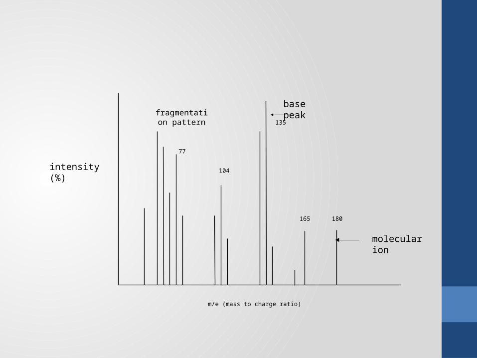
m/e (mass to charge ratio)
fragmentation pattern
intensity (%)
molecular ion
base peak
180
135
165
104
77
Interpreting mass spectra
1. Molecular weight• in most cases, some of the molecular ion remains unfragmented• it produces a peak in the spectrum which is the highest mass peak• this mass peak is most likely equal to the molecular weight of the compound• known as the molecular ion• example spectrum - 180• most intense peaks will have a peak 1 higher (due to presence of C-13)

• each molecule will always fragment in the same way• compounds may be positively identified by a comparison of their mass spectra• MS of very similar compounds are very similar• therefore, not as good a “fingerprint” as IR
2. Molecular fingerprint

• MS shows a peak for each isotope of a mixture• see the peaks due to both the 37Cl and 35Cl and these always occur in the natural
3:1 ratio• bromine shows peaks for 79Br and 81Br in 1:1 ratio
3. Isotope peaks
• a molecule of even number MW must contain either no nitrogen or an even number (or no) of N’s
• an odd MW signifies an odd number of Ns
• it doesn’t matter what other elements are in the compound
4. The “nitrogen rule”

• some functional groups fragment in a particular and consistent way• eg ethyl esters always lose the OCH2CH3 group• show a strong peak 45 less than the molecular ion
Exercise 6.1What loss would you expect to see from the molecular ion if the compound was a methyl ester?• 31 (CH3O)
5. Characteristic fragmentation losses
105 <= 15099 <= 144

Exercise 6.2(a)
FW 165
loss of 45 => possibly ethyl ester
odd FW: odd no. Ns
COOCH2CH3
NH2

Exercise 6.3(b)
FW 214 & 216
Presence of Br
even FW: 0 or even no. NsCOOCH3
Br

Instrumental components
SampleInlet
Fragmentation Chamber
MassAnalyser
Detector
highvacuum
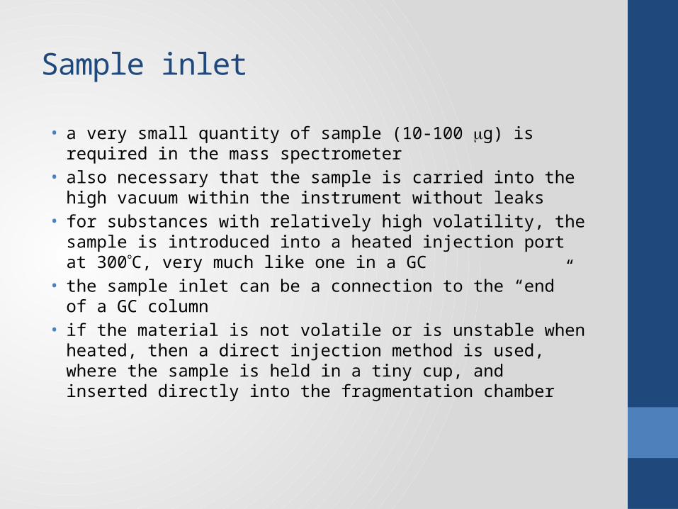
Sample inlet
• a very small quantity of sample (10-100 g) is required in the mass spectrometer
• also necessary that the sample is carried into the high vacuum within the instrument without leaks
• for substances with relatively high volatility, the sample is introduced into a heated injection port at 300C, very much like one in a GC
• the sample inlet can be a connection to the “end” of a GC column• if the material is not volatile or is unstable when heated, then a direct
injection method is used, where the sample is held in a tiny cup, and inserted directly into the fragmentation chamber

Ion source
• MS relies on the detection of charged fragments of the original molecules• the analyte must be ionised first• a number of methods for achieving this, including:
• electron impact – the most common; the only one you need to know • chemical ionisation• fast atom bombardment
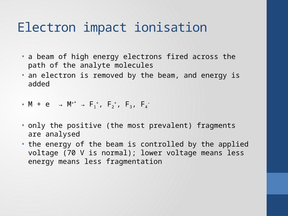
Electron impact ionisation
• a beam of high energy electrons fired across the path of the analyte molecules
• an electron is removed by the beam, and energy is added
• M + e → M+* → F1+, F2
+, F3, F4-
• only the positive (the most prevalent) fragments are analysed • the energy of the beam is controlled by the applied voltage (70 V is normal);
lower voltage means less energy means less fragmentation
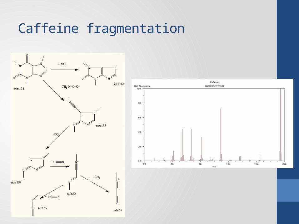
Caffeine fragmentation
Vacuum
• is very substantial: around 10-7 torr (1 atm is 760 torr)• requires two levels of vacuum pump: one to remove the bulk of the air, and
the ultra high vacuum pump to remove the last traces
Exercise 6.3• Why is such a high vacuum required?
• to remove contaminants (eg O2, N2)• to increase the stability of the fragments by reducing chance of collision
Mass analyser
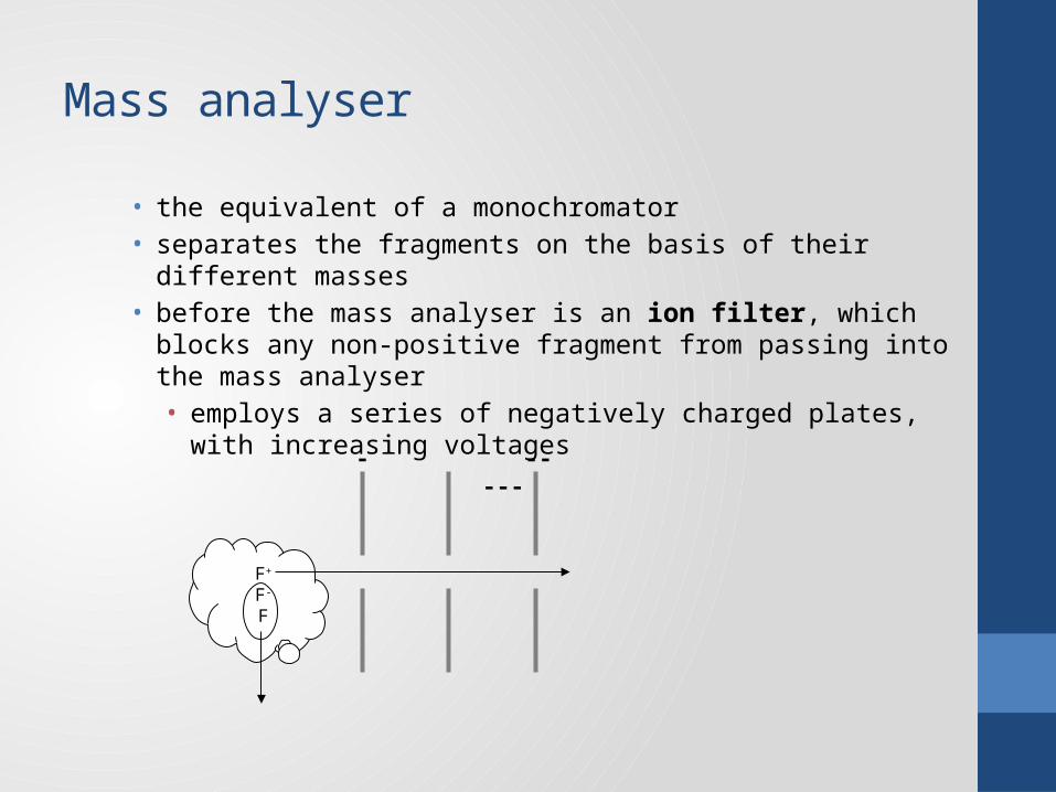
• the equivalent of a monochromator• separates the fragments on the basis of their different masses• before the mass analyser is an ion filter, which blocks any non-positive
fragment from passing into the mass analyser• employs a series of negatively charged plates, with increasing voltages
F+
F-
F
- -- ---
Magnetic sector mass analyser
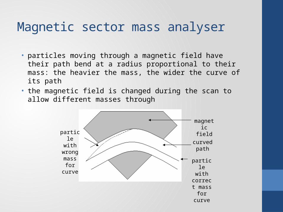
• particles moving through a magnetic field have their path bend at a radius proportional to their mass: the heavier the mass, the wider the curve of its path
• the magnetic field is changed during the scan to allow different masses through
magnetic field
curved path
particle with
wrong mass for
curve particle with
correct mass for
curve

Quadrupole mass analyser
• answers to Exercise 6.4• four metals poles aligned to create electrical fields that allow one mass
through at a given setting

Comparison
• the magnetic sector (MS) is physically larger than the quadrupole (Q) because of the large magnet
• the MS cannot scan as quickly as the Q• the MS costs more than the Q• the MS is capable of much better resolution (0.001 amu) than the Q (0.1
amu)
• Q is ideal as a detector for another instrument eg GC, HPLC, ICP (known as hyphenated instruments)
• MS is better for freestanding MS instruments

GC-MS
• the MS needs very small quantities, which equates to the amounts of analytes coming from capillary GC columns
• the carrier gas is a problem because it would swamp the MS• separated and removed by differences in momentum for light He
GC column
MS inlet
vacuum pump

How does the MS produce a chromatogram?
• at least once a second, the MS does a full mass scan• the electronics generate two measures from the detector:
• the mass spectrum which is stored• the total number of fragments (of all masses) in the scan – this is plotted
on the chromatogram• when only carrier gas is eluting, few fragments are generated• when a compound elutes, more fragments are generated, giving a peak• the chromatogram then looks the same as if a TC or FI detector had been
used
The great advantage of a MSD
• each point in the chromatogram is linked to a MS• this can be used to identify the compound (in conjunction with a spectral
library)• no need for spiking or RT comparison

Making the MSD selective
• setting the mass analyser to a fixed mass which is characteristic of the analyte (equivalent to quantitative analysis on UV-VIS)
• known as single (or selected) ion monitoring• no scanning involved (no ID possible, but not necessary as you know what
you are looking for)• peaks only generated by compounds producing a fragment of the specified
mass• simplifies chromatogram• increases sensitivity (all fragments of that mass are detected – in TIC, most
are lost)
Comparison with other GC detectors
• MS is as sensitive as FI
• MS is much more expensive than other detectors
• no other detectors can identify the peak
HPLC-MS
• the problem of removing the mobile phase is much harder with a liquid• must be instantaneous and must not “damage” analytes• solution was electrospray• uses a high voltage electrical field applied to a nebuliser• this vapourises the solvent and ionises the analytes at the same time• HPLC-MS is the standard “drugs-in-sport” testing method
ICP-MS
• same front end: plasma torch• not used to produce characteristic radiation, but a stream of ions• obviously different to GC- & HPLC-MS which analyse organic molecules• this analyses elements• no fragmentation occurs (these are atoms already!!)• very expensive• far more sensitive than any commercially available instrument (ng/L)• able to distinguish between isotopes of same element