a tightly controlled conditional knockdown system … in the presence of transposase. furthermore,...
TRANSCRIPT

A Tightly Controlled Conditional Knockdown SystemUsing the Tol2 Transposon-Mediated TechniqueTokuichi Iguchi1,2, Hideshi Yagi1, Chen-Chi Wang1, Makoto Sato1,2,3*
1 Division of Cell Biology and Neuroscience, Department of Morphological and Physiological Sciences, Faculty of Medical Sciences, University of Fukui, Fukui, Japan,
2 Research and Education Program for Life Science, University of Fukui, Fukui, Japan, 3 Faculty of Medical Sciences, Child Development Research Center, University of
Fukui, Fukui, Japan
Abstract
Background: Gene knockdown analyses using the in utero electroporation method have helped reveal functional aspects ofgenes of interest in cortical development. However, the application of this method to analyses in later stages of braindevelopment or in the adult brain is still difficult because the amount of injected plasmids in a cell decreases along withdevelopment due to dilution by cell proliferation and the degradation of the plasmids. Furthermore, it is difficult to excludethe influence of earlier knockdown effects.
Methodology/Principal Findings: We developed a tightly controlled conditional knockdown system using a newlyconstructed vector, pT2K-TBI-shRNAmir, based on a Tol2 transposon-mediated gene transfer methodology with thetetracycline-inducible gene expression technique, which allows us to maintain a transgene for a long period of time andinduce the knockdown of the gene of interest. We showed that expression of the endogenous amyloid precursor protein(APP) was sharply decreased by our inducible, stably integrated knockdown system in PC12 cells. Moreover, we induced anacute insufficiency of Dab1 with our system and observed that radial migration was impaired in the developing cerebralcortex. Such inhibitory effects on radial migration were not observed without induction, indicating that our system tightlycontrolled the knockdown, without any expression leakage in vivo.
Conclusions/Significance: Our system enables us to investigate the brain at any of the later stages of development or in theadult by utilizing a knockdown technique with the aid of the in utero electroporation gene transfer methodology.Furthermore, we can perform knockdown analyses free from the influence of undesired earlier knockdown effects.
Citation: Iguchi T, Yagi H, Wang C-C, Sato M (2012) A Tightly Controlled Conditional Knockdown System Using the Tol2 Transposon-Mediated Technique. PLoSONE 7(3): e33380. doi:10.1371/journal.pone.0033380
Editor: Alain Chedotal, Institut de la Vision, France
Received November 24, 2011; Accepted February 8, 2012; Published March 13, 2012
Copyright: � 2012 Iguchi et al. This is an open-access article distributed under the terms of the Creative Commons Attribution License, which permitsunrestricted use, distribution, and reproduction in any medium, provided the original author and source are credited.
Funding: This work was supported in part by the FUKUI BRAIN PROJECT, Research and Education Program for Life Science, University of Fukui, and Grants-in-Aidfor Scientific Research from the Ministry of Education, Culture, Sports, Science and Technology of Japan (KAKENHI 21390052 and 21700402). The funders had norole in study design, data collection and analysis, decision to publish, or preparation of the manuscript.
Competing Interests: The authors have declared that no competing interests exist.
* E-mail: [email protected]
Introduction
During the development of the cerebral cortex, neurons are
generated in the ventricular zone (VZ) and then migrate
outward to the cortical plate (CP), an event that is called
‘‘radial migration.’’ These neurons stop their movement and
settle into the six layers of the cortex in an inside-out pattern,
whereby early-born neurons are positioned in the deeper layers,
and later-born neurons are located in the more superficial
layers, migrating outward by passing the earlier born neurons
[1].
In the later stage of the cortical development, just after radial
migration ends, neurons that have settled in the cortex continue to
elongate their axons toward their targets. In mice, layer-specific
stereotyped projection patterns are established for each layer of the
cortex: layer V neurons, which are born around embryonic day
(E)12.5, mainly project to the subcortical regions as the subcortical
projection, whereas layer II/III pyramidal neurons, which are
born around E14.5, mainly project to the contralateral cortex as
the callosal projection [2,3].
In such studies of cortical development, in utero electroporation
gene transfer is frequently employed because it enables us to
perturb the in vivo gene expression in a convenient manner
compared to conventional techniques, such as making genetically
modified animals [4,5]. Using the in utero electroporation gene
transfer, it has been revealed that various molecules are involved
in the development of the cerebral cortex by controlling radial
migration. One such example is an unexpected activity of the
amyloid precursor protein (APP), for which involvement in
Alzheimer’s disease has been established, in radial migration by
controlling Disabled-1 (Dab1) activity downstream [6]. Dab1 is an
adapter protein that is crucial for the signal transduction of Reelin
signaling, which regulates neuronal migration during corticogen-
esis. A disruption in Dab1 results in the malformation of the
cerebral cortex in which lamination is inverted such that neurons
do not keep to the inside-out pattern [7,8].
Although in utero electroporation gene transfer is a powerful tool
for studying the events in early corticogenesis, there are some
difficulties in applying this technique to the later stages of
development, such as the formation of a neuronal network, which
PLoS ONE | www.plosone.org 1 March 2012 | Volume 7 | Issue 3 | e33380

includes axon path-finding and synaptogenesis, due to the
following reasons: (1) Gene expression from the injected plasmids
decreases along with development due to dilution by cell
proliferation and the degradation of the plasmids. (2) It is difficult
to exclude the influence of earlier gene manipulation. For
example, modified neuronal differentiation may influence the
later neuronal network formation. Therefore, it is desirable to
develop a technique that enables us to stably maintain a transgene
and to induce the expression of genes of interest by design.
Up to now, transposable elements, such as Sleeping Beauty,
piggyBac and Tol2, have been utilized for integrating a gene of
interest into the chromosome for steady expression [9–11]. For
example, the Tol2-flanking regions are integrated into the
chromosome in the presence of transposase. Furthermore, because
the Tol2 transposon, which originated in medaka fish, possesses an
ability to undergo transposition in many kinds of species, including
chick and mouse [12], it has been successfully applied to in ovo or in
utero electroporation gene transfer in model animals for stable gene
expression [13].
Although the tetracycline-controlled gene expression system, in
which transcription is reversibly turned on or off in the presence of
tetracycline, is one of the most frequently employed methods for
inducible gene expression, we sometimes experienced a leakage in
expression with this system in spite of the tetracycline control. A
recent study showed that Tol2-mediated gene transfer systems with
the tetracycline-induced expression system maintain controllable
gene expression during chick embryogenesis [14–16]. However, it
is unclear if the application of this system to induce the knockdown
of endogenous genes with tight expression control at a particular
point in mouse development can be expanded through the
maturation period and into adulthood. Because siRNA is more
stable than single-stranded RNA and siRNA within the RNA-
induced Silencing Complex (RISC) are re-used after the
degradation of the target mRNA, small amounts of siRNA are
enough to cause knockdown effects [17,18]. As a result, tightly
controlled expression is crucial for the evaluation of the effects of
gene knockdown. Therefore, generating tightly controlled induc-
ible expression systems is pivotal to elucidate the in vivo function of
a gene of interest using a knockdown approach without the
influence of earlier events.
In this study, by combining the tightly controlled inducible gene
expression cassette with the Tol2 system, we developed a new tool
that enables us to knockdown the gene of interest at any point
during development.
Results and Discussion
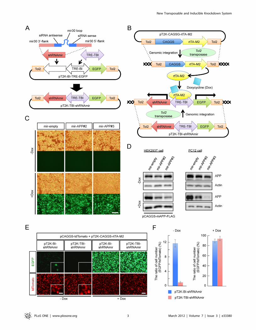
Construction of a Tol2 transposable vector for theinducible expression of the mir30-based knockdowncassette
For the purpose of conditional gene knockdown at any point
during development, we constructed the inducible knockdown
vector pT2K-TBI-shRNAmir based on the Tol2 transposable
vector pT2K-BI-TRE-EGFP by inserting the mir30 cassette
(shRNAmir) and exchanging the promoter (Figure 1A). pT2K-
BI-TRE-EGFP is originally developed for the stable integration
and conditional expression of transgenes in chick embryos [13],
which can be used to express EGFP and a gene of interest under
the control of a bidirectional tetracycline-responsive promoter that
consists of the tetracycline-responsive element (TRE-BI) between
two minimal cytomegalovirus (CMV) promoters. For better
control of inducible expression, we first replaced TRE-BI with
TRE-Tight-BI (TRE-TBI), which has a modified TRE that yields
high induction levels and low basal promoter activity compared
with TRE [19]. The shRNAmir cassette has a hairpin stem
composed of siRNA sense and antisense strands designed to
knockdown the target gene, a loop derived from human mir30 and
mir30 flanking sequences on both sides of the hairpin [20,21]. By
adopting this mir30-based shRNA expression cassette, conditional
shRNA expression from an RNA polymerase II promoter, such as
CMV, can be achieved.
pT2K-CAGGS-rtTA-M2, which expresses the modified reverse
tetracycline-controlled transactivator (rtTA-M2) [22], and pT2K-
TBI-shRNAmir are designed such that Tol2-flanking regions are
integrated into the chromosome in the presence of Tol2
transposase. The rtTA-M2 specifically binds to TRE-TBI in the
presence of Doxycycline (Dox), an analogue of tetracycline, and
activates transcription (Figure 1B). Thereby, shRNAmir is induced
simultaneously with EGFP from the Tol2-flanking region of the
pT2K-TBI-shRNAmir vector, which is integrated into the
chromosome, with tightly controlled expression.
We designed the knockdown cassette based on the miRNA
backbone for pT2K-TBI-shRNAmir. There are some advantages
in using the miRNA-based knockdown cassette: (1) Compared
with the conventional stem-loop-based shRNAs, miRNA-based
hairpins have been proven to exhibit lower cellular toxicity effects
[23,24]. (2) In addition, because the miRNA based cassette is
transcribed by RNA polymerase II, tissue or cell type specific
expression of the knockdown can be achieved with an appropriate
promoter. It has been reported that, while it is not inducible nor
an miRNA-based knockdown system, the combination of the Tol2
system with glial lineage-specific promoters drives the glial cell-
specific expression of transgenes in the mouse cerebral cortex
when transfected using in utero electroporation gene transfer [25].
Because a large number of mouse lines in which Cre recombinase
is expressed in a tissue or a cell type-specific manner have been
reported and are available [26], it is likely that the use of these
bioresources with specific promoters expands the usefulness of our
inducible knockdown system. For instance, this approach enables
knockdown in a specific cortical layer or in a specific neuronal
subtype.
Verification of knockdown effect against an endogenoustarget gene by shRNAmir expressed from a stablyintegrated knockdown cassette
We examined whether the pT2K-TBI-shRNAmir vectors were
integrated into the chromosomes. If the vectors were not
integrated into the chromosome, these vectors were re-distributed
into two daughter cells after mitosis. Eventually the number of
these vectors in individual cells should become very small. In
contrast, in the cells harboring such vectors in their chromosomes,
these vectors should be retained after cell division. We tested the
ability of this vector to integrate into the genome with APP as an
inducible knockdown target. We used pT2K-TBI-shRNAmir
vector as a control empty vector (mir-empty), and designed two
pT2K-TBI-shRNAmir constructs, pT2K-TBI-shRNAmir-APP#2
(mir-APP#2) and pT2K-TBI-shRNAmir-APP#3 (mir-APP#3),
which targeted different regions of the APP sequence.
We transfected mir-empty, mir-APP#2 or mir-APP#3 into
PC12 cells, which express APP endogenously, together with
pT2K-rtTA-M2, pCAGGS-T2TP (Tol2 transposase) and
pCAGGS-tdTomato for the visualization of transfected cells.
The cells were cloned and then subcultured every 4 to 5 days for
47 days until the expression of the transfection control, tdTomato,
became undetectable. Even after 47 days of culturing, almost all of
the cells still expressed EGFP upon induction by Dox, confirming
that these vectors were stably integrated into the genome
(Figure 1C).
New Transposable and Inducible Knockdown System
PLoS ONE | www.plosone.org 2 March 2012 | Volume 7 | Issue 3 | e33380
New Transposable and Inducible Knockdown System
PLoS ONE | www.plosone.org 3 March 2012 | Volume 7 | Issue 3 | e33380

We next determined whether mir-APP#2 and mir-APP#3
knocked down APP. Transient expression of either mir-APP#2 or
mir-APP#3 resulted in the reduction of APP in the presence of
Dox in HEK293T cells exogenously expressing APP following
transfection with pCAGGS-mAPP-FLAG. A stronger knockdown
effect was observed with mir-APP#3 (Figure 1D). Furthermore,
we examined the efficiency of knockdown induced by Dox in
PC12 cells in which the APP knockdown vectors had been
integrated into the genome. In the presence of Dox, APP was
knocked down suggesting that the chromosome-integrated pT2K-
TBI-shRNAmir vector was able to induce the knockdown of the
target gene. Consistent with the results in HEK293T cells, mir-
APP#3 gave a stronger activity of knockdown against APP
(Figure 1D). Tight control of the expression of the pT2K-TBI-
shRNAmir vector was also verified in HEK293T cells. The pT2K-
TBI-shRNAmir vector showed an approximate 12-fold reduction
in leakage compared with the pT2K-BI-shRNAmir vector in the
absence of Dox (Figs. 1E and 1F). Therefore, changing promoters
from TRE-BI to TRE-TBI contributes to the reduction of
undesired knockdown effects due to leakage of the expression
without induction.
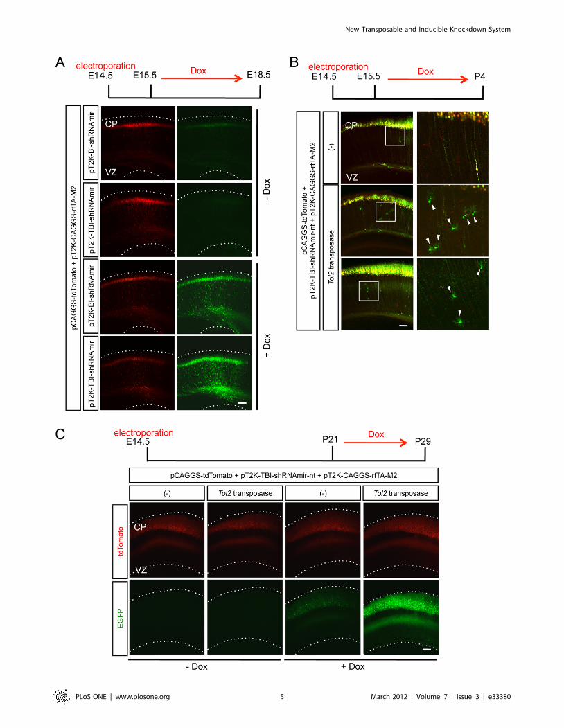
The pT2K-TBI-shRNAmir vector is integrated into thegenome, and its inducible expression is tightly controlledin the mouse cortex
Next, we asked whether our Tol2 inducible knockdown vector,
pT2K-TBI-shRNAmir, works in vivo, especially in the developing
cerebral cortex in mice. First, we tested whether the expression
from our pT2K-TBI-shRNAmir was tightly regulated. pT2K-BI-
shRNAmir or pT2K-TBI-shRNAmir was electroporated into
mouse cerebral cortices together with pT2K-CAGGS-rtTA-M2
and pCAGGS-tdTomato, and gene expression was then induced
by Dox administration. For both pT2K-BI-shRNAmir and pT2K-
TBI-shRNAmir, the expression of EGFP that had been induced
simultaneously with the shRNAmir knockdown cassette from the
bidirectional promoter was observed following Dox administra-
tion. However, we detected much weaker EGFP expression from
pT2K-TBI-shRNAmir without induction than that from pT2K-
BI-shRNAmir (Figure 2A). These results indicated that the
expression of pT2K-TBI-shRNAmir was controlled tightly in vivo,
similar to our in vitro results.
Next, we asked whether pT2K-TBI-shRNAmir was integrated
into the genome of the developing cortex in mice. If the Tol2-
flanked regions were integrated into the genome by the Tol2
transposase, EGFP expression would be observed even in the glial
cells in which transfected non-integrated vectors were otherwise
diluted due to cell proliferation. pT2K-TBI-shRNAmir-non-target
(pT2K-TBI-shRNAmir-nt), pT2K-CAGGS-rtTA-M2 and
pCAGGS-tdTomato were electroporated into the mouse cerebral
cortex with or without the Tol2 transposase expression vector, and
gene expression was induced by Dox at E15.5. Cortices were
observed at P4, by which point glial cells must have experienced
multiple rounds of cell divisions (Figure 2B). At P4, EGFP
expression was clearly confirmed in the glial cells with Tol2
transposase but not without Tol2 transposase (Figure 2B). These
results showed that Tol2 knockdown vectors were integrated into
the genome in the presence of Tol2 transposase. In addition, we
assessed whether the knockdown cassette is stably maintained
throughout development by observing the expression of EGFP,
which is under the control of the bidirectional promoter TRE-
TBI, after 4 weeks of postnatal life with Dox administration for a
week before the harvest. We did not detect any obvious leakage of
EGFP expression in the adult mouse cortex without induction. In
Dox-treated animals, a drastic induction of EGFP expression was
observed in the cortices that had been electroporated with the
transposase expression vector (Figure 2C).
Although plasmids were introduced into the neural progenitors
in the VZ by in utero electroporation gene transfer, these
progenitors, as well as recently recognized intermediate progen-
itors in the subventricular zone [27], undergo mitosis before finally
settling into the cortical plate. Because an episomally located
vector is likely to be diluted during mitosis, whereas a transposed
vector is not, it is probable that higher inducible expression was
observed in the cortical neurons of the adult brain when the Tol2
transposase vector was co-transfected (Figure 2C). Indeed, EGFP
expression was weaker in superficial neurons, which are likely to
experience more mitotic cycles, compared with those in deeper
layers in the absence of Tol2 transposase activity (Figure 2C, 3rd
column).
Inducible knockdown of a gene that is essential for thecortical development impairs radial migration
Although our observations showed that the combination of the
Tol2 inducible knockdown vector and the in utero electroporation
gene transfer method enables the induction of gene expression at
the time of interest, it remains unclear whether the inducible
knockdown is effective in vivo. We performed inducible knockdown
against Dab1, a molecule that plays an important role in radial
migration under the Reelin signaling pathway, in addition to APP
(Figure S1). First, the efficiency of the inducible knockdown
against the exogenously expressed Dab1 was examined in
HEK293T cells. Because pT2K-TBI-shRNAmir-Dab1#2 showed
a knockdown effect in a Dox-dependent manner, subsequent
experiments were performed with this vector (Figure 3A). To
reveal the knockdown efficiency against the endogenous Dab1 in
the cerebral cortex, we performed in utero electroporation in the
mouse cortex. Knockdown was then induced by the administra-
tion of Dox. Because the knockdown cassette expressed EGFP
Figure 1. The Tol2 transposable vector enables inducible knockdown from a stably integrated knockdown cassette. (A) A schematicdiagram of the pT2K-TBI-shRNAmir vector. The shRNAmir cassette was inserted into the pT2K-BI-TRE-EGFP vector, and TRE-BI was replaced with TRE-TBI. The shRNAmir cassette consisted of the hairpin stem, which is composed of siRNA sense and antisense strands designed for the knockdown ofthe target gene, a loop derived from human mir30, and mir30 flanking sequences on the 39 and 59 sides of the hairpin. (B) A schematic diagramshowing the principle of induction of knockdown from the genomically integrated shRNAmir cassette. The Tol2-flanked region of the plasmids wereexcised and integrated into the chromosome using Tol2 transposase. In the presence of Doxycycline (Dox), rtTA-M2 bound to TRE-TBI, and theexpression of both EGFP and the mir30-based knockdown cassette were induced under the control of TRE-TBI. (C) Expression of EGFP, induced fromthe each of pT2K-TBI-shRNAmir vectors (mir-empty, mir-APP#2 and mir-APP#3), was observed in almost all PC12 cells following Dox administration.The upper panels show the bright-field images. Scale bar, 100 mm. (D) Immunoblot analyses for evaluating the knockdown efficiency against APP.Actin was used as a loading control. (E) The basal expression (2Dox) and the induced expression (+Dox) of EGFP from the pT2K-BI-shRNAmir andpT2K-TBI-shRNAmir vectors in HEK293T cells. pCAGGS-tdTomato was co-transfected as a transfection control. Inset shows a higher magnification.Scale bars: 100 mm, inset 20 mm. (F) Ratio of the number of EGFP-positive cells to tdTomato-positive cells between the cells expressing pT2K-BI-shRNAmir and pT2K-TBI-shRNAmir with or without Dox. (mean 6 SEM, n = 3). Abbreviations of the vector name and their components are listed inthe table (Table S1).doi:10.1371/journal.pone.0033380.g001
New Transposable and Inducible Knockdown System
PLoS ONE | www.plosone.org 4 March 2012 | Volume 7 | Issue 3 | e33380
New Transposable and Inducible Knockdown System
PLoS ONE | www.plosone.org 5 March 2012 | Volume 7 | Issue 3 | e33380

from the bidirectional promoter, EGFP-expressed cells were
FACS-sorted, and quantitative real-time PCR was performed. In
the EGFP-positive cells, Dab1 expression was decreased signifi-
cantly compared with control cells (Figure 3B). It has been
reported that reduction of the Dab1 protein in the cerebral cortex
results in the malformation of the cortical lamination [8,28].
Therefore, we assessed the effect of the induction of Dab1
knockdown and the leakage of this knockdown cassette in the
absence of Dox. Radial migration was inhibited in the cortex
following the induction of the Dab1 knockdown, but not in brains
exposed to the control vector or in the absence of Dox (Figure 3C).
These results indicated that the Tol2 inducible knockdown vector
was tightly controlled and effectively knocked down the expression
of the endogenous gene without expression leakage.
Molecules that control neuronal migration, including regulators
of the cytoskeleton and cell adhesion molecules [29,30], also affect
other processes, such as axon projection and synaptogenesis
[31,32], after the cessation of migration. Application of the Tol2
inducible knockdown system described here allows us to
investigate gene functions in each event separately by controlling
the timing of the gene silencing.
Currently, the demand for the development of tools for
conditional genetic manipulation, especially in a small number of
neurons, is increasing. Young et al. create one such method, SLICK
(single-neuron labeling with inducible Cre-mediated knockout) that
utilizes drug inducible Cre-mediated knockout technology [33].
With SLICK, they can visualize a whole neuronal shape by co-
expressing YFP (yellow fluorescent protein) together with Cre,
which is also accomplished by our system with EGFP. However,
they knockout a gene of interest in an irreversible manner with
SLICK, whereas we knockdown a gene of interest in a reversible
manner, in which we can bring back the gene expression later.
Moreover, specific Cre-expressing neurons are targeted in SLICK,
whereas our system enables us to knockdown a gene in any
electroporated neurons. To facilitate functional genomics in the
nervous system, an appropriate methodology should be taken for
conditional genetic manipulation to the purpose. Our system has
the potential to be a tool with which to elucidate the in vivo molecular
mechanisms of later stages of development.
Materials and Methods
AnimalsICR mice were purchased from SLC (Hamamatsu, Japan). Noon
on the day when the presence of a vaginal plug was confirmed was
designated as embryonic day 0.5 (E0.5). For pups, the day of birth
was designated as postnatal day 0 (P0). All experiments were
conducted in accordance with the guidelines for animal experiments
of the University of Fukui, and all efforts were made to minimize
both the number of animals used and their suffering. The protocol
was approved by the Committee on the Ethics of Animal
Experiments of the University of Fukui (Permit Number: 23012).
Vector constructionsMouse full-length Dab1 (GenBank accession number
NM_177259) cDNA was cloned from E14.5 mouse brains by the
polymerase chain reaction (PCR). For pCAGGS-myc-mDab1, the
Dab1 cDNA was amplified by PCR with the primers 59-
AACCTCGAGATGTCAACTGAGACAGAACTTCAAG-39 and
59-GCTCTCGAGCTAGCTACCGTCTTGTGGACTTATATT-
ATC-39. The PCR product was cloned into the XhoI sites of the
pCAGGS-myc vector. Mouse full-length APP (GenBank accession
number NM_007471) cDNA was cloned from E14.5 mouse brains
by PCR. For pCAGGS-mAPP-FLAG, the APP cDNA was amplified
by PCR with the primers 59-AATAGATATCATGCTGCC-
CAGCTTGG-39 and 59- GAAGATATCGTTCTGCATTTGCT-
CAAAGAAC-39. The PCR product was cloned into the EcoRV sites
of the pCAGGS-FLAG-IRES-GFP vector. For the construction of
pT2K-BI-shRNAmir, the mir-30 cassette was excised from pCAG-
mir30 (Addgene plasmid 14758; Addgene, Cambridge, MA) [34]
with XbaI/HindIII sites and inserted into the EcoRV/ApaI site of
pT2K-BI-TRE-EGFP [13] with blunt-end ligation. The EcoRI sites
flanking the TRE sequence were then eliminated. For the
construction of pT2K-TBI-shRNAmir, the TRE-BI sequence was
removed from pT2K-BI-shRNAmir using the BamHI/NcoI site,
then blunted and self-ligated to reform the BamHI site. The TRE-
Tight-BI sequence was excised with BamHI/BglII from pTRE-
Tight-BI (Clontech, Palo Alto, CA), which lacks the EcoRI and XhoI
sites, and then inserted into the BamHI site of pT2K-BI-shRNAmir.
RNA interferenceThe mir30-based shRNA was synthesized by PCR with the
primers 59-CAGAAGGCTCGAGAAGGTATATTGCTGTT-
GACAGTGAGCG-39 and 59-CTAAAGTAGCCCCTTGAAT-
TCCGAGGCAGTAGGCA-39 using the following template
oligonucleotides: for shRNAmir-non-target (shRNAmir-nt), 59-
TGCTGTTGACAGTGAGCGATCTCGCTTGGGCGAGAG-
TAAGTAGTGAAGCCACAGATGTACTTACTCTCGCCCA-
AGCGAGAGTGCCTACTGCCTCGGA-39; for shRNAmir-A-
PP#2, 59-TGCTGTTGACAGTGAGCGCGCACTAACTTG-
CACGACTATGTAGTGAAGCCACAGATGTACATAGTCG-
TGCAAGTTAGTGCTTGCCTACTGCCTCGGA-39; for sh
RNAmir-APP#3, 59-TGCTGTTGACAGTGAGCGCGCTGA-
CAAGAAGGCCGTTATCTAGTGAAGCCACAGATGTAGA-
TAACGGCCTTCTTGTCAGCTTGCCTACTGCCTCGGA-
39; for shRNAmir-Dab1#1, 59-TGCTGTTGACAGTGAGCG-
CCCTCCTCCTCTTAGCACTAAATAGTGAAGCCACAGA-
TGTATTTAGTGCTAAGAGGAGGAGGATGCCTACTGC-
CTCGGA-39; for shRNAmir-Dab1#2, 59-TGCTGTTGACA-
GTGAGCGCAAGGATAAGCAGTGTGAACAATAGTGAA-
GCCACAGATGTATTGTTCACACTGCTTATCCTTTTGC-
CTACTGCCTCGGA-39. The PCR product was cloned into the
XhoI/EcoRI sites of the pT2K-TBI-shRNAmir vector. The
underlined sequences correspond to the sense and antisense target
sequences, respectively.
Cell culture, Transfection and Immunoblotting analysisCells were cultured in Dulbecco’s modified Eagle’s medium
(DMEM) supplemented with either 10% fetal bovine serum for
HEK293T cells (ATCC No. CRL-2828) or 10% fetal bovine
serum and 5% horse serum for PC12 cells (ATCC No. CRL-
1721). Plasmid DNAs were transfected into cells using the
Figure 2. Tol2 knockdown vectors were genomically integrated and the inducible expression was tightly controlled in vivo. (A)Representative neocortical sections showing the basal expression and induced expression of EGFP derived from the pT2K-BI-shRNAmir or pT2K-TBI-shRNAmir vectors. (B) The retention and expression of inducible knockdown vector in glial cells. In the presence of Tol2 transposase, EGFP wasobserved in the glial cells (arrowheads). The right panels are higher magnification views of the boxed regions in the left panels. (C) Representativeneocortical sections showing the retention and expression of the inducible knockdown vector in the adult cortex. CP, cortical plate; VZ, ventricularzone. Scale bars, 100 mm.doi:10.1371/journal.pone.0033380.g002
New Transposable and Inducible Knockdown System
PLoS ONE | www.plosone.org 6 March 2012 | Volume 7 | Issue 3 | e33380

LipofectamineTM 2000 transfection reagent (Invitrogen, Carls-
bad, CA). The cells were lysed with the lysis buffer (20 mM
HEPES-NaOH, pH 7.5, 100 mM NaCl, 3 mM MgCl2, 1 mM
dithiothreitol, 1 mM EGTA, 0.5% Nonidet P-40) supplement-
ed with 1% Protease Inhibitor Cocktail (Sigma, St. Louis,
MO). After centrifugation, the supernatant was mixed with
Figure 3. Inducible knockdown of a gene essential for cortical development impaired radial migration. (A) Immunoblot analysis of theinducible knockdown against the exogenously expressed Dab1 in HEK293T cells. Dab1 was decreased in the presence of the Tol2-inducibleknockdown vector pT2K-TBI-shRNAmir-Dab1#2 following the application of Dox. (B) Quantitative real-time PCR data showing the efficiency ofinducible knockdown against the endogenous Dab1 in cortical neurons. In the EGFP-positive cells that had been electroporated with pT2K-TBI-shRNAmir-Dab1#2, the expression level of Dab1 was significantly decreased compared with the control. (C) Inducible knockdown of Dab1 duringcortical development. The empty control vector did not inhibit neuronal migration. The induction of mir-Dab1#2 expression resulted in impairedradial migration in the presence of Dox but not without Dox. Scale bar, 100 mm.doi:10.1371/journal.pone.0033380.g003
New Transposable and Inducible Knockdown System
PLoS ONE | www.plosone.org 7 March 2012 | Volume 7 | Issue 3 | e33380

Laemmli sample buffer, boiled, and subsequently separated on
SDS-polyacrylamide gels. The separated proteins were trans-
ferred to polyvinylidene difluoride membranes. After blocking
for 30 min in 5% skim milk-TBST (50 mM Tris-HCl, pH 7.4,
150 mM NaCl, 0.1% Tween 20), the membranes were probed
for 1 h with the primary antibodies in 5% skim milk-TBST.
The membranes were then washed three times with TBST and
incubated with the secondary antibodies coupled to horserad-
ish peroxidase. Immunoblotting was visualized using an
Immobilon Western Chemiluminescent HRP Substrate (Milli-
pore, Bedford, MA) on a LAS-3000 mini imaging system
(Fujifilm, Tokyo, Japan). The following antibodies (and
dilutions) were used: the mouse anti-myc antibody (1:1000)
(Santa Cruz Biotechnology, Santa Cruz, CA), anti-FLAG
antibody (1:1000) (Sigma, St. Louis, MO), anti-actin antibody
(1:500) (Sigma, St. Louis, MO) and the mouse anti-APP
antibody (1:1000) (Millipore, Bedford, MA).
Semi-quantitative analysisFor the semi-quantitative analysis of the ratio of EGFP-positive
cells to tdTomato-positive cells, images were obtained with a
fluorescence microscope (Axio observer A1; Carl-Zeiss, Oberko-
chen, Germany), and the number of EGFP-positive and
tdTomato-positive cells were counted using NIH Image J software.
In utero electroporationPlasmids were transfected by in utero electroporation using
previously described methods with some modifications [4,5,35].
Briefly, on E14.5, pregnant female ICR mice were anaesthetized
with an intraperitoneal injection of 2,2,2-tribromoethanol (200–
300 mg/kg body weight) before the experiments. Approximately
1–2 ml of plasmid solution (1–5 mg/ml) was injected into the lateral
ventricle of the embryos. Relative ratio of plasmids in mixed
solution is as follows. pCAGGS-tdTomato: pT2K-CAGGS-rtTA-
M2: pT2K-BI-shRNAmir or pT2K-TBI-shRNAmir: pCAGGS-
T2TP (Tol2 transposase) = 1:5:10:10. After soaking the uterine
horn with PBS, the head of the embryo was pinched with a
forceps-type electrode (CUY650P3 for E12.5 or CUY650P5 for
E14.5; Nepa gene, Chiba, Japan), and five cycles of square electric
pulses (35 V, 50 ms for E12.5 or 45 V, 50 ms for E14.5) at
intervals of 950 ms were delivered using an electroporator
(CUY21EDIT; Nepa Gene, Chiba, Japan). For the analysis of
inducible expression in the cerebral cortex, the brains were fixed
with 4% paraformaldehyde in 0.1 M phosphate buffer, pH 7.4,
cut coronally into 100 mm slices with a Vibratome (DTK-1000;
DSK, Kyoto, Japan), and imaged on a laser-scanning confocal
microscope (LSM 5 PASCAL; Carl-Zeiss, Oberkochen, Ger-
many).
FACS sorting and quantitative real-time PCRpT2K-TBI-shRNAmir or pT2K-TBI-shRNAmir-Dab1#2 was
electroporated with pT2K-CAGGS-rtTA-M2 into the mouse
cerebral cortex at E14.5. The cells were collected at E18.5 using
FACS (FACS Aria II, BD Bioscience, San Jose, CA). Doxycycline
(Dox) was administrated to mice at 2 mg/ml in a 5% sucrose
solution in drinking water during the experiment from E14.5 to
E18.5. To make cDNA for real-time PCR from the total RNA of
FACS-sorted cells, reverse transcription was performed using
reverse transcriptase (ReverTra AceTM; Toyobo, Osaka, Japan).
Real-time PCR reactions were performed on an ABI StepOne-
PlusTM Real-Time PCR System (Applied Biosystems, Foster city,
CA) using the SYBR Green PCR Master Mix (ThunderbirdTM;
Toyobo, Osaka, Japan). The thermal cycling was performed using
the following cycle conditions: the initial denaturation step at 95uCfor 60 s, followed by 40 cycles at 95uC for 15 s, and 60uC for 60 s.
The experiments were carried out in triplicate. The relative
quantification of gene expression compared to a control was
determined using the DDCt method. Changes in gene expression
were normalized to the internal control gene PRS18. The
following gene-specific primers were used for real-time PCR
analysis: for PRS18, 59-AACGGTCTAGACAACAAGCTG-39
and 59-AGTGGTCTTGGTGTGCTGAC-3; for b-actin, 59-
ATGCTCCCCGGGCTGTAT-39 and 59-CATAGGAGTCCT-
TCTGACCCATTC-3; and for Dab1, 59-AGCTCAAGGGTG-
TTGTTGCT-39 and 59- TGTGATGTCCTTCGCAATGT-3.
Supporting Information
Figure S1 Knockdown effects of shRNA- and miRNA-based knockdown vectors against APP on the radialmigration using the various promoters. APP was knocked
down using the shRNA- or miRNA-based knockdown vectors
(Text S1) in the cerebral cortex. (A) Immunoblot analyses of the
knockdown against the exogenously expressed mouse APP in
HEK293T cells. (B) Mouse cortices were electroporated with
vectors at E14.5 and observed at E18.5. Only the knockdown
vector with mU6pro-shAPP#2 inhibited neuronal migration. (C)
Immunoblot analyses of the knockdown efficiency against the
exogenously expressed mouse APP in HEK293T cells using
shRNAmir vector with various promoters. (D) CMV-driven or
mU6-driven knockdown vectors were transfected at E14.5 and
cortices were observed at E18.5. There was no significant change.
Scale bars, 100 mm.
(TIF)
Table S1 The nomenclature of the vectors and compo-nents.
(PDF)
Text S1 Supporting methods for vector constructions.
(DOC)
Acknowledgments
We thank T. Bando, H. Yoshikawa and S. Kanae for technical assistance,
T. Taniguchi for secretarial assistance, and Y. Oka, M. Komada, M-J. Xie,
K. Kuroda for critical reading of the manuscript. We are also grateful to Y.
Takahashi for the kind gift of pT2K-CAGGS-rtTA-M2 and pT2K-BI-
TRE-EGFP, K. Kawakami for pCAGGS-T2TP, and C. Cepko for pCAG-
mir30.
Author Contributions
Conceived and designed the experiments: TI MS. Performed the
experiments: TI CCW. Analyzed the data: TI HY MS. Contributed
reagents/materials/analysis tools: TI HY. Wrote the paper: TI MS.
References
1. Marın O, Rubenstein JLR (2003) Cell migration in the forebrain. Annu Rev
Neurosci 26: 441–483.
2. Fame RM, MacDonald JL, Macklis JD (2011) Development, specification, and
diversity of callosal projection neurons. Trends Neurosci 34: 41–50.
3. Molyneaux BJ, Arlotta P, Menezes JRL, Macklis JD (2007) Neuronal subtype
specification in the cerebral cortex. Nat Rev Neurosci 8: 427–437.
4. Saito T, Nakatsuji N (2001) Efficient gene transfer into the embryonic mouse
brain using in vivo electroporation. Dev Biol 240: 237–246.
New Transposable and Inducible Knockdown System
PLoS ONE | www.plosone.org 8 March 2012 | Volume 7 | Issue 3 | e33380

5. Tabata H, Nakajima K (2001) Efficient in utero gene transfer system to the
developing mouse brain using electroporation: visualization of neuronalmigration in the developing cortex. Neuroscience 103: 865–872.
6. Young-Pearse TL, Bai J, Chang R, Zheng JB, LoTurco JJ, et al. (2007) A critical
function for beta-amyloid precursor protein in neuronal migration revealed by inutero RNA interference. J Neurosci 27: 14459–14469.
7. Sheldon M, Rice DS, D’Arcangelo G, Yoneshima H, Nakajima K, et al. (1997)Scrambler and yotari disrupt the disabled gene and produce a reeler-like
phenotype in mice. Nature 389: 730–733.
8. Howell BW, Hawkes R, Soriano P, Cooper JA (1997) Neuronal position in thedeveloping brain is regulated by mouse disabled-1. Nature 389: 733–737.
9. Ivics Z, Hackett PB, Plasterk RH, Izsvak Z (1997) Molecular reconstruction ofSleeping Beauty, a Tc1-like transposon from fish, and its transposition in human
cells. Cell 91: 501–510.10. Kawakami K, Shima A, Kawakami N (2000) Identification of a functional
transposase of the Tol2 element, an Ac-like element from the Japanese medaka
fish, and its transposition in the zebrafish germ lineage. Proc Natl Acad Sci U S A97: 11403–11408.
11. Wang W, Lin C, Lu D, Ning Z, Cox T, et al. (2008) Chromosomal transpositionof PiggyBac in mouse embryonic stem cells. Proc Natl Acad Sci U S A 105:
9290–9295.
12. Kawakami K, Noda T (2004) Transposition of the Tol2 element, an Ac-likeelement from the Japanese medaka fish Oryzias latipes, in mouse embryonic
stem cells. Genetics 166: 895–899.13. Sato Y, Kasai T, Nakagawa S, Tanabe K, Watanabe T, et al. (2007) Stable
integration and conditional expression of electroporated transgenes in chickenembryos. Dev Biol 305: 616–624.
14. Yokota Y, Saito D, Tadokoro R, Takahashi Y (2011) Genomically integrated
transgenes are stably and conditionally expressed in neural crest cell-specificlineages. Dev Biol 353: 382–395.
15. Hou X, Omi M, Harada H, Ishii S, Takahashi Y, et al. (2011) Conditionalknockdown of target gene expression by tetracycline regulated transcription of
double strand RNA. Dev Growth Differ 53: 69–75.
16. Watanabe T, Saito D, Tanabe K, Suetsugu R, Nakaya Y, et al. (2007) Tet-oninducible system combined with in ovo electroporation dissects multiple roles of
genes in somitogenesis of chicken embryos. Dev Biol 305: 625–636.17. Meister G, Tuschl T (2004) Mechanisms of gene silencing by double-stranded
RNA. Nature 431: 343–349.18. Shi Y (2003) Mammalian RNAi for the masses. Trends Genet 19: 9–12.
19. Agha-Mohammadi S, O’Malley M, Etemad A, Wang Z, Xiao X, et al. (2004)
Second-generation tetracycline-regulatable promoter: repositioned tet operatorelements optimize transactivator synergy while shorter minimal promoter offers
tight basal leakiness. J Gene Med 6: 817–828.20. Zeng Y, Wagner EJ, Cullen BR (2002) Both natural and designed micro RNAs
can inhibit the expression of cognate mRNAs when expressed in human cells.
Mol Cell 9: 1327–1333.
21. Paddison PJ, Cleary M, Silva JM, Chang K, Sheth N, et al. (2004) Cloning of
short hairpin RNAs for gene knockdown in mammalian cells. Nat Methods 1:
163–167.
22. Urlinger S, Baron U, Thellmann M, Hasan MT, Bujard H, et al. (2000)
Exploring the sequence space for tetracycline-dependent transcriptional
activators: novel mutations yield expanded range and sensitivity. Proc Natl
Acad Sci U S A 97: 7963–7968.
23. McBride JL, Boudreau RL, Harper SQ, Staber PD, Monteys AM, et al. (2008)
Artificial miRNAs mitigate shRNA-mediated toxicity in the brain: implications
for the therapeutic development of RNAi. Proc Natl Acad Sci U S A 105:
5868–5873.
24. Bauer M, Kinkl N, Meixner A, Kremmer E, Riemenschneider M, et al. (2009)
Prevention of interferon-stimulated gene expression using microRNA-designed
hairpins. Gene Ther 16: 142–147.
25. Yoshida A, Yamaguchi Y, Nonomura K, Kawakami K, Takahashi Y, et al.
(2010) Simultaneous expression of different transgenes in neurons and glia by
combining in utero electroporation with the Tol2 transposon-mediated gene
transfer system. Genes Cells 15: 501–512.
26. Madisen L, Zwingman TA, Sunkin SM, Oh SW, Zariwala HA, et al. (2010) A
robust and high-throughput Cre reporting and characterization system for the
whole mouse brain. Nat Neurosci 13: 133–140.
27. Molnar Z, Vasistha NA, Garcia-Moreno F (2011) Hanging by the tail:
progenitor populations proliferate. Nat Neurosci 14: 538–540.
28. Morimura T, Ogawa M (2009) Relative importance of the tyrosine
phosphorylation sites of Disabled-1 to the transmission of Reelin signaling.
Brain Res 1304: 26–37.
29. Tullio AN, Bridgman PC, Tresser NJ, Chan CC, Conti MA, et al. (2001)
Structural abnormalities develop in the brain after ablation of the gene encoding
nonmuscle myosin II-B heavy chain. J Comp Neurol 433: 62–74.
30. Jossin Y, Cooper JA (2011) Reelin, Rap1 and N-cadherin orient the migration of
multipolar neurons in the developing neocortex. Nat Neurosci 14: 697–703.
31. Hodges JL, Newell-Litwa K, Asmussen H, Vicente-Manzanares M, Horwitz AR
(2011) Myosin IIb activity and phosphorylation status determines dendritic spine
and post-synaptic density morphology. PLoS One 6: e24149.
32. Ranscht B (2000) Cadherins: molecular codes for axon guidance and synapse
formation. Int J Dev Neurosci 18: 643–651.
33. Young P, Qiu L, Wang D, Zhao S, Gross J, et al. (2008) Single-neuron labeling
with inducible Cre-mediated knockout in transgenic mice. Nat Neurosci 11:
721–728.
34. Matsuda T, Cepko CL (2007) Controlled expression of transgenes introduced by
in vivo electroporation. Proc Natl Acad Sci U S A 104: 1027–1032.
35. Nagano T, Morikubo S, Sato M (2004) Filamin A and FILIP (Filamin A-
Interacting Protein) regulate cell polarity and motility in neocortical subven-
tricular and intermediate zones during radial migration. J Neurosci 24:
9648–9657.
New Transposable and Inducible Knockdown System
PLoS ONE | www.plosone.org 9 March 2012 | Volume 7 | Issue 3 | e33380