sentinella mini‐symposium moderators: alessandro testori and sergi vidal‐sicart (non‐cme...
Upload: cancer-metastasis-through-the-lymphovascular-system-biology-treatment-7th-intl-symposium
Post on 13-Feb-2017
250 views
TRANSCRIPT
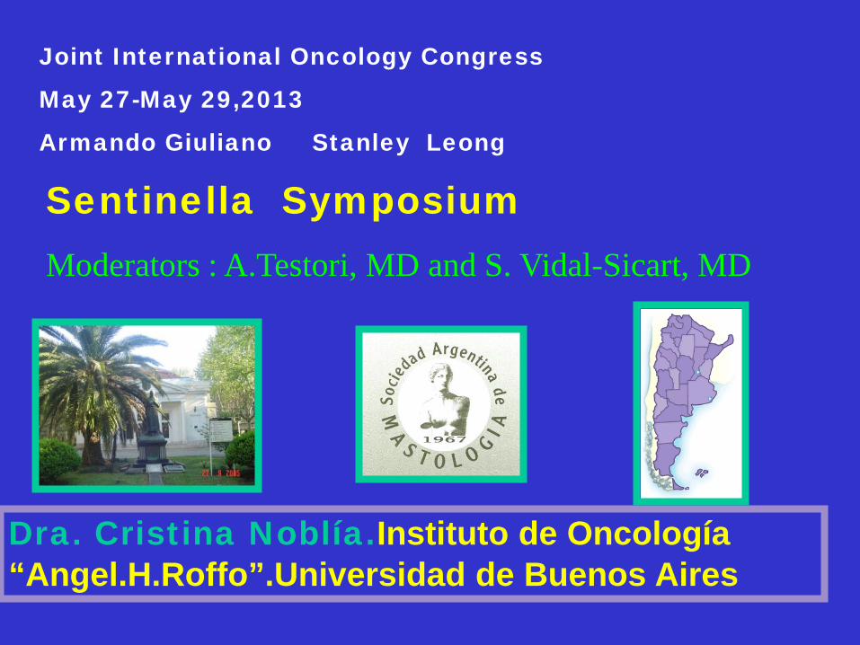
Dra. Cristina Noblía.Instituto de Oncología “Angel.H.Roffo”.Universidad de Buenos Aires
Joint International Oncology Congress
May 27-May 29,2013
Armando Giuliano Stanley Leong
Sentinella Symposium Moderators : A.Testori, MD and S. Vidal-Sicart, MD
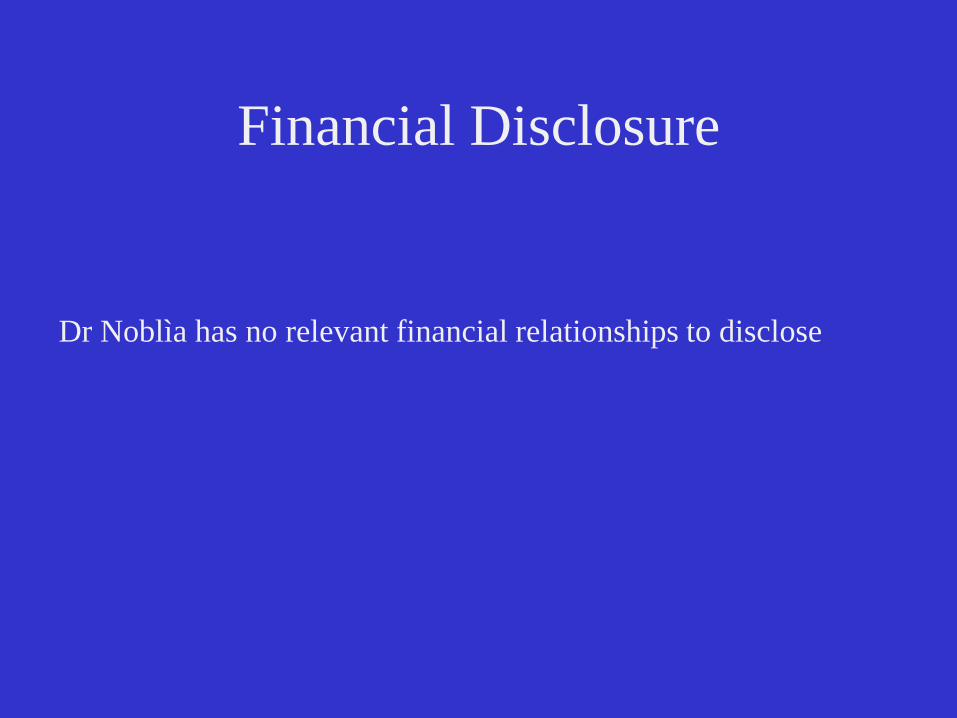
Financial Disclosure
Dr Noblìa has no relevant financial relationships to disclose

Breast Surgery Departament Instituto Instituto de de Oncología Oncología “Angel H. “Angel H. Roffo Roffo ” UBA ” UBA
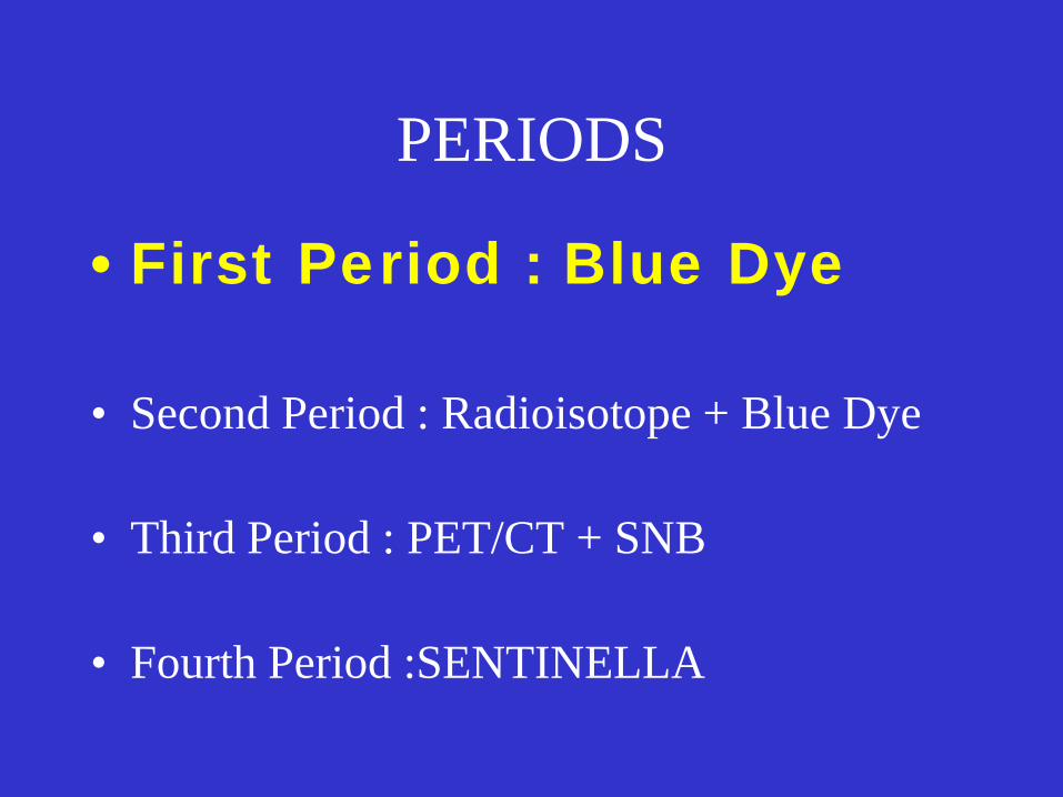
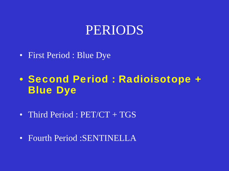
PERIODS
• First Period : Blue Dye • Second Period : Radioisotope + Blue Dye • Third Period : PET/CT + SNB • Fourth Period :SENTINELLA

FIRST PERIOD • BLUE DYE (PATENT BLUE). • 3cc Peritumoral • Massage for 5 minutes.

Transverse axillary incision just inferior to the hair-bearing region of the axilla.
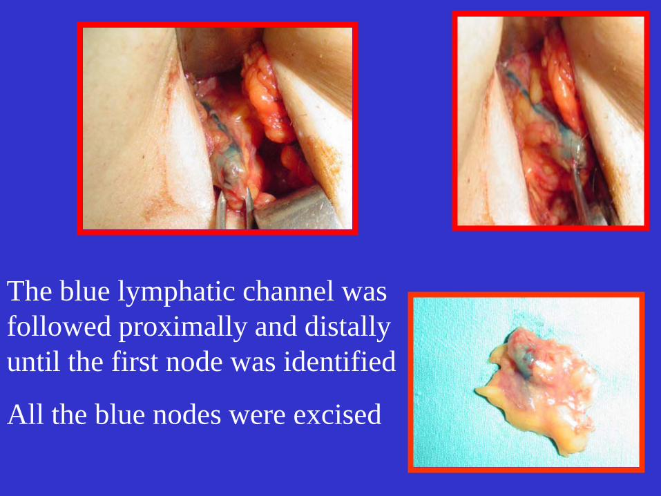
The blue lymphatic channel was followed proximally and distally until the first node was identified
All the blue nodes were excised
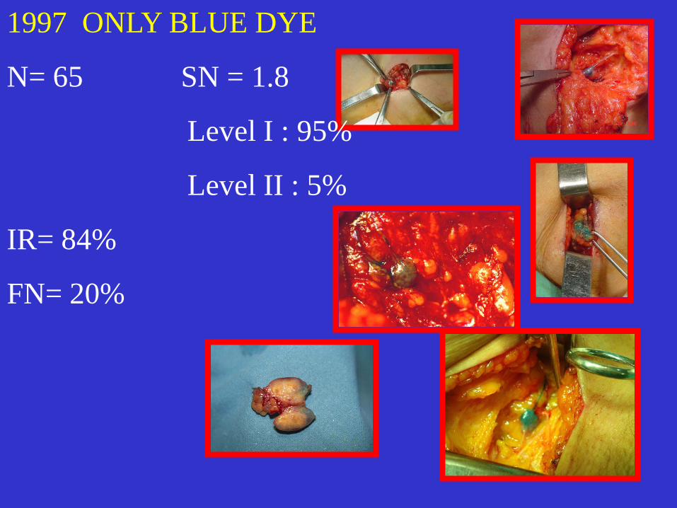
1997 ONLY BLUE DYE
N= 65 SN = 1.8
Level I : 95%
Level II : 5%
IR= 84%
FN= 20%
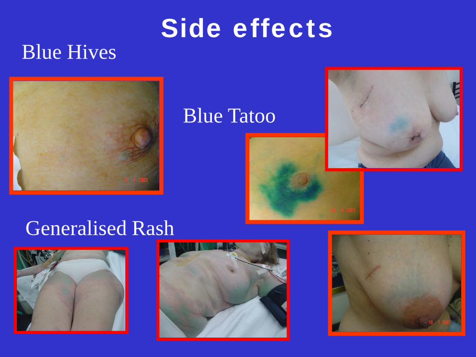
Side effects Blue Hives
Blue Tatoo
Generalised Rash
PERIODS
• First Period : Blue Dye • Second Period : Radioisotope +
Blue Dye • Third Period : PET/CT + TGS • Fourth Period :SENTINELLA

SECOND PERIOD (2000) • Radioisotope + lymphoscintigraphy
•Gamma Probe •Patent blue
Tc 99 labelled human albumin colloide particles in 0.2 ml saline
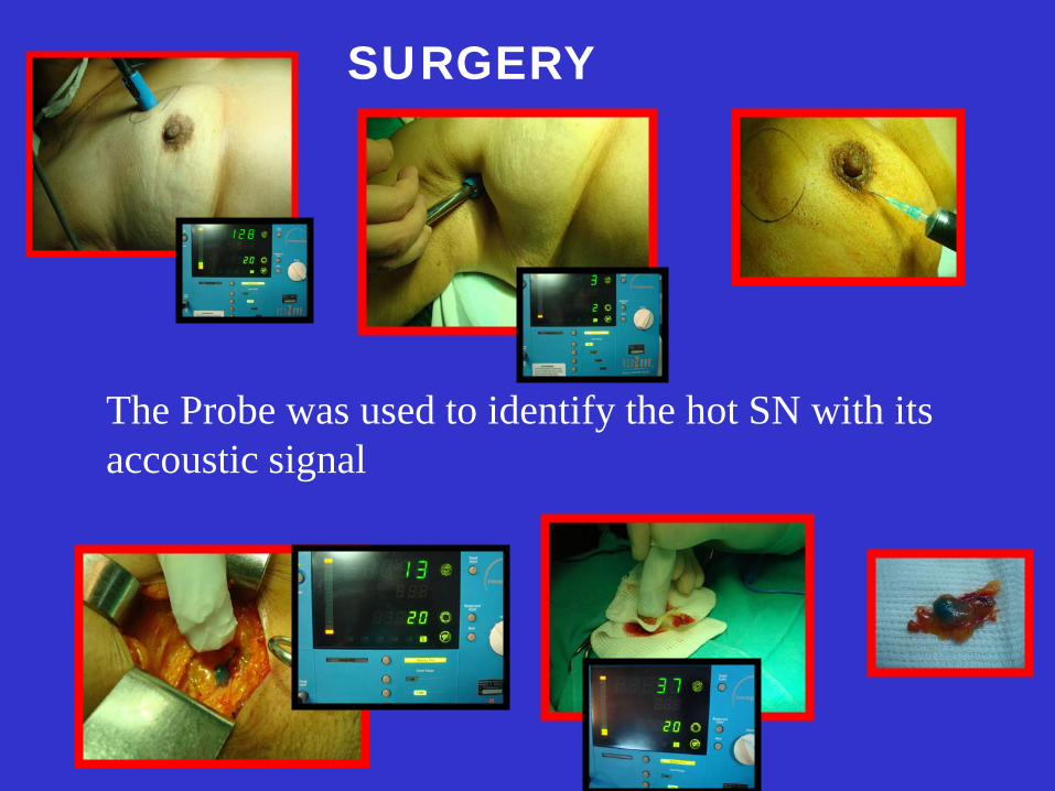
SURGERY
The Probe was used to identify the hot SN with its accoustic signal
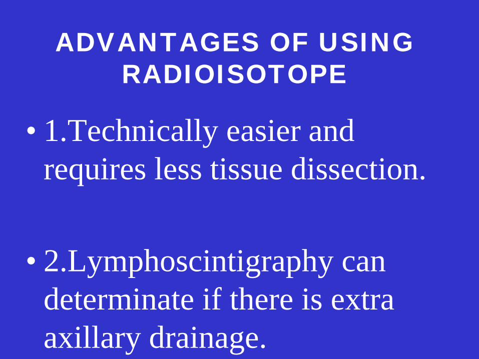
ADVANTAGES OF USING RADIOISOTOPE
• 1.Technically easier and requires less tissue dissection.
• 2.Lymphoscintigraphy can
determinate if there is extra axillary drainage.
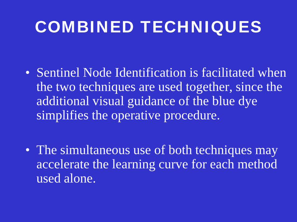
COMBINED TECHNIQUES
• Sentinel Node Identification is facilitated when the two techniques are used together, since the additional visual guidance of the blue dye simplifies the operative procedure.
• The simultaneous use of both techniques may
accelerate the learning curve for each method used alone.
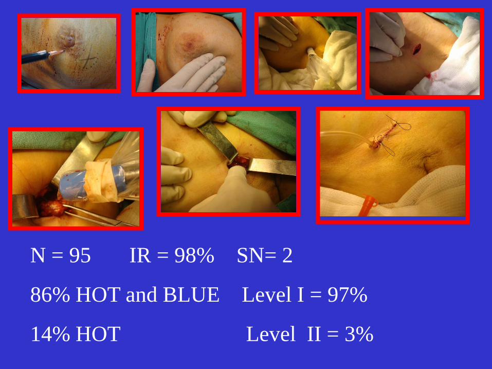
N = 95 IR = 98% SN= 2
86% HOT and BLUE Level I = 97%
14% HOT Level II = 3%

No axillary dissection
IR : 95%
FN : 5%
PERIODS
• First Period : Blue Dye • Second Period : Radioisotope + Blue Dye • Third Period : PET/CT + TGS • Fourth Period :SENTINELLA
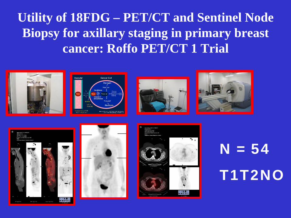
Utility of 18FDG – PET/CT and Sentinel Node Biopsy for axillary staging in primary breast
cancer: Roffo PET/CT 1 Trial
N = 54
T1T2NO
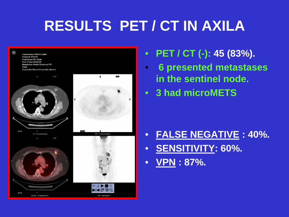
RESULTS PET / CT IN AXILA
• PET / CT (-): 45 (83%). • 6 presented metastases
in the sentinel node. • 3 had microMETS
• FALSE NEGATIVE : 40%. • SENSITIVITY: 60%. • VPN : 87%.
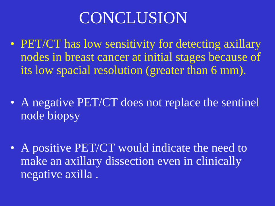
CONCLUSION • PET/CT has low sensitivity for detecting axillary
nodes in breast cancer at initial stages because of its low spacial resolution (greater than 6 mm).
• A negative PET/CT does not replace the sentinel
node biopsy • A positive PET/CT would indicate the need to
make an axillary dissection even in clinically negative axilla .

PERIODS
• First Period : Blue Dye • Second Period : Radioisotope + Blue Dye • Third Period : PET/CT + TGS • Fourth Period :SENTINELLA
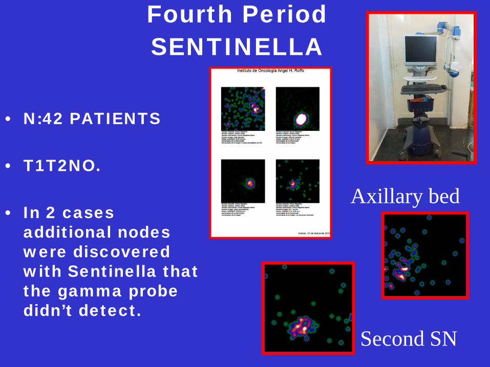
Fourth Period SENTINELLA
• N:42 PATIENTS • T1T2NO. • In 2 cases
additional nodes were discovered with Sentinella that the gamma probe didn’t detect.
Axillary bed
Second SN
SENTINELLA
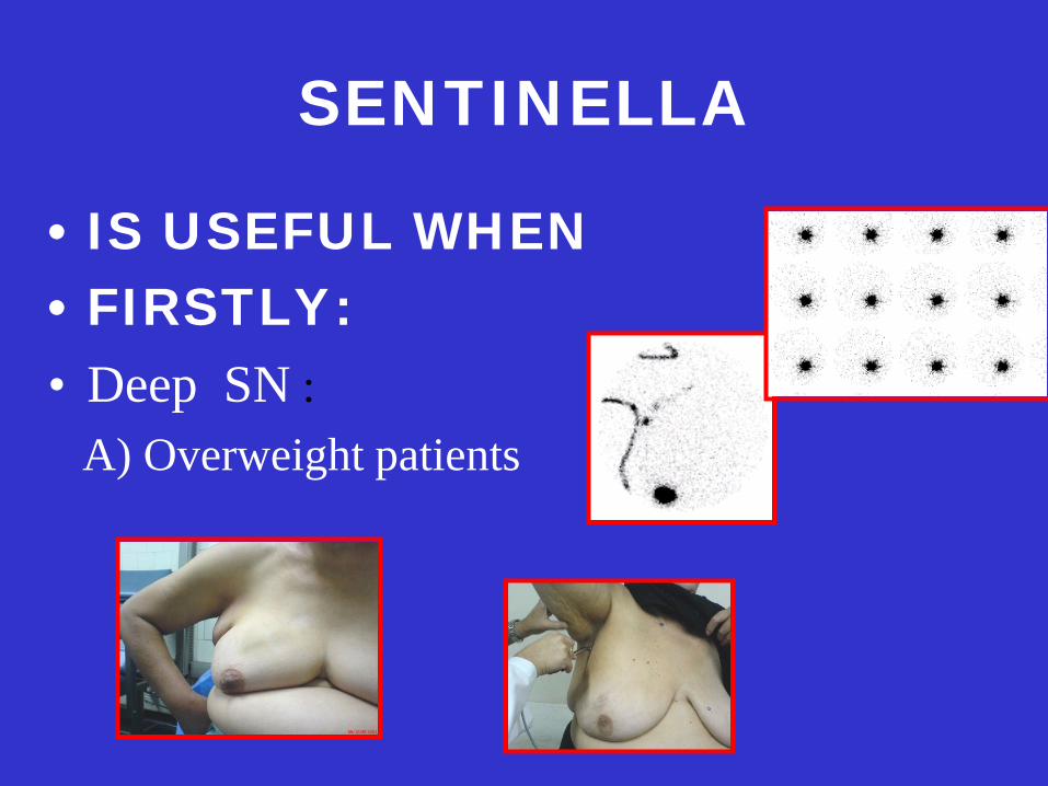
• IS USEFUL WHEN • FIRSTLY: • Deep SN : A) Overweight patients

Deep Node
b) Sentinel Node in Internal Mammary Chain
IN SITU EX VIVO
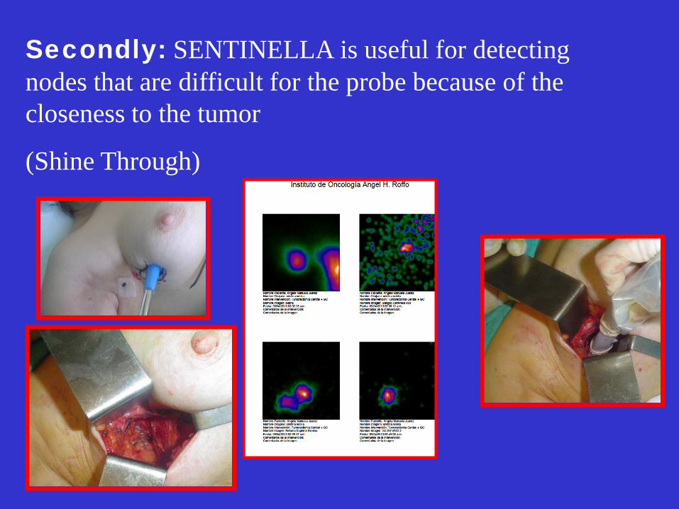
Secondly: SENTINELLA is useful for detecting nodes that are difficult for the probe because of the closeness to the tumor
(Shine Through)
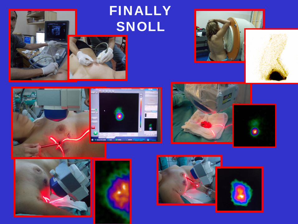
FINALLY SNOLL
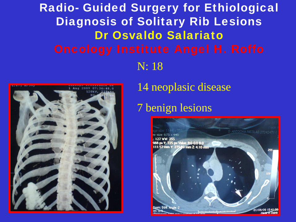
Radio- Guided Surgery for Ethiological Diagnosis of Solitary Rib Lesions
Dr Osvaldo Salariato Oncology Institute Angel H. Roffo
N: 18
14 neoplasic disease
7 benign lesions
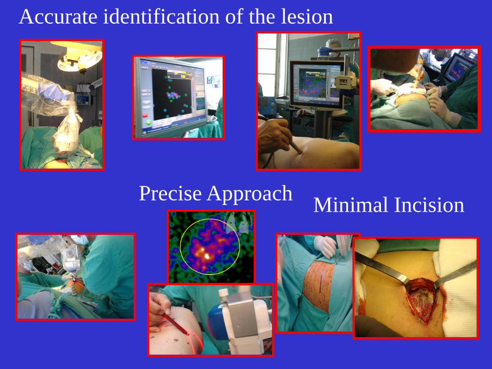
Accurate identification of the lesion
Precise Approach Minimal Incision
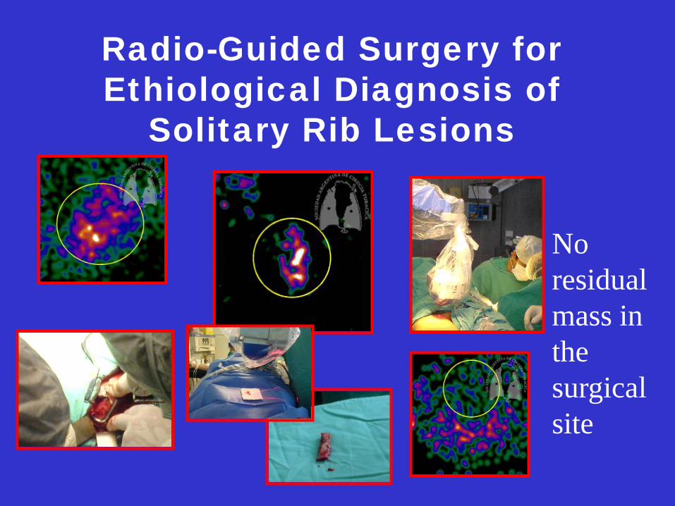
Radio-Guided Surgery for Ethiological Diagnosis of
Solitary Rib Lesions
No residual mass in the surgical site
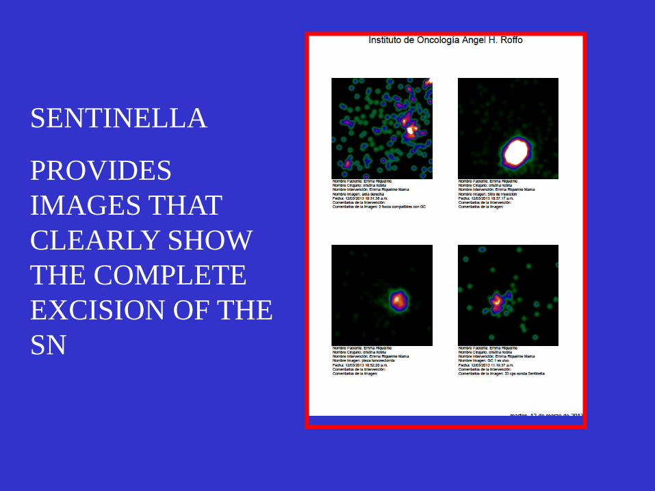
SENTINELLA
PROVIDES IMAGES THAT CLEARLY SHOW THE COMPLETE EXCISION OF THE SN

Departamento de Mastología: Dr.Cresta Morgado Dra.Cristina Noblía
Dr. González Dr. Armanasco
Dra. Azar Dra. Montoya
Dr. Ipiña
Departamento de Medicina Nuclear:
Dra. Parma Dra. Zarlenga Dra.Armesto
Dra San Martin Gabriela.
Dra Lidia Katz Departamento de Anatomía
Patológica: Dra. Gorostidi
Departamento de Oncología:
Dra. Mickiewicz Dra. Alvarez