sentinella mini‐symposium moderators: alessandro testori and sergi vidal‐sicart (non‐cme...
Upload: cancer-metastasis-through-the-lymphovascular-system-biology-treatment-7th-intl-symposium
Post on 13-Feb-2017
249 views
TRANSCRIPT

Intraoperative sentinel node imaging in breast cancer
Is this the way forward?
Presented by D.B.Ghosh MS,FRCS(Edin),FRCS(G.Surg) FEBS(Breast & Surgical Oncology)
Consultant Breast & Oncoplastic Surgeon

∗ This study was performed independently and none of the authors received any fees or reimbursement from the manufacturers of Sentinella
∗ None of the authors or their family have any financial interest in the company that manufactures Sentinella.
Disclosure of Conflict of Interest

SLN
“Any node receiving direct
lymphatic drainage from
the primary tumour”
Donald Morton
Sentinel Node biopsy Is an established as standard of care in
management of early breast cancer
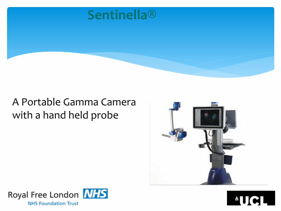
A Portable Gamma Camera with a hand held probe
Sentinella®

Pioneering study from UK to compare Sentinella® with the conventional gamma camera (cGC) in terms of:
∗ accuracy and speed
∗ sensitivity and resolution
∗ evaluate it as an intraoperative imaging modality for SLN detection that can reliably replace the cGC
Aims

∗ Sentinella® was compared with the conventional gamma camera (cGC) in laboratory settings
∗ Sentinella was used intraoperatively and its accuracy and sensitivity was compared with Lymphoscintograms and Hand held gamma probe
Methods

Evaluation of Sentinella in the Lab
∗ A simulator containing seeds10 kBq, 100kBq, 500kBq of radiocolloid Tc, mimicking SLN, were used
∗ Seeds were placed at several depths in the axilla, at 3 cm, 5cm and 8 cm from the skin

• Images were obtained with Sentinella and Gamma Camera
• The cGC was placed 20 cm away from the radioactive seeds

∗ Spatial resolution was measured by calculating the full width half maximum (FWHM) of a line profile measured perpendicular to the image of a capillary tube filled with high activity concentration technetium-99m pertechnetate.

∗ For the Sentinella gamma camera spatial resolution was measured at the centre and at the edge of the field of view

The three images above are of 10 MBq seed(low radioactivity) at 3 , 5 & 8 cms depth at 1minute.The decreasing counts picked up with increasing distance can be seen

∗ 68 Sentinella® images and 34 cGC images obtained from the simulated axilla
∗ Sentinella® resolution is comparable with the cGC for objects close to the camera i.e. ~ 5 cm, but reduces rapidly as it’s moved away from the camera
∗ For distances up to about 7 cm the Sentinella® with the blue collimator is more sensitive than the cGC
Results

∗ Sentinella detects high radioactivity (500 kBq) faster than cGC (1 vs 2.5min)
∗ In cases of low radioactivity (10kBq) Sentinella® was equally accurate and faster than cGC, when placed close to the skin
∗ Identification of different number of beads with varying radioactivity was similar in Sentinella and cGC
Results

∗ All patients undergoing SLNB underwent standard imaging with cGC
∗ This was followed by intraoperative mapping using Sentinella
∗ Sentinella was also compared with a standard hand held probe and images recorded when the hand held probe was silent to activity
Clinical Evaluation


∗ Number of patients studied = 150
∗ Sentinella correlated 100% with images
∗ Sentinella scans detected more nodes as compared to cGC (1.25 vs 2.2) p<0.01
Results

∗ Sentinella picked up extra nodes in 5/150 cases due to visual residual activity when the hand held probe was silent
∗ 2 /150 cases picked up histologically positive nodes not detected by cGC and resulted in axillary clearance & change in management

∗ Sentinella is accurate and fast in detecting radioactivity in the axilla
∗ The anatomical shape of its collimator allows the operator to place it adjacent to the axilla, thus increasing its sensitivity in cases of low radioactivity
∗ Our independent tests and initial patient data confirms the excellent sensitivity and specificity of the machine in localisation of radioactive nodes
Conclusion

∗ This potentially increases the identification of the SLN and reduces false negative SLNB
∗ Reduces time of patient and personnel for the use of Conventional Lymphoscintigrams
∗ It can resolve the problem of centres that do not have on site nuclear medicine department in the UK setting.
Conclusion

∗ Vania Stafyla ∗ David McCool ∗ Fred Wickham ∗ Andrew O’Brien
Acknowledgements

Thank You
Your Questions