clinical anatomy, embryology and imaging page 1 bms...
TRANSCRIPT

Clinical Anatomy, Embryology and Imaging Page 1 BMS 6115C Version 6/4/2007
Clinical Anatomy, Embryology and Imaging Page 2 BMS 6115C Version 6/4/2007
General Course Syllabus You will be given a ten question multiple-choice examination on the first day of lecture to generally assess whether you read the syllabus!!! A score will not be calculated into your grade, but a 100% score will provide a bonus point on the first in-term examination. This syllabus is a guide for participation in the course and certain information may be changed during the implementation of the course. Every effort will be made to announce any changes in class or laboratory sessions, but please periodically review course Blackboard site for any changes.
Overview Human Anatomy, Embryology and Imaging Course – BMS 6115
Goals. Human Anatomy, Embryology and Imaging (BMS 6115) is a 10 week long course and runs concurrently with the Doctoring 1 Course. Because this is the first basic science course presented in the curriculum, its prime goal is to provide the students with a basic understanding of the gross anatomy, embryology and radiologic imaging of the entire body which will serve as a solid foundation for the remainder of the student's medical education and future profession. Second, this course was designed to prepare the student for the applications of anatomy and embryology to the clinical sciences, and for the application of radiologic imaging toward diagnosis of clinical disorders. Students will be stimulated to utilize learning resources such as faculty, textbooks, journals and FSU-COM computer resources. The course will also promote the development of student-directed problem solving skills to recognize an existing knowledge base and any gaps in that knowledge base that must be learned in order to understand the applications of clinical anatomy to clinical reasoning. Working in small groups studying clinical cases and studying in the laboratory will promote a team approach to learning. Students will utilize a variety of digital imaging programs that will supplement learning that occurs in the laboratory setting, lectures, small-group sessions and personal study time. As a side benefit, this course will introduce the student to anatomical terminology commonly used in medicine today. The anatomic knowledge learned in this course will be both applied and reinforced in later courses in the curriculum. Course Objectives: The student will be able to: Knowledge:
1. Demonstrate a foundation of knowledge in normal anatomy, embryology, cross-sectional anatomy and radiologic imaging of the human body.
2. Apply anatomical knowledge to solving clinical problems. 3. Demonstrate knowledge of the anatomical differences in the human body from birth to senescence; 4. Identify the limits of their anatomical knowledge when trying to apply it to understanding clinical
problems, and locate needed information in a timely manner. 5. Recognize the anatomy and laboratory findings related to variations, pathology, previous surgery and
human life cycle from gestation to the elderly patient.

Clinical Anatomy, Embryology and Imaging Page 3 BMS 6115C Version 6/4/2007
Skills:
6. Utilize a variety of resources (faculty, textbooks, computers, internet, etc.) to locate anatomic, embryologic, and/or radiologic information in order to understand how it relates to clinical problems.
Attitudes/Behaviors:
7. Work together as a professional team in the anatomy laboratory and in the case-based small-group sessions.
8. Engage in self-evaluation and evaluate peer performance during the laboratory and small-group experiences of the course.
COMPETENCY & LEARNING OBJECTIVES AND EXAMPLES OF OUTCOME MEASURES IN THE CLINICAL ANATOMY, EMBRYOLOGY AND IMAGING BMS 6115C
The following are the general institutional learning objectives that have been organized by the competencies established by the College of Medicine.
KEY Institutional Learning Objective General Competency K=Knowledge S=Skills AB=Attitudes/Behaviors
Outcome Measure(s) Examples
AB1
Display the personal attributes of compassion, honesty, and integrity in relationships with patients, families, communities, and the medical profession.
Observational during case-based small-group sessions
AB3
Exhibit appropriate value for the sensitive nature of the doctor/ patient relationship and the importance of compassionate communication which is sensitive to the patient's familial, cultural, and spiritual circumstances.
Observational during case-based small-group sessions
AB6
Demonstrate social awareness and commitment to the welfare of underserved communities (rural, urban underserved, and elderly).
NA
AB7 Demonstrate awareness of the health care needs of aging patients and a willingness to care for the elderly.
Observational during case-based small-group sessions.
S15
Adopt a comprehensive, integrated approach to patient care incorporating relevant biomedical, psychological, and social/cultural factors.
Observational during case-based small-group sessions
Professional values, attitudes, and behaviors
AB9
Demonstrate respect for the roles of other healthcare providers and of the need to collaborate with others in caring for individual patients and in promoting public health and community service.
Observational during case-based small-group sessions

Clinical Anatomy, Embryology and Imaging Page 4 BMS 6115C Version 6/4/2007
KEY Institutional Learning Objective General Competency K=Knowledge S=Skills AB=Attitudes/Behaviors
Outcome Measure(s) Examples
K10
Describe and discuss the implications of basic ethical principles, including confidentiality, informed consent, truth telling, and justice, for the care of patients.
Observational during case-based small-group sessions
Moral reasoning and ethical conduct
AB4
Demonstrate professionalism and high ethical standards in all aspects of medical practice, specifically competence, honesty, integrity, compassion, respect for others, professional responsibility and social responsibility.
Observational during case-based small-group sessions, laboratory sessions and team teaching.
KEY Institutional Learning Objective General Competency K=Knowledge S=Skills AB=Attitudes/Behaviors
Outcome Measure(s) Examples
S1
Demonstrate the ability to elicit accurate, comprehensive, and focused medical histories by employing techniques that facilitate the patient's sharing of information.
Observational during case-based small-group sessions
S7
Demonstrate the ability to educate patients about their health problems and to motivate them to adopt health promoting behaviors.
Observational during case-based small-group sessions
S8
Demonstrate the ability to build rapport and to employ active listening and relationship enhancing behaviors (e.g., empathic responding).
NA
S13
Demonstrate the ability to communicate compassionately and effectively, both verbally and in writing, with patients, their families, colleagues, and others with whom physicians must exchange information in carrying out their responsibilities.
Observational during case-based small-group sessions
Communicating with patients, families, and colleagues
AB2 Exhibit well-developed interpersonal skills in providing information and comfort to patients and their families.
Observational during case-based small-group sessions
KEY Institutional Learning Objective General Competency K=Knowledge S=Skills AB=Attitudes/Behaviors
Outcome Measure(s) Examples
Application of basic biomedical and behavioral sciences to patient care
K1
Recognize the scientific basis of health, disease, and medicine in the management of common and high impact medical conditions in contemporary society.
Observational during case-based small-group sessions

Clinical Anatomy, Embryology and Imaging Page 5 BMS 6115C Version 6/4/2007
K2
Describe the development, structure and function of the healthy human body and each of its major organ systems at the macroscopic, microscopic, and molecular levels.
Observational during case-based small-group sessions. Performance during lab session, large group session, quizzes and major examinations
K3
Recognize and discuss the implications of altered structure and function (pathology and pathophysiology) of the body and its major organ systems that are seen in various diseases and conditions.
Performance during lab sessions, weekly clinical cases, cadaver autopsy reports
K4
Identify changes in the structure and function of the human body associated with the aging process and be able to distinguish normal changes associated with aging from those that denote disease.
Performance during lab sessions, weekly cases, cadaver autopsy reports
K5 Describe the molecular basis of diseases and maladies and the way in which they affect the body (pathogenesis).
NA
K7
Describe normal human psychosocial development across the lifespan and recognize deviations requiring further evaluation and intervention.
NA
K12
Discuss the application of psychodynamic, behavioral, and social-cognitive theories of human thought and behavior in describing and analyzing patient behavior.
NA
S9
Demonstrate the effective use of pharmacotherapeutic agents and other therapeutic modalities, while teaching patients the importance of preventative medicine, health promotion, and wellness.
Performance during case-based small group sessions and cases
KEY Institutional Learning Objective General Competency K=Knowledge S=Skills AB=Attitudes/Behaviors
Outcome Measure(s) Examples
S2
Demonstrate the ability to conduct both effective and accurate comprehensive and focused physical examinations and know when each is most appropriate.
NA
Essential clinical skills
S10 Demonstrate appropriate technique for performing routine procedures as specified below: assist or apply cast/splint (n=2); observe a circumcision (n=1); perform and interpret an EKG (n=3); perform an elliptical excision of a skin lesion (n=1); place a Foley catheter in a male or female (n=4); incise and drain an abscess (n=3); perform endotracheal
NA

Clinical Anatomy, Embryology and Imaging Page 6 BMS 6115C Version 6/4/2007
intubation on a model or assist with patient intubation (n=1); place an IV line (n=2); perform a lumbar puncture (n=1); place an NG Tube (n=1); perform a Pap Smear (n=5); perform a vaginal delivery (n=1); perform a simple closure of a surgical skin wound (n=10). Perform arterial puncture (n=1).
S19 Demonstrate appropriate technique for performing Basic Life Support and Advanced Life Support.
NA
K13
Demonstrate knowledge of the functional approach to managing chronic conditions, including knowledge of the impact of chronic illness on function.
NA
S16
Demonstrate the ability to assess functional status and to plan and implement interventions to increase functional ability or limit disability.
NA
S6
Demonstrate the ability to employ a comprehensive, multidisciplinary approach to the care of patients that integrates biomedical and psychosocial considerations.
NA
KEY Institutional Learning Objective General Competency K=Knowledge S=Skills AB=Attitudes/Behaviors
Outcome Measure(s) Examples
K6
Describe basic biobehavioral and clinical science principles used to analyze and solve problems related to the diagnosis, treatment, and prevention of disease.
Performance during lab sessions, small group sessions, large group interactive sessions, quizzes and examinations
S3
Demonstrate the appropriate use of laboratory tests and radiographic studies in making diagnostic and treatment decisions.
Performance during lab sessions, small group sessions, large group interactive sessions, quizzes and examinations
S4
Demonstrate the ability to evaluate the patient's medical problems and to formulate accurate hypotheses to serve as the basis for making diagnostic and treatment decisions.
Performance in small-group sessions, case wrap ups and cadaver autopsy report
Problem solving and critical thinking
S5
Demonstrate the ability to formulate and implement a plan of care for both the prevention and treatment of disease, enhancement of the patient’s functional capabilities, and the relief of symptoms and suffering.
Performance in small-group sessions and case wrap up sessions
KEY Institutional Learning Objective General Competency K=Knowledge S=Skills AB=Attitudes/Behaviors
Outcome Measure(s) Examples
Lifelong learning and information management
K11 Describe strategies to support lifelong learning via both print and electronic sources to assist in making diagnostic
Observation and performance in case-based small-group sessions and case wrap up

Clinical Anatomy, Embryology and Imaging Page 7 BMS 6115C Version 6/4/2007
and treatment decisions (e.g., practice guidelines) and to remain current with advances in medical knowledge and practice (e.g., medical information data bases).
S11
Demonstrate the ability to acquire new information and data and to critically appraise its validity and applicability to one's professional decisions, including the application of information systems technologies for support of clinical decision-making.
Performance in case-bases small group sessions and cadaver autopsy reports
S12 Demonstrate the ability to organize, record, research, present, critique, and manage clinical information.
Performance in cadaver autopsy report
KEY Institutional Learning Objective General Competency K=Knowledge S=Skills AB=Attitudes/Behaviors
Outcome Measure(s) Examples
K8
Describe the role of family, community and culture as factors influencing patient presentations, interpretations of illness episodes and adherence to treatment recommendations.
NA
K9 Recognize the implications of cultural, social, economic, legal, and historical contexts for patient care.
Performance in case-based small-group sessions and case wrap up
K14 Demonstrate knowledge of the unique health care needs of ethnically diverse populations and communities.
NA
Social, cultural, and community context of health, illness, and care
AB8 Demonstrate awareness of the unique health care needs of ethnically diverse populations and communities.
Performance in case-based small-group sessions and case wrap up
KEY Institutional Learning Objective General Competency K=Knowledge S=Skills AB=Attitudes/Behaviors
Outcome Measure(s) Examples
AB5
Exhibit a capacity for self-evaluation, moral reflection, and ethical reasoning to form the basis for a self-directed, lifelong engagement in the responsible, committed, compassionate practice of medicine.
Observation and performance during lab sessions, small group sessions, large group interactive sessions, quizzes, self/peer evaluations and examinations
Personal awareness
S17 Recognize abilities and limitations; know when to request assistance.
Observational assessment by faculty and staff.
KEY Institutional Learning Objective General Competency K=Knowledge S=Skills AB=Attitudes/Behaviors
Outcome Measure(s) Examples
S14
Demonstrate the ability to work effectively as part of a health care team, and appreciate the contributions of other health care professionals and agencies to the health of the individual and the health of the community.
Observation of performance in lab sessions, small-group sessions, exchange of dissection and imaging information, cadaver autopsy report
Organizations, systems, and quality improvement
AB10 Demonstrate a respect for the roles of NA

Clinical Anatomy, Embryology and Imaging Page 8 BMS 6115C Version 6/4/2007
other healthcare providers and of the need to collaborate with others in caring for individual patients and in promoting public health and community service.
K15
Demonstrate basic knowledge of the global health care delivery system in the community including physicians, hospitals, outpatient centers, home health agencies and the role of community agencies in that system.
NA
AB11
Demonstrate an understanding of the role of the physician in working with community agencies for the benefit of his/her patients.
NA
S18
Demonstrate an understanding of the principles and method of Practice-Based Learning and Improvement that involves investigation and evaluation of one's own patient care, appraisal and assimilation of scientific evidence, and improvements in patient care.
Performance in case-based small group sessions, lab sessions and cadaver autopsy report
University Curriculum Committee Approved Academic Honor Policy and ADA Statement ACADEMIC HONOR POLICY: The Florida State University Academic Honor Policy outlines the University’s expectations for the integrity of students’ academic work, the procedures for resolving alleged violations of those expectations, and the rights and responsibilities of students and faculty members throughout the process. Students are responsible for reading the Academic Honor Policy and for living up to their pledge to “. . . be honest and truthful and . . . [to] strive for personal and institutional integrity at Florida State University.” (Florida State University Academic Honor Policy, found at http://www.fsu.edu/~dof/honorpolicy.htm.) AMERICANS WITH DISABILITIES ACT: Students with disabilities needing academic accommodation should: (1) register with and provide documentation to the Student Disability Resource Center; and (2) bring a letter to the instructor indicating the need for accommodation and what type. This should be done during the first week of class. This syllabus and other class materials are available in alternative format upon request. For more information about services available to FSU students with disabilities, contact the Student Disability Resource Center 97 Woodward Avenue, South Florida State University Tallahassee, FL 32306-4167 (850) 644-9566 (voice) (850) 644-8504 (TDD) [email protected] http://www.fsu.edu/~staffair/dean/StudentDisability/

Clinical Anatomy, Embryology and Imaging Page 9 BMS 6115C Version 6/4/2007
FSU COM ATTENDANCE POLICY COM Philosophy We believe that: Professionalism is a major component of our medical curriculum. We believe students should conduct themselves appropriately in the various educational acti vities of the curriculum. This conduct includes coming to educational activities on-time, using the laptop computers only for course work during the educational activity, and not disrupting the class if late. The faculty should also demonstrate professionalism, by starting and ending all scheduled educational activities on time and providing a course schedule with clearly explained course policies in the course syllabus. Any changes in the schedule should be given to the students in a timely manner. Students will be accountable and personally responsible for attending all educational activities (small groups, labs, clinical experiences, examinations, lectures, computer sessions, etc.). Unexcused absences reflect negatively on the goals and objectives of the medical curriculum and demonstrate unprofessional behavior by the respective student. We owe it to our state legislature and the citizens of the State of Florida to provide a quality educational program that meets the needs of our students in preparing them for the M.D. degree. Attendance Policy Students are expected to attend all scheduled activities. Students are expected to be on time. Being on time is defined as being ready to start at the assigned time. If a student has an emergency that prevents her/him from attending a scheduled activity, s/he is to call and notify the Office of Student Affairs (Year 1/2) or the Regional Campus Dean (Year 3/4) and request that they inform the supervisors/professors/clerkship faculty/education director for that activity. If at all possible, the student should also call and at a minimum, leave a message with one of the course/clerkship directors. It is important that students realize that their absence or tardiness negatively impacts a number of other people. Attendance, including tardiness, is part of the student’s evaluation for professionalism. Negative evaluations may result in decreased grades and in severe cases, referral to the Student Evaluation and Promotion Committee.

Clinical Anatomy, Embryology and Imaging Page 10 BMS 6115C Version 6/4/2007
Procedure for Notification of Absence Year 1/2 If the student knows in advance of an upcoming legitimate absence, the “Advance Notification of Absence from Educational Activity(ies)” form should be completed with signatures from the student, the Associate Dean for Student Affairs, the course faculty member and the Course Director. The form will be filed in the Office of Student Affairs. The implications for the absence (e.g., remediation, course grade adjustment, make-up exam, etc.) will be given to the student by the course director and final decisions regarding these actions shall rest with the course director. If the absence occurs due to an unforeseen emergency, the student should contact the course director and the Associate Dean for Student Affairs immediately to report the absence including the reason for the absence. The implications for the absence (e.g., remediation, course grade adjustment, make-up exam, etc.) will be given to the student by the course director and final decisions regarding these actions shall rest with the course director. Year 3/4 If the student requests an absence in advance, the “Advance Notification of Absence from Educational Activity(ies) form should be completed, signed by the student and given to the regional campus dean. The Regional Campus Dean, after consultation with the Education Director and the Clerkship Director, will make the final decision regarding the student’s request and give the student the implications for the absence (e.g., remediation, course grade adjustment, make-up exam, etc.). Final decisions regarding implications for the student’s grade shall rest with the education director. The clerkship director will notify the faculty member of the decision. The form will be filed in the Office of Student Affairs. If the absence occurs due to an unforeseen emergency, the student should contact the clerkship director and the Regional Campus Dean immediately to report the absence including the reason for the absence. The Regional Campus Dean, after consultation with the education director and the clerkship director will make the final decision regarding implications of the student’s absence. The implications for the absence (e.g., remediation, course grade adjustment, make-up exam, etc.) will be given to the student by the campus dean. Final decisions regarding implications for the student’s grade shall rest with the education director. The clerkship director will notify the faculty member of the decision. The form will be filed in the Office of Student Affairs. As a general rule there will be no excused absence from a required rotation except in cases of emergency. Under extenuating circumstances excused absence from a required rotation may be allowed for purposes of a residency interview if it is determined by the campus dean that the student has no alternative. Remediation Policy for Absences from Examinations, Quizzes, Small Group Sessions, Laboratory Sessions, Clinical Learning Center Sessions, Preceptor visits, and Clerkship Call

Clinical Anatomy, Embryology and Imaging Page 11 BMS 6115C Version 6/4/2007
The remediation policies for absences from examinations, quizzes, small group sessions, laboratory sessions and clerkship call are:
1. POLICY ON MISSED EXAMINATIONS: Students are required to take major in-term and
final examinations. According to the curriculum committee a student can only be excused from an examination by a course/education director decision based on the personal situation of the student. The course/education director will determine the time of the exam make-up session. Also, according to the curriculum committee decision and the existence of the FSU-COM honor code, the student will be given the same examination given to the other students.
2. POLICY ON MISSED QUIZZES: Students are required to take scheduled and
unscheduled quizzes in the courses. A student can only be excused from a quiz by a course director decision based on the personal situation of the student. The student must make arrangements with the course/education director to make up a missed quiz. Also, according to the curriculum committee decision and the existence of the FSU-COM honor code, the student will be given the same quiz given to the other students.
3. POLICY ON MISSED SMALL GROUP SESSIONS, LABORATORY SESSIONS,
CLINICAL LEARNING CENTER SESSIONS, PRECEPTOR VISITS, AND CLERKSHIP CALL: The student should contact the course director, small group leader or education director for instructions on remediation of the missed session and material covered.
To obtain an excused absence the student must contact the Office of Student Affairs who will initiate a process to determine whether the absence can be excused. This request must be submitted in a timely manner to allow the process to occur and a decision made for the student request. Emergency requests will also be handled by the Office of Student Affairs. Remediation Policy for Students Who Fail a Course
Remediation of courses/clerkships will be planned and implemented by a combined decision of the Evaluation and Promotion Committee in collaboration with the course/education director. Un-excused Absences
It will be the responsibility of the course/education directors to clearly state in their respective course/clerkship syllabi the implications for having an un-excused absence from a scheduled educational or examination activity in a course or clerkship. For BMS 6115, students with more than 2 such absences in the summer term will not receive academic credit for the course and a grade of “F” will be submitted to the Registrar. Students who have an unexcused absence from an examination or a quiz will lose the entire score (points) awarded for that examination or quiz, and the final grade for the course will reflect this loss.

Clinical Anatomy, Embryology and Imaging Page 12 BMS 6115C Version 6/4/2007
Course Components Anatomy Laboratory: The laboratory experience will consist of highly interactive, small group activities designed to integrate structure identification with anatomical relationships and clinical significance. A significant portion of the course will be devoted to a dissection lab (four, two hour sessions per week). Student lab teams will be divided into a red and blue team. Each week one of the student lab teams will study the human cadaver, and the other team will study cross-sectional imaging and radiology of the entire body by anatomical regions. The understanding of anatomical relationships will be reinforced with the use of cross-sectional, radiographic and digital imaging. To accent the clinical significance of the dissections, students will have anatomy faculty, teaching assistants (year two students), and clinical faculty ( Geriatrics, Family Medicine, Pathology General Medicine, Surgery, etc.) present to reinforce clinical applications of the anatomical information, to understand the geriatric anatomy that is in the laboratory and to assist in recognizing common pathology and surgeries found on the cadavers. Digital video demonstrations of actual dissections and use of the ADAM Interactive Anatomy Program will be presented weekly during a Friday session to confirm the anatomy students studied during each week. For example the blue team from each assigned lab group will be responsible for dissecting a region of the body that week. The red team students in the group will be responsible for studying the osteology, cross-sectional anatomy and radiology of the same region of the body. All students in the group will share their learning with the others in the lab group toward the end of the week. For example, the students dissecting will demonstrate the anatomy on the cadaver dissection and discuss clinical correlations related to that region of the body. The student team who studied the osteology of that same region will demonstrate their learning to others in the lab group. The student team studying cross-sectional imaging of that same region will demonstrate their learning to the others in the group. The faculty and teaching assistants will be available during the laboratory hours to facilitate learning by all students. Students not actively dissecting during lab hours and assigned to study osteology, radiology and/or cross-sectional anatomy can do so in the study room adjacent to the anatomy labs or in their respective community areas. The anatomy lab study room is equipped with models, skeletons, computers, anatomy software, a computer and LCD projector. The anatomy labs and student study rooms are available to students 24 hours a day, seven days a week. Lectures: Lectures will be offered weekly to the entire class. In general, speakers will focus the content of their lectures around major anatomical concepts and introducing clinical relationships aimed at stimulating active student participation. The lectures are intended to be very interactive between students and faculty. In order for this type of dialogue to occur, the student must read the assigned material prior to attending a lecture in order to intelligently discuss issues or ask for clarification about a concept. The lecture is not intended to present all information the student will be evaluated on. The textbooks will be the benchmark for the level of detail examined upon for each anatomical region. Radiology concepts will be presented to stress the value of modern radiologic imaging techniques to clinical problem solving. The lectures are not meant to be data dumps of information. Students in medical school should now transition from pre-med courses where faculty primarily presented the content into a learning mode that requires integration of discussion in large group lectures, the textbook readings, laboratory experiences and clinical correlations. Radiology & Cross-sectional Imaging: The objective of the radiology cross-section component of the course is not to train radiologists. The objective is to develop an understanding of the relationships of three

Clinical Anatomy, Embryology and Imaging Page 13 BMS 6115C Version 6/4/2007
dimensional anatomy to basic normal findings in radiology imaging. Radiology imaging related to the anatomical area being covered during each week will be assigned for self study using the required radiology atlas. Lists of structures to identify will be posted at the beginning of the week to guide students about the level of anatomical detail they should be looking for in the images. Students will be required to do self-study during the week using the imaging assigned in the required atlas, and in the cross sectional anatomy program. Representative examples of structures, organs, vessels, etc. that one should be able to identify will be presented prior to the week of radiology and cross-sectional study. This will assist students in understanding the level of detail to focus on in their self study time. Students should be prepared to discuss the imaging at the weekly radiology session that occurs at the end of the week. Students will be randomly chosen to participate in the sessions and describe the anatomy they see in the images presented dur ing the session. Your required textbook in radiology will provide you with all the necessary imaging for your self study of radiology. The cross-sectional anatomy tutor and Symbrio embryology program are located at the following web site, https://mcintranet.med.fsu.edu/sites/courses/anatomy/default.aspx and will be your resource for studying cross sections of the male and female body and correlating it with CT and MR radiologic imaging along with embryology animations. You will have to change your screen resolution to run the program – the web site explains how to do this. You will have access to the ADAM Interactive Anatomy program in the anatomy study room, the library and in each of the student communities. The lab is equipped with an ultrasound unit. We will provide opportunities for all students to use an ultrasound unit to visualize anatomy on themselves and each other. This will be related to their anatomical study on the cadaver. Our goal is to provide a basic understanding of how ultrasound images are produced and how they compare to findings from dissections. Students will be able to download the imaging to share with students and faculty. Clinical cases in small group sessions : This course will also incorporate the use of small-group case-based sessions to apply the anatomy the students have learned in the previous week of the course. There will be usually two small-group sessions each week that are 1 hour in length. The cases will correlate with the anatomical region that was studied the previous week. Students, working in small groups, and under the guidance of faculty and teaching assistants, will discuss the information in the case. From this information they will attempt to apply their anatomical knowledge to understanding the clinical problem. They will identify problems and develop a hypothesis list of what may be causing the clinical problem. Occasionally, they may even identify learning issues they have to get information about in order to move forward in the case. These activities will further emphasize the relevance of anatomy on clinical practice and will prepare the students for the process of continuous medical education as future physicians. There will be a panel discussion at the end of the week where students and faculty interact to wrap up the weekly cases with the entire class present. Attendance to all small group sessions is mandatory. Self-Study: Blocks of time are unscheduled each day for independent, self-directed use of faculty resources, educational materials such as videotaped demonstrations, interactive software, the Internet, and even text books. Available Resources The college of medicine has a variety of textbooks and digital texts available to you at the library web site. The college also has made available the Gold Standard Multimedia web site http://www.med.fsu.edu/library/gsm.asp. You are encouraged to visit the Clinical Human Embryology, Cross-sectional Anatomy, Human Anatomy and Radiologic components of this site. You will find imaging and text references including self testing components. A cross-sectional anatomy tutor will be available for you to study normal cross-sectional anatomy using the Visible Human data from the National Library of Medicine at: https://mcintranet.med.fsu.edu/sites/courses/anatomy/default.aspx.

Clinical Anatomy, Embryology and Imaging Page 14 BMS 6115C Version 6/4/2007
Assessment: Student performance on all activities will result in an accumulation of points which will determine the student's status for the course and grades will be presented as; A, A-, B+, B, C+, and C. The final grade in the courses will be determined by norm referencing. Depending on the activity, the points awarded will be based on student participation/preparedness and test scores. Practice exams may be presented on Blackboard by the faculty lecturers for the week on a weekly basis related to the anatomical region for students to assess their progress. A weekly quiz, for credit, will be given on material from the previous week on the following Monday (except on the week of the midterm exam). There will also be a Weekly Anatomy Laboratory Test (WALT) given on material from the previous week during the first lab of each week (except on the week of the midterm exam). The midterm and final tests will be given in the form of both laboratory practical exams and written exams. Students will also take the National Board of Medical Examiners Subject Examination in Anatomy and Embryology at the end of the course. Class rankings are NOT reported at the end of the course, only the grade category for each student. Individual students can obtain grades can be accessed from the course Blackboard site under the Student Tools section and My Grades subsection. Grades will be reported after all quizzes and in-term examinations and laboratory practicals.

Clinical Anatomy, Embryology and Imaging Page 15 BMS 6115C Version 6/4/2007
Required/ Recommended Textbooks & Atlases
Title Author Edition Required/Recommended Essential Clinical Anatomy, Lippincott Williams and Wilkins: ISBN: 078176274X
Moore, Keith, L., and Agur, Anne, M. R.
3rd Required
Essential Anatomy Dissector- Lippincott, Williams & Wilkins: ISBN: 0781732832
Hansen, J. T. 2nd Required
Langman’s Essential Medical Embryology, Lippincott Williams and Wilkins ISBN: 0781755719
Sadler, T. W. 1st Required
If you have not had any previous embryology courses, you may want to obtain the text listed below instead of the essentials of embryology text. Langman’s Medical Embryology Lippincott Williams and Wilkins ISBN:0781794854
Sadler, T. W. 10th Alternate required
Imaging Atlas of Human Anatomy Mosby ISBN: 0723432212 You can also get the following if you like working with the radiology imaging on your computers- Imaging Atlas of Human Anatomy – CD ROM ISBN-13: 978-0-323-03411-1 or ISBN-10: 0-323-03411-X
Weir, J., and Abrahams, P.H. Weir, J., and Abrahams, P.H.
3rd
August 2005
Required
Anatomy in Diagnostic Imaging - Harcourt Publishers; ISBN: 072169358x
Fleckenstein & Tranum-Jensen 2nd Recommended as an alternate
Choice of one only : (a) Grant's Atlas of Anatomy - Lippincott, Williams & Wilkins; ISBN: 081742552
Agur, A.M.R. and Lee, M.J. 11th Only 1 of these three atlases is
required
(b) Netter's Atlas of Human Anatomy – Icon Learning Systems ISBN007116
Netter, F.H. 3rd
(c)McMinn’s Color Atlas of Human Anatomy – Elsivier Science Limited ISBN: 0723432120
Abrahams, P.H., Marks Jr., S.C., Hutchings, R
5th
Other reference texts recommended, but not required
Clinically Oriented Anatomy - Lippincott Williams & Wilkins: ISBN: 0683061410
Keith L. Moore & Arthur Dalley
5th Recommended Reference
Book Color Atlas of Anatomy: A Photographic Study of the Human Body
Johannes W. Rohen, Chihiro Yokochi and Elke Lutjen-Drecoll
5th Recommended Reference
Clinical Anatomy, Embryology and Imaging Page 16 BMS 6115C Version 6/4/2007
Lippincott, Williams & Wilkins: ISBN: 0781731941
Atlas
What else do you need for the course??
1. dissecting kit (optional – we supply basic tools) 2. lab coat or scrubs 3. eye protection – this can be glasses or safety glasses
Optional items you might purchase that will help you in the laboratory 1. plastic baster for each table to remove fluid from cadaver cavities 2. plastic apron
We will provide surgical gloves for the laboratories
Page 17
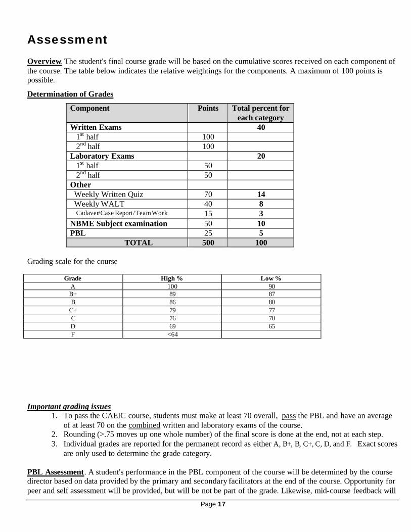
Assessment Overview. The student's final course grade will be based on the cumulative scores received on each component of the course. The table below indicates the relative weightings for the components. A maximum of 100 points is possible. Determination of Grades
Component Points Total percent for each category
Written Exams 40 1st half 100 2nd half 100 Laboratory Exams 20 1st half 50 2nd half 50 Other Weekly Written Quiz 70 14 Weekly WALT 40 8 Cadaver/Case Report/Team Work 15 3 NBME Subject examination 50 10 PBL 25 5 TOTAL 500 100
Grading scale for the course
Grade High % Low % A 100 90 B+ 89 87 B 86 80
C+ 79 77 C 76 70 D 69 65 F <64
Important grading issues
1. To pass the CAEIC course, students must make at least 70 overall, pass the PBL and have an average of at least 70 on the combined written and laboratory exams of the course.
2. Rounding (>.75 moves up one whole number) of the final score is done at the end, not at each step. 3. Individual grades are reported for the permanent record as either A, B+, B, C+, C, D, and F. Exact scores
are only used to determine the grade category. PBL Assessment. A student's performance in the PBL component of the course will be determined by the course director based on data provided by the primary and secondary facilitators at the end of the course. Opportunity for peer and self assessment will be provided, but will be not be part of the grade. Likewise, mid-course feedback will

Page 18
be given to each student at the beginning of the fifth week by the facilitator to give the student an idea of how their performance is perceived, and to give adequate time for improvement, if warranted. Students will also give peer evaluation of their group members and a self evaluation of their performance. The purposes and criteria for facilitators to evaluate students of the small group exercise are to:
• encourage team work • develop critical thinking skills • develop self-directed, life long learning skills • develop a professional attitude • develop better communicative skills
Therefore, the student performance in the PBL will be judged on both group interaction and professionalism rather than focusing solely on cognitive skills. The following criteria will be considered:
• Professional Behavior • Group Interaction & Communication • Preparedness • Knowledge Acquisition & Problem Solving Skills • Attendance
A score of 25 is reserved for the exceptional student. An example would be someone who possesses superior leadership qualities, whose opinions are highly respected and sought by the group and facilitator, who puts the group's function above his/her own personal acclaim, and demonstrates superior use of content knowledge in the understanding of the clinical cases. A score of 20 reflects a student who is performing exactly at the level expected at that stage of their medical education. Only students performing at or above the expected level (i.e. 20)will be eligible to receive an “A” grade in the course. In other words, a student may have the percentage points for an “A” grade, but receive a “B+” if they score below 20 in small group. The PBL part of the course will contribute to the course grade. In addition, the written exams will contain questions that will test the application of the relevant clinical anatomy of the cases. Students will also be given the opportunity to evaluate the facilitators at the end of the course.
Written Exams
Block Examinations . The mid-term and final exam will each carry a 20% value towards the final course grade. Two major examinations will be given in the course on the same day as the practical examinations : please refer to course schedule for the examination dates. Like the laboratory exams, the midterm written exam will cover the material encountered during the first five weeks. The second block exam will cover material from the last five weeks of the course and is not comprehensive. Each block exam will consist of approximately 100 multiple choice type questions. Many written questions will emphasize the clinical application of anatomy and will often be based on clinical scenarios. Information from all course activities is considered testable material for the written exams. Students will NOT be allowed to keep their examinations. The final examination is not comprehensive. The approximate percentage for the sources of the written exam questions are as follows:
1. Lecture-guided topics, 75-85% 2. Assigned reading not lectured upon, 5-10% 3. PBL Cases and Clinical Discussions, 5-10% 4. Integration of X-sectional, radiographic imagery and pathology, 5-10%

Page 19
• Weekly Quizzes: There will be a weekly quiz given that will include 10 questions on material covered in the previous week’s lectures and laboratory sessions. The quizzes will be administered every Monday at the start of class. Each student will be required to take the examination. There will be nine weekly quizzes. The weekly quizzes will count for 14% of the final grade. Two of the lowest scored quizzes will not be counted, so the grade will be actually based on 7 weekly quizzes.
NBME Subject Examination: This is a comprehensive examination testing knowledge in anatomy and embryology. The grade will be calculated by taking the highest score on the examination equaling 100% and the remaining scores factored according to that value. This examination will count for 10% of the final grade
Laboratory Assessment
Laboratory Block Exams . The primary evaluation of the student's anatomical knowledge over the laboratory activities will be through two practical exams, one at mid-term and one at the end of the course. The practicals consist of 50 questions consisting of basic identification and association type questions, plus a bonus question worth one point. Approximately 40 structures are tagged on the cadavers, models and skeletons, and the content level is comparable to most of the BOLDED TEXT structures in the dissector. About 10 questions will be test knowledge about normal radiology and cross-sectional anatomy. The two practicals are of equal value and each worth 10 percent of the final grade. The final practical is not comprehensive.
Weekly Anatomy Lab Tests (WALT). Each dissection table will be quizzed on a weekly basis except for the first week and the week of the mid-term examination. The grade received will apply to each person at the table. The purpose of the exercise is to encourage the completion of the dissections, promote team work and to provide an appreciation for anatomical differences between cadavers.. During the first lab of the week each table will receive a list of 5 structures related to the previous week’s dissections to be tagged on their cadaver. The structures to be identified will be checked by the instructor or TA and a score assigned by the faculty or teaching assistant member for the laboratory group. Students will be encouraged to view the tagged structures at other tables. This activity is 8% of the course grade. Evaluation of teamwork of red and blue lab activities. You will complete a peer evaluation of your group members’ participation in both the red and blue teams activities. You will also do a peer evaluation of your own performance. Cadaver/Case Report. Each dissection table will be required to keep a report on their cadaver. The students at each dissection table should note any significant findings in the report such as evidence of surgical procedures, trauma or injury, anatomic variation, cause of death and pathology. The report form contains boxes for drawings. The report will be checked on a weekly basis (usually on Monday) by a faculty member and will be worth 3% of the final grade The form for the report will be provided on the course web site. Your group will give a presentation of your cadaver autopsy report at the end of the course. Your group will hypothesize the cause of death of your cadaver based on your observations. Fellow students and faculty can ask you questions about your presentation, so be prepared for this possibility. You will also include a one page case report summary that describes, based on your observations on the cadaver and the cause of death, a case report about the cadaver based on the information you have obtained in your cadaver report. The completed cadaver/case report must be turned in on last day of formal classes. The reports must be submitted electronically.
Page 20
The report should include the table number and names of the group members. You will all complete peer evaluations of your group and a self evaluation related to working to complete this cadaver autopsy report.
Course and Faculty Evaluation:
Mid Course Evaluations: A random sample of students will be asked to participate in a Mid-Course evaluation by the Office of Medical Education. This will assist the course director in being able to make any mid course adjustments based on the student feedback. End of Course Evaluation. A random sample of students will be required to complete an evaluation of the course administered by the Office of Medical Education at the end of the course. Student evaluations will be kept anonymous to the course director. Comments are of particular interest for improvement of the course. Students must complete the evaluation in order to have their course grade recorded. Evaluation of Faculty – Students will be asked to complete the FSU SPOT/SUSSAI faculty evaluation forms for major course faculty and the FSU COM faculty evaluation form.
Student evaluations of the course, lecturers, facilitators peers and self are required in order for grades to be released to the FSU Registrar.

Page 21
Faculty & Staff Course Director Andrew F. Payer, Ph.D. Professor and Year One Director College of Medicine Room 2300F Phone 850-644-7501 FAX 850-644-5766 [email protected] Course Faculty
Edward Klatt, M.D. Professor and Year Two Director Morton Levitt. M.D. MHA Professor Dr. Jacob Vanlandingham, Ph.D. Assistant Professor
James Cavanagh, M.D. 75 Bellac Rd. 850-562-1533
Course Teaching Assistants
1. Cooke, Sarah 2. Doster, Jamie 3. Gadbois, Brian 4. Knobbs, Amanda 5. Hall, Megan 6. Lee, Phillip 7. McCall, Christina 8. Porter, Fernando 9. Rachals, Cara 10. Schwartz, Holly 11. Sochet, Anthony 12. Swanson, Steffanie

Page 22
Order of Weekly Content Areas
WEEK REGION 1 Thorax 2 Back & Upper Extremity 3 Abdominal Wall & Cavity 4 Abdominal Cavity
completed
5 Neck & Head 6 Head, Skull & Brain 7 Orbit, Pharynx, Nasal &
Oral cavities
8 Larynx and Ear 9 Pelvis & Perineum
10 Lower Extremity
Page 23

Page 24
Protocol For The FSU-COM Human Anatomy
Laboratory
Dr. Andrew Payer is the member of the State of Florida Anatomical Board representing the FSU-COM and responsible for the confidentiality and dignity for the remains of the individuals who willed their bodies to the state of Florida. Lab activity 1. Access. The anatomy lab will be open 24 hours a day, 7 days a
week during the semester. After hours the anatomy lab can be accessed by the card reader.
2. All students, faculty and approved guests must sign “Pledge of Respect” form
3. Authorized Personnel. Only COM medical students, faculty and other health-related personnel and facility workers are permitted access to the lab. FSU badges are the best form of I.D. All unauthorized persons will be told to leave immediately. After scheduled course hours, campus police regularly patrol the area and will escort trespassers from the lab and report the person(s) responsible for the unauthorized entry to appropriate authorities for corrective purposes. Immediate family members and health-oriented guests of medical student's must first receive authorization from Dr. Payer before being allowed entry into the lab. The lab doors should not be opened for anyone "knocking" other than for an authorized person (i.e. student forgetting their card). Visitation is NOT permitted during scheduled dissection periods. During any visit of authorized guests they should avoid all opened cadaver tanks. Minors will NOT be admitted except as part of an organized tour. It is the responsibility of all authorized personnel , faculty and students, to enforce these rules. It is the LAW that donors to the Florida Anatomical Board are guaranteed the respect and confidentiality in the spirit by which their gift was donated to our institution. Any disrespect to the cadavers will be dealt with accordingly.
4. According to Florida law, removal of any cadaver parts,
whatsoever, from the laboratory is a crime of g rave robbery. 5. NO photographs are to be taken of the cadavers or anything in the
laboratory, except for images necessary for cadaver autopsy reports.

Page 25
6. NO eating, drinking or smoking is allowed in the laboratory or
amphitheater. 7. NO radios or tape players are allowed in the laboratory, unless
used with earphones.
8. Personal protection in the lab:
§ No open toe shoes in the lab § Recommend wearing scrubs or lab coats. Some prefer an
additional plastic apron for protection from fluids § Recommend two layers of gloves (one vinyl and one latex § Wear glasses or protective goggles § Material Safety Data Sheets of chemicals used in the laboratory are
available in the lab § Use dust mask when using electric bone saws § Do not wear open toed shoes or sandals
9. First aid for cuts in the lab: First aid kits are available in the lab § Remove gloves and wash cut area § Cover with sterile bandage § Put on clean gloves
10. All lab coats, dissecting equipment and books should be stored in
the locker room or in the cadaver tank. Anything left out after regular lab sessions will be thrown out during daily lab cleaning. Do not wear dissection clothing or gloves outside of the anatomy laboratory.
11. Skeletons are available in the lab. Do not remove them from their
stands or take them apart. 12. Disarticulated bones are also available, and should not be removed
from the lab. Report any broken bone specimens to a faculty member for repair/replacement.
13. The antiseptic soap for washing hands is located on the sinks and
locker rooms. 14. Rule to Remember No not try to catch a dropped tool or retrieve a
tool dropped in the tank. In case of injury in the lab during regular lab sessions, notify a faculty member. If an injury occurs after regular lab hours, go to the emergency room.
Page 26
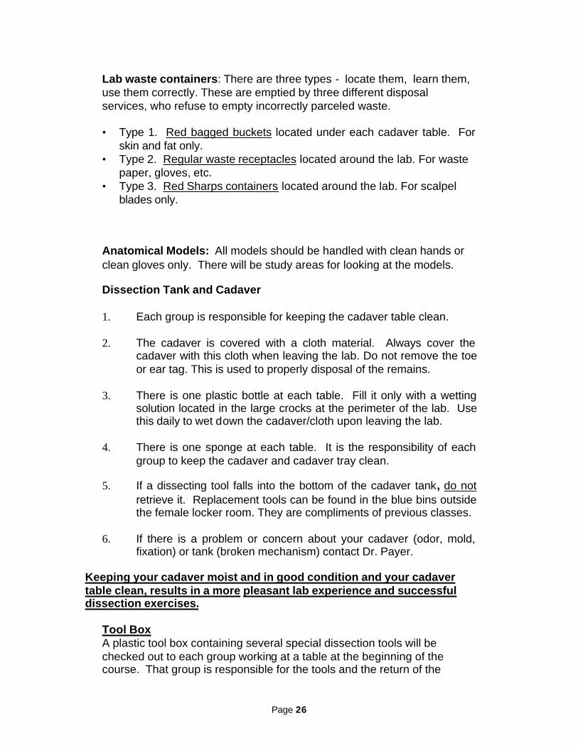
Lab waste containers: There are three types - locate them, learn them, use them correctly. These are emptied by three different disposal services, who refuse to empty incorrectly parceled waste. • Type 1. Red bagged buckets located under each cadaver table. For
skin and fat only. • Type 2. Regular waste receptacles located around the lab. For waste
paper, gloves, etc. • Type 3. Red Sharps containers located around the lab. For scalpel
blades only. Anatomical Models: All models should be handled with clean hands or clean gloves only. There will be study areas for looking at the models. Dissection Tank and Cadaver 1. Each group is responsible for keeping the cadaver table clean. 2. The cadaver is covered with a cloth material. Always cover the
cadaver with this cloth when leaving the lab. Do not remove the toe or ear tag. This is used to properly disposal of the remains.
3. There is one plastic bottle at each table. Fill it only with a wetting
solution located in the large crocks at the perimeter of the lab. Use this daily to wet down the cadaver/cloth upon leaving the lab.
4. There is one sponge at each table. It is the responsibility of each
group to keep the cadaver and cadaver tray clean. 5. If a dissecting tool falls into the bottom of the cadaver tank, do not
retrieve it. Replacement tools can be found in the blue bins outside the female locker room. They are compliments of previous classes.
6. If there is a problem or concern about your cadaver (odor, mold,
fixation) or tank (broken mechanism) contact Dr. Payer.
Keeping your cadaver moist and in good condition and your cadaver table clean, results in a more pleasant lab experience and successful dissection exercises.
Tool Box A plastic tool box containing several special dissection tools will be checked out to each group working at a table at the beginning of the course. That group is responsible for the tools and the return of the

Page 27
complete set in clean condition at the end of the course. Failure to do so will result in the withholding of the course grade and prevention of registration for the next term. There will be a charge to the group for any lost/unreturned tools. (PRICE LIST ENCLOSED BELOW).
TOOLS G IVEN TO EACH TABLE ARE TO BE RETURNED
AT THE END OF THE COURSE IN CLEAN CONDITION
TOOL BOX REPLACEMENT PRICE LIST Rib Shears ------------------------------------------------------------------------------------------------------------------$72.00 Bones hears ----------------------------------------------------------------------------------------------------------------$50.00 Bone Chisel ----------------------------------------------------------------------------------------------------------------$26.00 Bone saw --------------------------------------------------------------------------------------------------------------------$60.00 Rawhide Mallet ----------------------------------------------------------------------------------------------------------$40.00 Periosteal Elevator -----------------------------------------------------------------------------------------------------$15.00 Tool Box --------------------------------------------------------------------------------------------------------------------$15.00 UNRETURNED, LOST, OR BROKEN TOOLS WILL RESULT IN WITHHOLDING OF GRADES AND FUTURE REGISTRATION UNTIL EITHER RETURNED/PAID BY RESPONSIBLE DISSECTION TABLE/GROUP.

Page 28
Prologue
I WONDER WHAT HE WAS LIKE?
Andrew F. Payer1 It was Monday morning, my weekend was a disaster, my secretary was late, my schedule was full for the day, and I was really feeling sorry for myself. The door to my office swung open wide and a bright-eyed young man entered radiating with happiness and a lively step. He asked me if I was the doctor who ran the body donation program for the medical school. I acknowledged that I was in charge of such a program, but expressed that it might be a bit early for him to be thinking about donating his body for medical education and research. He smiled and said that he was only 28 years old, but that his time was very near. I gave him the official form to complete for my records. As he was filling out the form, he proceeded to tell me that he had a terminal form of brain tumor. He was going in for surgery this coming Friday for the “last time” and was told he would not survive much longer. Suddenly, he switched the topic and began asking about me and how things were going at work and home. He was politely amused to find out that I raised tortoises. It was evident that he sensed my “sad” state of mind and tried to cheer me up. He began to tell me more about himself. He was spending time visiting terminally ill patients at the hospital and trying to help them
1 Andrew F. Payer, Ph.D., associate professor of anatomy and neurosciences at The University of Texas Medical Branch at Galveston, and director of UTMB’s Willed-Body Program. Reprinted with permission of the author.
through their own ordeals. He was very worried about his mother, and wanted to ensure that all would be taken care of so she would not have to deal with the final details. He asked if he could use my phone to confirm his meeting with his lawyer. “You know how messy the legal details can get in settling the affairs of one's estate.” I told him that he was handling this situation much better than me. He smiled and said, “I have had my bad moments, but as I look back, I was very lucky to have had a life filled with the love of my family and friends. I have accepted my fate and am just luckier than most to know when the event will most likely happen.” We continued to talk. Emotions began to swell up inside me. My terrible day was not as bad as I originally thought. I caught myself feeling selfish because I wanted to go outside to feel the sea breeze, go home and kiss my wife, work out the problems I had with a faculty colleague, and tell my secretary to take the day off! As he walked down the hall from my office, his step and movements were as lively and happy as when he entered. He turned with a smile and said: “I would like to ask one favor When those medical students are looking down at my remains and getting the nerve up to make the first cut, tell them that I was a nice person who loved life and his fellow man. Tell them that they should not feel bad about what they are about to do. Finally, tell them that they got a damn good specimen and they should be able to learn a lot of anatomy from me!” He died peacefully at home several weeks later. That fall was the beginning of medical school for a new group of students. I was at the dissecting table that held his remains as the four medical students looked at their cadaver and pondered about what to do next. I heard one of them ask another: “I wonder what he was like”.

Page 29
Furthermore
Delese Wear is associate professor of behavioral sciences, Northeastern Ohio Universities College of Medicine, Rootstown, Ohio.
An often-cited axiom in medical education is that a medical student's first patient is a dead one. Because death and dying are essential issues in the Behavioral Sciences/Human Values in Medicine curriculum here at Northeastern Ohio Universities College of Medicine, we began to examine how we might work with our anatomy colleagues to provide some reflective experiences for medical students as they began their often-difficult dissection experience. The emotions evoked by cutting into a dead body are complicated, and are inevitably tangled up with the students' own feelings about and experiences with death. Because of our department's strong interest in literature and medicine, we decided that perhaps poetry might be a bridge to the anatomy lab -cadaver poetry, that is. We selected several poems written about the cadaver or the dissection experience itself, including Diane Roston's "On Studying Anatomy," which is reprinted here. Of course, precious curriculum time is necessary: how do we find it? Our department chairperson gave up one hour of large-group lecture during the early weeks of the term for behavioral sciences faculty to meet with small groups of twenty students each. Several of the faculty had never taught literature or used poetry in their teaching, so we met as a department beforehand to discuss what one might do in this one-hour class. Most of us decided to open up the class with a general discussion of how the students' dissections were going, rather than have them read the poetry first. Discussion was directed toward what students thought about the lived lives of their cadavers: how did they come to think that? what were the physical clues, if any? how did they feel about it? This proved to be an opening for reading aloud Roston's poem and a poem called "Anatomy Lesson" by Jack Coulehan, both of them musings about cadavers: their unknown lives, the stories that brought them to the lab, and more difficult questions of meaning in one's life and death. Discussion was abundant, and many - not all - students disclosed how the experience was unfolding for them, disclosures evoked by the personalized, richly described, contextualized lived lives of the cadavers in the poems. Coulehan describes his cadaver, Ernest's "three-days growth of beard," which made one student think and wonder about the painted fingernails of her cadaver: when had she painted them? how soon before her death? had she known she was dying? Sally Harris Sange, MD, a medical student when she wrote her poem "Disclosure," summarized best what poetry in the anatomy lab might mean as she wrote a poem imagining her cadaver alive as a future patient:
When I meet you in my examining room, pulsing breathing, limping a bit, complaining of a headache, perhaps, chest pain, God help me if I remember nothing but the arteries. …… Let me remember how deep your privacy dwells.
What else do we hope that this experience might mean to medical students? We hope that it will offer one of many opportunities for students to reflect on professional distance. We also hope that it is one of many experiences in which students look beyond science for insights and clues as they try to understand humanness - a lifetime search that just may have some of its origins in the cadaver lab. DELESE WEAR, PhD

Page 30
On Studying Anatomy By Diane M. Roston What is before me in these rags of skin, human fragments guttered on a metal table . . . should be as much the subject of poetry as the pooling of shadow in a brook or the subtle changes in a woman's face. - Charles LeBaron, Gentle Vengeance She knew down to her bones that everything that lives wants to go the limit. She lived to bellow naked on a dry dirt road split fast by black skid messages that she rode out each hot noon. The messages always read the same, scarred in every crevice of her body's day: leather, fancy feathers, strong perfume strutted all night, then at high sun, stripped away. She was a mama, wild mama. Gave birth to a night-black motorcycle bird, sucked and licked it clean until it angled like a hawk. Mounted it, and rode fast. One day she rode so fast she split the sun, that faithful high noon blood, and with a joyful bellow, soared naked, jubilant, to a gleaming ninety-mile-an-hour tomb. Now, student, to anatomy: cleave and mark this slab of thirty-one-year-old Caucasian female flesh, limbs, thorax, cranium, muscle by rigid muscle dissemble this motorcycle victim's every part (as if so gray a matter never wore a flashing ruby dress).
VOLUME 68 m NUMBER 2 a FEBRUARY 1993
Dr. Roston was a medical student at the University of Wisconsin Medical School when she wrote this poem, which won third place in the William Carlos Williams Poetry Competition in 1983. "On Studying Anatomy," @ 1983 by Northeastern Ohio Universities College of Medicine, is reprinted with permission from Poetry: A Collection of Poems Written by Medical Students. Lisa R. Dittrich, of the Ac-ademic Medicine staff, is the editor of "furthermore.,,
137
Page 31
A Piece of My Mind
John
We first met in a Frankensteinian manner-two first-year medical students approaching a galvanized casket with a crank protruding from one end, knowing that inside lay something human. We were not alone. Eighty other neophytes approached 40 other coffins with the same mixed feelings: forced jocularity interspersed with breath holding. There was a feeling much different from the days of dissecting cats or dogs in premedical days-one we could not identify other than uneasiness or discomfort.
As the casket was opened, there lay John. We knew he was John because of the shipping tag tied to his wrist. Months in the cadaver tank had faded all but "John." The remainder may have been "Doe," or perhaps his real last name. –
It took us a few days to overcome the revulsion at his leathery-brown skin and his compressed features that came from floating in the tank with his fellowman. Floating? Perhaps packed would better suit the circumstances. But we knew he was human because he had ten fingers and ten toes and a cartilage that we could identify as a nose. Every day from September until June we cranked him out of the abyss, unwrapped his bedsheet (brought from home), which was really his shroud. When the day's dissection was done, he was rewrapped, sprinkled with water to keep him pliant,.lowered again in the confines of his tomb, only to be disturbed again at the next dissection session. The fragments of the day's work were scraped away to the common grave of 50 cadavers.
Edited by Roxanne K. Young, Assistant Editor.
Since we had not as yet been exposed to pathology, the cause of John's death was not apparent to us. A more complete autopsy, however, had never been performed. First the abdomen, next the thorax, neck, and skull, to be followed by the extremities. Each fiber was meticulously dissected until merely the bony framework of a particular area remained. But what ended his life remained a mystery.
Our feelings were mixed. There could be no love for him as one human loves another. There was a muted respect because of what he was teaching us. We hated him when the professor, grade book in hand, ingloriously reached into John's cavities-and wordlessly asked that we identify a muscle, a nerve, a foramen, a blood vessel. Our relief was complete when John's structures and our answer satisfied the inquisition.
Doomed to obscurity, saved from a nameless grave, macabre in the timeless interval until burial of skeleton and gristle, leaving no estate. But the knowledge that John revealed to us became a priceless stone in the foundation of our medical knowledge.
Ah, John. If we could all do as much in death as you. R. E. Bower, MD Richlands, Va
We welcome contributions to A PIECE OF MY MIND from readers. Submissions should be addressed to Roxanne K. Young, The Journal of The American Medical Association, 535 N Dearborn St, Chicago, IL 60610.

Page 32
INTRODUCTION TO GROSS PATHOLOGY FOR ANATOMY STUDENTS
As you perform the dissection of your cadaver, you will most likely encounter some abnormal (pathological) anatomy. This handout outlines some general principles of gross observation of lesions, and describes some features of common pathologic processes you may encounter. You will learn more about pathology and pathophysiology in subsequent courses; however, it is important at this stage to begin to develop keen powers of observation. 1. VIEWING THE SPECIMEN AS A WHOLE: Your first task should be to IDENTIFY the organ. The second is to IDENTIFY and CHARACTERIZE any abnormalities (lesions). When you are examining an organ in a cadaver and can make use of normal anatomic relationships, the first part is easy. When you are examining a pathologic specimen that has been removed from the body, this may not be so simple. To identify and describe the organ as a whole you should observe the following features: SIZE: What are the dimensions of the specimen you are examining? Is it the entire organ, or just a section of it? If just a portion is provided to you, what plane is the slice taken from? Can you extrapolate from the size of the section to the size of the intact organ? And lastly, is the size normal or abnormal for that organ? Dimensions that are relevant differ depending on the type of organ. For example, for the heart, it is customary to weigh the organ as a whole, and measure the thickness of the right and left ventricular walls. These measurements will tell you if there is too much or too little cardiac muscle present; the former situation indicates hypertrophy of the muscle, as seen in hypertension, the latter indicates atrophy of the muscle or loss of muscle due to myocardial infarctions or other processes. By contrast, a solid organ such as the spleen is best described in terms of weight and/or overall dimensions. Several conditions can lead to enlargement of the spleen, including portal hypertension, tumors, acute or chronic infection. SHAPE: The shape of an organ will provide clues as to its identity. For example, if you are provided with a tubular segment of tissue, it can only be part of the gastrointestinal tract, the genitourinary tract, or the vascular system. Specific features of the inner lining and the wall itself can help you distinguish between these--e.g. small intestine has very distinct transverse mucosal folds, as opposed to a large elastic artery such as aorta, which would normally have a smooth inner surface (intima). Solid organs will have different tell-tale shapes as well; a kidney sectioned saggitally looks like a bean, the liver is very large and has two major lobes, the thyroid is shaped like a butterfly, etc. It is also important to notice abnormalities in shape, once you have identified the organ. For example, a heart from a patient with congestive heart failure will appear more rounded (globular) than normal. This is because the chamber becomes dilated. CAPSULAR SURFACE: It is important to distinguish between an anatomically relevant surface of the organ (capsule or other covering), and the surface that has been exposed by the pathologist or anatomist by cutting open the organ (cut surface, see below). Capsules of organs should normally be smooth, glistening, thin, and nearly transparent. Some abnormalities you may observe include thickening of the capsule, focal lesions (see below), wrinkling of the capsule (indicating shrinkage of the organ), rough
Page 33
shaggy material on the capsule (fibrin or collagen, indicating recent or remote inflammatory process), or a nodular “cobblestone” appearance, that indicates extensive scarring in the underlying organ. ANATOMIC RELATIONSHIPS: These relationships, if any are preserved in the specimen you are presented with, can help you identify the organ, but may also indicate pathologic processes. For example, radiation therapy to the uterus for cervical cancer may cause fibrosis (scarring) that results in plastering together of the cervix, the bladder, and the rectum. Sometimes, abnormal connections (fistulas) may be formed between these organs. Scarring processes in general, if they involved the outer surfaces of organs, may result in “adhesions”, bands of scar tissue that connect separate structures, or disparate parts of the same structure (e.g. loops of bowel, or visceral and parietal pleural surfaces). 2. EXAMINING THE CUT SURFACE: The CUT SURFACE should be examined and described in terms of COLOR, ARCHITECTURE, and CONSISTENCY. Any abnormal areas (LESIONS) should be described using a similar scheme. COLOR can sometimes be useful in characterizing the organ or the lesion. The normal brown/tan/red color of many tissues is largely a function of the presence of blood. Material that appears darker than normal (brown, black or gray in fixed specimens, red or brown in fresh specimens) is often blood. Conversely, one reason an organ or lesion might be paler that surrounding areas is lack of blood (e.g. infarct). Fat also imparts a pale yellow appearance. Carbon pigment (black) may be seen in lungs and thoracic lymph nodes of smokers or city dwellers, pus usually appears white to yellow, bile appears green to yellow, etc. ARCHITECTURE: The cut surface of an organ will be the best clue as to its identity. For example, the cut surface of the kidney will demonstrate the cortex and medullary pyramids very clearly. The absence or blurring of these regions in a kidney signifies pathology (e.g. diffuse infiltration by tumor, or swelling as may be seen in acute tubular necrosis). The cut surface of glandular tissues like pancreas or salivary gland has multiple little lobules that are joined together by collagen (interlobular septa). It is important to notice any focal (e.g. holes, masses, discolorations, etc. )or diffuse abnormalities of the underlying tissue architecture. CONSISTENCY: Much can be learned about a specimen by palpating it (with gloves on!). For example, normal lung is spongy; pneumonia will make the lung feel very firm and solid (“consolidation”). Scar tissue (fibrosis) will cause tissue to feel tougher than normal, cancer is often hard, like a rock. Processes that cause death of the tissue (necrosis) may lead to softening (liquefaction) of the specimen in areas. 3. DESCRIBING AND CHARACTERIZING THE LESIONS: LESIONS should be described according to size, shape, number, color, and consistency (see above). Once the salient features of the lesions have been noted, follow the steps below to arrive at a tentative diagnosis. Determine whether the lesion(s) is FOCAL or DIFFUSE. FOCAL lesions are discrete, stand out from surrounding unaffected tissue, and may have well defined margins. Examples of focal lesions are infarcts, abscesses, granulomas, benign neoplasms, and some malignant neoplasms. DIFFUSE lesions lack discrete margins, tend to be more subtle than focal lesions, and may involve the entire organ.

Page 34
Examples of diffuse lesions are organ hypertrophy, leukemic infiltration, inflammatory processes, or diffuse scarring processes. If the lesion is FOCAL (or MULTIFOCAL, i.e. several discrete, well demarcated lesions): Does it reflect the blood supply of the organ or part of the organ? Infarcts (areas of tissue death due to interruption of the blood supply) are typically “wedge-shaped” with the apex of the wedge at or near a blocked blood vessel, and the base at the capsular surface. The affected area encompasses the region supplied by that artery. Is it cavitary? (Is the lesion a hole or softened region?) If so, is the wall of the cavity thin or thick? Thin walled cavities may include benign cysts, or abscesses. Thicker walls indicate scarring and host reaction of a chronic infection, and are typical of granulomas, as seen in tuberculosis. Is it expansile? Expansile masses have “pushing borders” and compress the adjacent tissues, forming a “pseudocapsule” of attenuated tissue around itself. These lesions can sometimes be “shelled out” of the organ. This is a feature of benign neoplasms (e.g. uterine fibroids (leiomyomas)) Is it infiltrative? Infiltrative lesions, in contrast to expansile ones, have an intimate relationship with surrounding tissue and cannot be shelled out. Close inspection of the border of the lesion may reveal irregular tongues of tumor extending into the tissue. This is a feature of malignant neoplasms (cancers). If the lesion is DIFFUSE: Does it involve overall enlargement of the organ? Examples of diffuse pathologic processes that result in organomegaly (enlargement) are: hypertrophy, chronic passive congestion, and infiltration by leukemia. Can the cardinal signs of inflammation be seen? Swelling, congestion of blood vessels, and infiltration of the tissue by white blood cells are features of inflammatory processes, such as pneumonia. Swelling is manifested by bulging of the cut surface, blurring of normal architectural features, or more firm consistency of the tissues. Vascular congestion will make tissue appear darker in color, and infiltration by white blood cells may make tissue appear paler and firmer (indurated) than normal. Inflammation may also show up on the capsular surface as a shaggy, soft material (fibrin, fibrinous exudate). Lung is a common place to see these changes. Is there generalized scarring? A sequel of inflammatory or degenerative processes is scarring (fibrosis). This can be most readily recognized by observing the capsular surface for areas of retraction or a cobblestone appearance. The cut surface will show deranged architecture, areas of tough, gristle-like tissue, or general increase in firmness. A scarred organ is often smaller than normal. Examples are benign nephrosclerosis in the kidneys (common result of aging and hypertension), cirrhosis of the liver, and interstitial pulmonary fibrosis in the lung.
Page 35
Clinical Anatomy, Embryology and Imaging Examination Protocol FOR WRITTEN EXAMS : The course directors should inform all students, that if they were 15 minutes late to an exam, it would be considered an unexcused absence. The student(s) will have to contact the Student Affairs Office. FOR PRACTICAL EXAMS: All students must arrive promptly at their assigned time or they will not be permitted to take the practical. A course coordinator or the course director will announce the following items to the students. 1. Students will enter the examination room ten-minutes prior to the scheduled time of the examination(s). 2. Students will be instructed to write their name on the test and answer sheet, and bubble in their name and personal
identification number prior to starting the examination(s). 3. All answers must be recorded on the answer sheet in order to receive credit. 4. If a restroom break is necessary, take your test and answer sheet to the proctor(s)
5. All Question and Answer sheets will be collected at the end of the examination by the proctor. 6. Warnings:
A. Prior to the end of the exam(s), students will be given a 30-minute warning and will be reminded that all answers must be recorded on the computer answer sheet in order to receive credit.
B. Prior to the end of the exam(s), students will be given a 10-minute warning. 7. At the end of the exam, all students will be instructed to:
A. Put their pencils down B. Take their answer sheet and in some cases their test to the proctor stationed in the foyer. C. Failure to comply is considered irregular behavior and will be noted.
8. BRING YOUR SHARPENED PENCILS