estrategias de aplicacao_polimeros
TRANSCRIPT

This journal is c The Royal Society of Chemistry 2013 Chem. Soc. Rev., 2013, 42, 1147--1235 1147
Cite this: Chem. Soc. Rev.,2013,42, 1147
Design, functionalization strategies and biomedicalapplications of targeted biodegradable/biocompatiblepolymer-based nanocarriers for drug delivery
Julien Nicolas,* Simona Mura, Davide Brambilla, Nicolas Mackiewicz andPatrick Couvreur
Design and functionalization strategies for multifunctional nanocarriers (e.g., nanoparticles, micelles,
polymersomes) based on biodegradable/biocompatible polymers intended to be employed for active
targeting and drug delivery are reviewed. This review will focus on the nature of the polymers involved
in the preparation of targeted nanocarriers, the synthesis methods to achieve the desired
macromolecular architecture, the selected coupling strategy, the choice of the homing molecules
(vitamins, hormones, peptides, proteins, etc.), as well as the various strategies to display them at the
surface of nanocarriers. The resulting morphologies and the main colloidal features will be given as well
as an overview of the biological activities, with a special focus on the main in vivo achievements.
Institut Galien Paris-Sud, UMR CNRS 8612, Univ Paris-Sud, Faculte de Pharmacie, 5 rue Jean-Baptiste Clement, F-92296 Chatenay-Malabry cedex, France.
E-mail: [email protected]; Fax: +33 1 46 83 55 11; Tel: +33 1 46 83 58 53
Julien Nicolas
Dr Julien Nicolas graduatedfrom the Ecole Superieure deChimie Organique et Minerale(ESCOM), France, in 2001. Hecompleted his PhD in 2005under the supervision of Prof.Bernadette Charleux at theUniversity Pierre and MarieCurie, Paris, where he studiednitroxide-mediatedpolymerization in homogeneousand aqueous dispersed media.He then joined the group of Prof.David M. Haddleton at the
University of Warwick, UK, for a postdoctoral fellowship to designpolymer–protein bioconjugates by controlled/living radicalpolymerization. In 2007, he was appointed permanent CNRSresearcher in the group of Prof. Patrick Couvreur, University Paris-Sud, France, where his current research activities are focused oncontrolled/living radical polymerization techniques and on thesynthesis of novel biomaterials and functionalized, biodegradablenanoparticles for active targeting and cell imaging purposes. He is(co)author of more than 50 peer review articles in internationaljournals, 4 patents and 7 book chapters.
Simona Mura
Dr Simona Mura gained thedegree in PharmaceuticalChemistry and Technology in2005. In 2009, she was awardedher PhD in Chemistry andTechnology of Drug at theUniversity of Cagliari, Italy,working on the design andin vitro evaluation of novelvesicular systems for the topicaldelivery of drugs. In 2008 shejoined the group of Pr. EliasFattal (UMR CNRS 8612) atUniversity Paris-Sud, Chatenay-
Malabry, France, as a post-doctoral research assistant to study thelung toxicity of biodegradable nanoparticles designed forpulmonary administration of drugs. In 2011 she was appointedAssociate Professor within the framework of the { CNRS-HigherEducation chairs c program, in the group of Pr. Patrick Couvreur(UMR CNRS 8612) at University Paris-Sud, Chatenay-Malabry,France. Her research focuses on the assessment of thetherapeutical activity of novel nanoparticulate systems andbiopolymers for the treatment of severe neoplastic diseases.
Received 16th July 2012
DOI: 10.1039/c2cs35265f
www.rsc.org/csr
Chem Soc Rev
REVIEW ARTICLE
Publ
ishe
d on
13
Dec
embe
r 20
12. D
ownl
oade
d by
Uni
vers
idad
e Fe
dera
l do
Para
na o
n 12
/07/
2013
21:
06:0
8.
View Article OnlineView Journal | View Issue

1148 Chem. Soc. Rev., 2013, 42, 1147--1235 This journal is c The Royal Society of Chemistry 2013
1. Introduction
In the recent years, significant achievements have been witnessed inthe field of nanotechnology, especially in material science,electronics, photonics, supramolecular assemblies and drugdelivery. In particular, the medical application of nanotechnologies,usually termed nanomedicine,1–6 has given a crucial impulse to thedevelopment of various types of drug-loaded nanocarriers, such asliposomes, nanoparticles, micelles etc. A great deal of effort is nowfocused on the engineering of nanoparticulate systems able to serve
as efficient diagnostic and/or therapeutic tools against severediseases, such as cancer, infectious or neurodegenerative dis-orders.7–24
Among the numerous classes of materials employed for drugdelivery purposes, colloidal systems based on polymers haveattracted much attention due to the flexibility offered bymacromolecular synthesis methods, the almost infinite diversityof polymers in terms of nature, properties and composition, andtheir ease of functionalization.3,7–10,12–15,25–27 Polymer-based,engineered colloids represent one of the most promising oppor-tunities for in vivo diagnosis and treatment of many diseases.Indeed, the possibility to load them with a drug (which can alsobe covalently linked to the polymer) and to functionalize themwith specific ligands, intended to be recognized by receptorsoverexpressed at the surface of particular cells (e.g., cancer cells,brain endothelial cells etc.), paved the way to the so-called activetargeting.25,27–28
The successful design of targeted nanocarriers is usuallygoverned by the nature of the polymer and the ligand, as well asby the selected coupling reaction. So far, a plethora of ligationstrategies have been developed, each of them exhibiting benefitsand drawbacks. Hence, these constructions represent a subtleequilibrium between organic chemistry, macromolecular synthesis,physico-chemistry and pharmaceutical science.
The present review will detail the different steps to constructfunctionalized nanoparticulate systems based on biodegradable/biocompatible polymers for targeted drug delivery (Fig. 1). Moreprecisely, it will focus on the nature of the polymer employed forthe preparation of targeted nanocarriers, the synthesis method
Davide Brambilla
Dr Davide Brambilla obtained hisMSc in Experimental Medicinewith honors at the University ofMilano-Bicocca, Italy, in 2008.Then he moved to France in thegroup of Patrick Couvreur at theUniversity of Paris-Sud where hecompleted his PhD in 2012. SinceJune 2012 he has been a post-docin the group of Drug Formulationand Delivery headed by Prof.Jean-Christophe Leroux at theSwiss Federal Institute ofTechnology (ETH Zurich). In
2012 he got the ETH-VPFW/Marie Curie Post-Doctoral Fellowship.His main research interest is focused on the design of smart, brain-targeted delivery systems against Alzheimer’s disease.
Nicolas Mackiewicz
Dr Nicolas Mackiewiczgraduated from the EcoleCentrale Marseille (ECM),France, in 2004. He obtained hisPhD in 2007 under thesupervision of Prof. CharlesMioskowski at the Institute ofBiology and Technologies (CEA),Paris, where he studied carbonnanotubes functionalization andpotential new applications.Then, he joined the group of DrFrederic Duconge at the Instituteof Biomedical Imaging (CEA), for
a postdoctoral fellowship to design polymerized micelles forin vivo diagnosis. In 2009, he moved to the group of Prof. PatrickCouvreur, University Paris-Sud, France, for another postdoctoralfellowship in collaboration with the pharmaceutical companySanofi on the synthesis of novel biodegradable nanoparticles foractive targeted drug delivery and imaging. In 2012, he moved toSwitzerland as a R&D project manager at Vestergaard-Frandsen,to work in the field of disease control textiles to prevent and fightpests associated diseases such as malaria.
Patrick Couvreur
Prof. Patrick Couvreur is FullProfessor of Pharmacy at theUniversity Paris-Sud and holderof the chair of ‘‘InnovationTechnologique’’ (2009–2010) atthe ‘‘College de France’’. He isappointed as a Senior Member ofthe ‘‘Institut Universitaire deFrance’’. Prof. Couvreur’scontributions in the field of drugdelivery and targeting are highlyrecognized around the world.The major scientific contributionof Patrick Couvreur to the
Pharmaceutical Sciences is also recognized by numerousinternational awards, including the ‘‘2004 PharmaceuticalSciences World Congress Award’’ (Kyoto, Japan), the prestigious‘‘Host Madsen Medal’’ (Beijing, 2007), and very recently the‘‘European Pharmaceutical Scientist Award’’ (EuropeanFederation of Pharmaceutical Sciences). In 2009, he was alsoawarded by the ‘‘Prix Galien’’, the highest French distinction intherapeutics. He has been appointed as a member of fouracademies (Academie des Technologies, Academie de Medecine,Academie de Pharmacie in France, and Academie Royale deMedecine in Belgium).
Review Article Chem Soc Rev
Publ
ishe
d on
13
Dec
embe
r 20
12. D
ownl
oade
d by
Uni
vers
idad
e Fe
dera
l do
Para
na o
n 12
/07/
2013
21:
06:0
8.
View Article Online

This journal is c The Royal Society of Chemistry 2013 Chem. Soc. Rev., 2013, 42, 1147--1235 1149
to achieve the desired macromolecular architecture, theselected coupling strategy, the choice of the homing devices(vitamins, hormones, peptides, proteins, etc.), as well as thevarious strategies to display them at the surface of nanocarrierswithout altering their colloidal properties. The resultingmorphologies and the main colloidal features will be given aswell as a general discussion about the biological activities, witha special focus on the main achievements that have beenreported in vivo.
2. Morphology and preparation ofnanocarriers
Nanoparticulate systems are colloidal-sized particles, withdiameters ranging from 1 to 1000 nm. A wide variety ofnanocarriers composed of different materials including lipids,polymers and inorganic materials have been developed, resultingin delivery systems that vary in their physicochemical propertiesand thus in their applications (Fig. 2). The following sections willdescribe the most used polymer-based nanoparticulate systemsfor drug delivery and targeting purposes.
2.1. Nanocarrier morphologies
In this review, we will focus our attention on polymer-basednanocarriers. In this regard, three classes of polymer-based
colloidal systems will be discussed: (i) nanoparticles, (ii) polymericmicelles and (iii) polymersomes. The reader who would like moredetails about all other nanoparticulate systems (e.g., liposomes,solid lipid nanoparticles, etc.) is referred to the adequatereviews.29,30 It is noteworthy that due to intense research effortin the field, most of these systems can be easily engineered, whichmakes them easily adjustable in terms of size, surface charge,drug loading, release mechanism, etc.
2.1.1. POLYMERIC MICELLES. Polymeric micelles belong to agroup of nanosized colloids that can be formed by self-assemblyof amphiphilic block copolymers in aqueous solution.31 Thehydrophobic core region serves as a reservoir for hydrophobicdrugs, whereas the hydrophilic shell region stabilizes the hydro-phobic core, making the particle an appropriate candidate fori.v. administration.32 They typically present the so-called core–shell morphology and exhibit average diameters in the 5–100 nmrange.33 Contrary to nanoparticles, polymeric micelles arecharacterized by a critical micelle concentration (CMC). There-fore, upon dilution below the CMC, micelles disassemble intofree unimers.34 Nonetheless, polymeric micelles mainly showlow CMC values, which make them relatively insensitive todilution, thus leading to enhanced circulation times.32 Micellesappear to be the most advanced nanoparticulate systems forclinical trials33 and proved already to be a relevant approach forthe delivery of hydrophobic drugs and DNA, with the possibilityto be functionalized by many different ligands.35
2.1.2. NANOPARTICLES. Nanoparticles (NPs) are solid colloidalsystems in which the drug is physically dispersed, dissolved, orchemically bounded to the polymer chains.36 Depending on themethod employed for their preparation, either nanospheres ornanocapsules can be obtained. Nanospheres are matrix-likesystems in which the drug is dispersed within the polymerchains. On the contrary, nanocapsules are vesicular systemswhich are formed by a drug-containing liquid core (aqueous orlipophilic) surrounded by a single polymeric membrane.37
Nanocapsules may thus be considered as a ‘‘reservoir’’ system.The advantage of NPs results from the ability to incorporatehydrophobic drugs at concentrations greater than their intrinsicwater solubility.38 Polymeric nanoparticles offer a very widerange of possibilities to greatly modify their composition, theirsurface (in order to have an impact on the drug loading), theircirculation time or the drug release.25
2.1.3. POLYMERSOMES. Polymersomes are reservoir-like systemsbut are opposed to nanocapsules regarding the nature of thepolymer membrane, which is composed, in that case, of self-assembled amphiphilic block copolymers. They are biomimeticanalogs of natural phospholipids and can be formed with sizesranging from tens of nanometers to tens of micrometers withrelatively high control of the size distribution.39 The hydrophobicblocks of each copolymer tend to associate with each other inorder to minimize direct exposure to water, whereas the hydro-philic blocks face inner and outer aqueous solutions and therebydelimit the two interfaces of a typical bilayer membrane.40 Themembrane is the key feature of this kind of nanocarrier. It servesto partition aqueous volumes with different compositions andconcentrations, based on its selective permeability to hydrophobic
Fig. 2 Lipid and polymer-based nanoparticulate systems used in the field of drugdelivery and active targeting.
Fig. 1 Design of targeted nanoparticulate systems.
Chem Soc Rev Review Article
Publ
ishe
d on
13
Dec
embe
r 20
12. D
ownl
oade
d by
Uni
vers
idad
e Fe
dera
l do
Para
na o
n 12
/07/
2013
21:
06:0
8.
View Article Online

1150 Chem. Soc. Rev., 2013, 42, 1147--1235 This journal is c The Royal Society of Chemistry 2013
and hydrophilic molecules.41 Polymersomes have the advantageof allowing for both the encapsulation of hydrophilic componentsin their aqueous cavities and encapsulation of hydrophobicmolecules within their membranes.42 The successful use oftargeted polymersomes for the delivery of a wide variety of drugswill be detailed in the following chapters of this review.
2.2. Preparation methods
Several strategies can be employed in order to produce targetednanocarriers for drug delivery. A (co)polymer can be modifiedwith a targeting ligand either before nanocarrier formation orafter by coupling it at the surface of preformed nanocarriers.The choice of the preparation method for nanocarriers mainlydepends on the employed polymeric materials. For the self-assemblyof (co)polymers in aqueous solution, the emulsion–solventevaporation process and the nanoprecipitation technique areoften described. When natural polymers such as albumin (BSAand HSA), gelatin or gliadin are employed, the desolvationprocess is the preferred method.
2.2.1. SELF-ASSEMBLY OF PREFORMED (CO)POLYMERS. Self-assemblyof preformed (co)polymers is the most convenient method toprepare various nanoparticulate morphologies (e.g., nano-particles, micelles). Self-assembly of block copolymers insolution is driven by the different affinity, also referred to asblock selectivity, of the solvent to each block of the copolymer.The specific size and morphology of such self-assembled structuresis driven largely by thermodynamic forces.43 Among the differentmethods that have been proposed so far, two of them are widelyused: (i) the emulsion–solvent evaporation process and (ii) thenanoprecipitation technique (also called the solvent diffusionmethod).
The emulsion–solvent evaporation process is based on theemulsification (e.g., ultra-sounds, microfluidizer etc.) of non-water-miscible organic solutions of a hydrophobic drug and preformedpolymers into an aqueous phase containing surfactants in order toobtain nano-sized organic solvent droplets that serve as a templatefor nanocarrier assembly. The organic solvent is subsequentlyremoved under reduced pressure (Fig. 3).44–46 To encapsulatehydrophilic drugs, a double emulsion (water-in-oil-in-water) isformed with the drug dissolved in the internal aqueous phase.
Although the emulsion-evaporation process is a widelyemployed technique, it usually leads to poor encapsulationyields of hydrophilic drugs.7 For that matter, the technique
has to be adapted with a double emulsion process. Severalfactors can affect the release rate of the entrapped drug such asthe nanocarrier size (larger particles have a smaller initial burstrelease and longer sustained release than smaller particles) andthe drug loading (the higher the drug loading is, the greater theburst and the faster the release rate).47
The nanoprecipitation technique requires two solvents thatare miscible. Ideally, both the polymer and the drug must besoluble in the first one (the solvent), but not in the secondsystem (the non-solvent). Nanoprecipitation occurs by a rapiddesolvation of the polymer when the polymer solution is addedto the non-solvent (Fig. 4).48–50 Indeed, as soon as the polymer-containing solvent has diffused into the dispersing medium,the polymer precipitates, involving immediate drug entrap-ment. The rapid nanoparticle formation is governed by theso-called Marangoni effect, which is due to interfacial turbu-lences that take place at the interface of the solvent and thenon-solvent and result from complex and cumulated phenomenasuch as flow, diffusion and surface tension variations. In contrastto emulsion–solvent evaporation, no surfactants are necessaryand a broad variety of benign solvents, such as DMSO or acetone,can be used.
Another widely-employed nanocarrier formulation pathwayfrom preformed (co)polymer is the dialysis method. In thisprocess, the (co)polymer is dissolved in a water-miscibleorganic solvent and introduced into a dialysis tube. Nanocarriersare then formed upon dialysis against water.51
In order to form nanoparticles from polysaccharides, thegelation method is usually employed.52 In this process, poly-saccharides are dissolved in water or in weak acidic medium(chitosan) and added dropwise under constant stirring tocounterion solutions. Due to the complexation between oppo-sitely charged species, polysaccharides undergo ionic gelationand precipitate into spherical (nano)particles. The nano-particles are removed by filtration, washed with distilled waterand dried. The gelation method is very simple and is performedunder mild conditions. In addition, reversible physical cross-linking by electrostatic interaction instead of chemical cross-linking avoids the potential toxicity of reagents and otherundesirable side effects.
The strong influence of the preparation method of poly-meric micelles on the particle size, drug loading and releasehas been highlighted by Tyrell et al.53 Yokoyama et al. studiedFig. 3 Preparation of polymeric nanocarriers by emulsion–solvent evaporation.
Fig. 4 Preparation of polymeric nanocarriers by nanoprecipitation.
Review Article Chem Soc Rev
Publ
ishe
d on
13
Dec
embe
r 20
12. D
ownl
oade
d by
Uni
vers
idad
e Fe
dera
l do
Para
na o
n 12
/07/
2013
21:
06:0
8.
View Article Online
This journal is c The Royal Society of Chemistry 2013 Chem. Soc. Rev., 2013, 42, 1147--1235 1151
the drug loading efficiency of camptothecin in poly(ethyleneglycol)-block-poly(aspartic acid) (PEG-b-Asp) micelles accordingto three different preparation methods: dialysis, oil-in-water(O/W) emulsion and solution casting. The drug loadings werefound to increase from 1% to 26% to 58% respectively. Whensonication was applied in conjunction with the dialysis andemulsion methods, the drug loading efficiencies increased upto 45% and 37%, respectively.54 The solvent can also have animpact on the drug loading for a given polymer. By using asolvent evaporation process for the loading of amiodarone inpoly(e-caprolactone)-block-poly(ethylene glycol) (PCL-b-PEG)micelles, it was shown that acetone gave a significantly higherdrug loading, compared to chloroform (87% vs. 67%).55
For the preparation of polymersomes, a complete studyreported by Mora-Huertas et al. gave a detailed overview ofthe different parameters influencing the particle size, drugloading and other factors.56 For this kind of nanocarriers,nanoprecipitation is valued for the simplicity of its procedure,low cost, reproducible carrier size and high encapsulationefficiency. However, when the emulsion-evaporation methodis employed, parameters such as the type and the volume of theorganic and aqueous phases, as well as the nature and quantityof surfactants and polymers have relevant effects on particlesize distribution. In addition, this parameter can be controlledby the intensity and duration of the emulsification process. Thedrug loading efficiency is mainly directed by the chemicalnature of the drug itself and in particular its polarity. Forinstance, hydrophilic drugs can reach maximum encapsulationvalues of about 10%, whereas hydrophobic drugs can reach anencapsulation efficiency higher than 70%.56 The drug release isgoverned by numerous factors, such as: (i) the conditions of thepreparation method; (ii) the concentration and the physico-chemical characteristics of the active substance (particularly itssolubility and oil/water partition coefficient); (iii) the nature,the degradability, the molecular weight and the concentrationof the polymer, the polymer solid microstructure whenre-precipitated; (iv) the nature of the oil; (v) the nanocapsulesize and (vi) the conditions of the in vitro release test (mediumpH, temperature, contact time, among others).
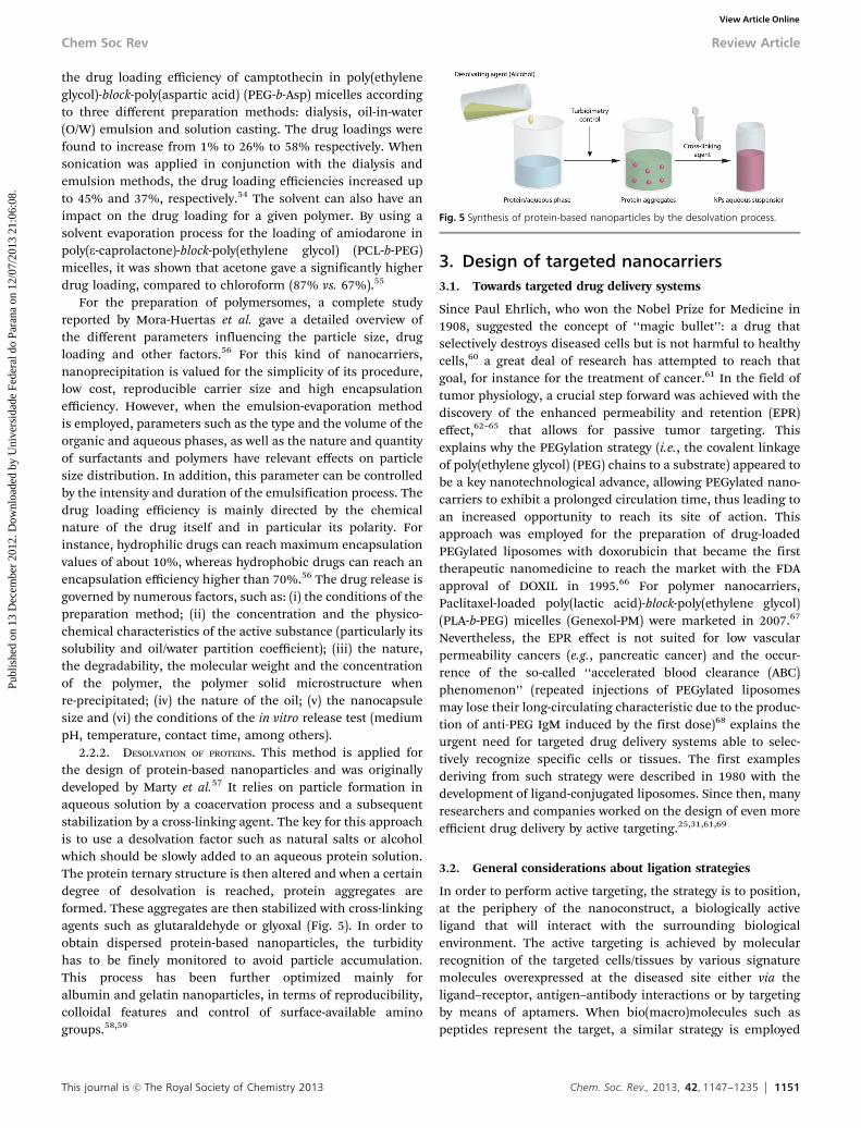
2.2.2. DESOLVATION OF PROTEINS. This method is applied forthe design of protein-based nanoparticles and was originallydeveloped by Marty et al.57 It relies on particle formation inaqueous solution by a coacervation process and a subsequentstabilization by a cross-linking agent. The key for this approachis to use a desolvation factor such as natural salts or alcoholwhich should be slowly added to an aqueous protein solution.The protein ternary structure is then altered and when a certaindegree of desolvation is reached, protein aggregates areformed. These aggregates are then stabilized with cross-linkingagents such as glutaraldehyde or glyoxal (Fig. 5). In order toobtain dispersed protein-based nanoparticles, the turbidityhas to be finely monitored to avoid particle accumulation.This process has been further optimized mainly foralbumin and gelatin nanoparticles, in terms of reproducibility,colloidal features and control of surface-available aminogroups.58,59
3. Design of targeted nanocarriers3.1. Towards targeted drug delivery systems
Since Paul Ehrlich, who won the Nobel Prize for Medicine in1908, suggested the concept of ‘‘magic bullet’’: a drug thatselectively destroys diseased cells but is not harmful to healthycells,60 a great deal of research has attempted to reach thatgoal, for instance for the treatment of cancer.61 In the field oftumor physiology, a crucial step forward was achieved with thediscovery of the enhanced permeability and retention (EPR)effect,62–65 that allows for passive tumor targeting. Thisexplains why the PEGylation strategy (i.e., the covalent linkageof poly(ethylene glycol) (PEG) chains to a substrate) appeared tobe a key nanotechnological advance, allowing PEGylated nano-carriers to exhibit a prolonged circulation time, thus leading toan increased opportunity to reach its site of action. Thisapproach was employed for the preparation of drug-loadedPEGylated liposomes with doxorubicin that became the firsttherapeutic nanomedicine to reach the market with the FDAapproval of DOXIL in 1995.66 For polymer nanocarriers,Paclitaxel-loaded poly(lactic acid)-block-poly(ethylene glycol)(PLA-b-PEG) micelles (Genexol-PM) were marketed in 2007.67
Nevertheless, the EPR effect is not suited for low vascularpermeability cancers (e.g., pancreatic cancer) and the occur-rence of the so-called ‘‘accelerated blood clearance (ABC)phenomenon’’ (repeated injections of PEGylated liposomesmay lose their long-circulating characteristic due to the produc-tion of anti-PEG IgM induced by the first dose)68 explains theurgent need for targeted drug delivery systems able to selec-tively recognize specific cells or tissues. The first examplesderiving from such strategy were described in 1980 with thedevelopment of ligand-conjugated liposomes. Since then, manyresearchers and companies worked on the design of even moreefficient drug delivery by active targeting.25,31,61,69
3.2. General considerations about ligation strategies
In order to perform active targeting, the strategy is to position,at the periphery of the nanoconstruct, a biologically activeligand that will interact with the surrounding biologicalenvironment. The active targeting is achieved by molecularrecognition of the targeted cells/tissues by various signaturemolecules overexpressed at the diseased site either via theligand–receptor, antigen–antibody interactions or by targetingby means of aptamers. When bio(macro)molecules such aspeptides represent the target, a similar strategy is employed
Fig. 5 Synthesis of protein-based nanoparticles by the desolvation process.
Chem Soc Rev Review Article
Publ
ishe
d on
13
Dec
embe
r 20
12. D
ownl
oade
d by
Uni
vers
idad
e Fe
dera
l do
Para
na o
n 12
/07/
2013
21:
06:0
8.
View Article Online

1152 Chem. Soc. Rev., 2013, 42, 1147--1235 This journal is c The Royal Society of Chemistry 2013
by using molecules or antibodies with specific affinity towardthe targeted peptides.
The ligand can either be linked to the (co)polymer prior toself-assembly into the resulting ligand-decorated nanocarrier ordirectly coupled at the surface of preformed nanocarriers inaqueous solutions. The choice of the synthetic route is usuallygoverned by the size of the ligand to be attached to: whereassmall ligands (e.g., small organic molecules or small peptides)can be linked either to the copolymer prior to nanocarrierformation or at the surface of preformed nanocarriers,the linkage of bulky ligands (e.g., polypeptides, proteins orantibodies) occurs, but not exclusively,70 via the second pathwayfor self-assembly and hydrophilic/lipophilic balance reasons.This pathway is also preferred because the secondary structureof proteins and antibodies can be denatured by solubilization inorganic solvents.71
An undeniable advantage of conjugating the ligand prior tonanocarrier formation relies on the possibility to control thereaction yield and to thoroughly characterize the resultingconjugate by routine methods. Consequently, by its simplemix with the non-conjugated (co)polymer, it is then possibleto tune the density of ligands on the surface of the nano-carriers, and therefore to control the binding efficiency for agiven target. This is of high importance regarding the so-calledmultivalency feature of the nanocarriers.72 However, a draw-back of this method lies in the fact that the physicochemicalproperties of the original copolymer will be altered, especiallywith (bio)macromolecules. Indeed, after ligands conjugation,the hydrophilic/hydrophobic balance will be changed togetherwith the charge and therefore the conditions to obtain nano-carriers will need to be adapted.
When conjugation occurs after the nanocarrier formation,some difficulties may arise from its purification and chemicalcharacterization. Indeed, purification processes such as centri-fugation, dialysis or filtration can degrade/modify the nano-carriers. Besides, proving the covalent linkage will necessitatemore advanced surface characterization methods (e.g., XPS,ToF-SIMS) whereas NMR can appear limited.73 Surface charac-terization using surface plasmon resonance (SPR) spectroscopycan also be employed but will lead to indirect proofs and will notgive the possibility to distinguish between covalent linkage orsimple adsorption.
Several studies compared both approaches using smallpeptides as a ligand, but led to opposite results.74,75
3.3. Ligands employed for targeted delivery
A broad range of ligands have been used for targeted nano-carriers and belong to the families of small molecules, carbo-hydrates, peptides, proteins or antibodies (Table 1).
3.3.1. SMALL MOLECULES. Molecular ligands are often readilyavailable, inexpensive, easy to handle, to be chemically mod-ified and to be characterized. Among biologically active smallmolecules, vitamins such as folic acid (FA, vitamin B9),76,77 andbiotin (vitamin B7)78 are widely employed for the targeting ofcancer cells. Indeed, FA binds with a low affinity to the reducedfolate carrier virtually present in all cells, but with a high
affinity (in the nanomolar range) to the glycosylphosphatidyli-nositol-linked folate receptor, which exhibits very limited dis-tribution but is overexpressed at the surface of many types ofcancer cells including ovarian, breast, brain, and lung cancercells.79 The expression levels in tumors have been reported tobe 100–300 times higher than those observed in normaltissues.80 In the case of biotin, which is a cell growth promoter,its content in tumors is significantly higher than in normaltissue. Due to the rapid proliferation of cancer cells, a higherquantity of biotin is needed, and therefore, biotin receptors areoften overexpressed on their surface.81 Biotin has also theadvantage of binding to (strept)avidin with an extremely highaffinity constant and this feature is commonly employed to link(strep)avidin-antibody constructs at the surface of biotinylatednanocarriers. Cobalamin (vitamin B12), known to promote theintestinal adsorption of associated molecules, was recentlyemployed for the development of an oral delivery system of insulinin order to bypass subcutaneous administration drawbacks.82
Other small molecules such as curcumin, selegilin, selectinor alendronate have also been used as targeting ligands.Curcumin, a major component of the yellow curry spiceturmeric, presents potential anti-inflammatory, antioxidant,anti-ischemic, antiarthritic and anticancer properties.83 It isalso able to bind the b-amyloid peptide (Ab) and to disaggregateAb plaques, as well as to prevent fibril and oligomer formation,which is of great interest in the field of Alzheimer’s disease.84–86
Selegiline, a methamphetamine derivative able to selectivelyinhibit monoamine oxidase-B (MAO-B), is clinically employedfor the treatment of Parkinson’s disease87 and has showninteresting properties for management of AD.88 Moreover,selegiline also displays high affinity for the amyloid peptide,a key molecule in AD etiology. Selectin was used to targetP-selectin and E-selectin, whose expression levels on endo-thelial cells are highly increased during inflammation.89,90
Targeting of hydroxyapatite for treatment of bone disease wasachieved using alendronate as ligand.91
Although the use of small molecules as biologically activeligands is a convenient route towards targeted nanocarriers,their main downside comes from their relatively nonselectiveinteraction.92 For instance, folate can indeed bind to some non-target tissues. In addition, vitamins are present in diets and canbe found in significant levels in body fluids. Therefore, the freevitamin can compete for binding with the targeted nano-carriers. Structure–activity relationships of Ab-aggregationusing curcumin derivatives revealed that subtle changes inthe curcumin structure led to severe variations of activity.93
3.3.2. CARBOHYDRATES. Similarly to small molecules, carbo-hydrates are often readily available, inexpensive to manufactureand sugar chemistry is sufficiently well-known to allow theirefficient modification and characterization. Most carbohydratemolecules used for targeting purposes (e.g., galactose, lactose,mannose) bind specifically to asialoglycoprotein receptors(ASGPR), which are membrane lectin receptors commonlyfound in liver cells.94 In this view, carbohydrates are interestingligands to target hepatic and cervical cancer cells.95,96 Inter-action of glucose and lactose with lectins was investigated to
Review Article Chem Soc Rev
Publ
ishe
d on
13
Dec
embe
r 20
12. D
ownl
oade
d by
Uni
vers
idad
e Fe
dera
l do
Para
na o
n 12
/07/
2013
21:
06:0
8.
View Article Online

This journal is c The Royal Society of Chemistry 2013 Chem. Soc. Rev., 2013, 42, 1147--1235 1153
Table 1 Structures/schemes of biologically active ligands used for targeted nanocarrier synthesis
Ligand Structure/scheme Pathology Receptor(s)/target(s) Ref.
Small molecules
Folic acid (FA), alsoknown as vitamin B9 Cancer Folate receptor 76, 77
Biotin, also knownas vitamin B7 Cancer Biotin receptor
(MCF7, M109 cells) 78
Cobalamin (VB12) Intestine adsorption Ileocyte of intestine 82
Selegiline Neurodegenerativediseases (PD, AD) Amyloid-beta peptide 189
Curcumin Cancer, AD Amyloid-beta peptide 85, 86
Selectin Pathologicalinflammation Activated endothelium 89, 90
Alendronate Bone diseases Hydroxyapatite (majorcomponent of bones and teeth) 91
ACUPA S,S,-2-[(5-amino-1-carboxypentyl)-ureido]pentanedioic acid
Cancer PMSA 187
Carbohydrates
Galactose Cancer Asialoglycoprotein receptor 95
Galactosamine Cancer Asialoglycoproteins receptor 95
Chem Soc Rev Review Article
Publ
ishe
d on
13
Dec
embe
r 20
12. D
ownl
oade
d by
Uni
vers
idad
e Fe
dera
l do
Para
na o
n 12
/07/
2013
21:
06:0
8.
View Article Online
1154 Chem. Soc. Rev., 2013, 42, 1147--1235 This journal is c The Royal Society of Chemistry 2013
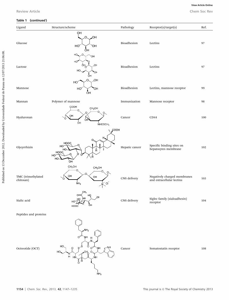
Table 1 (continued )
Ligand Structure/scheme Pathology Receptor(s)/target(s) Ref.
Glucose Bioadhesion Lectins 97
Lactose Bioadhesion Lectins 97
Mannose Bioadhesion Lectins, mannose receptor 99
Mannan Polymer of mannose Immunization Mannose receptor 98
Hyaluronan Cancer CD44 100
Glycyrrhizin Hepatic cancer Specific binding sites onhepatocytes membrane 102
TMC (trimethylatedchitosan) CNS delivery Negatively charged membranes
and extracellular lectins 103
Sialic acid CNS delivery Siglec family (sialoadhesin)receptor 104
Peptides and proteins
Octreotide (OCT) Cancer Somatostatin receptor 108
Review Article Chem Soc Rev
Publ
ishe
d on
13
Dec
embe
r 20
12. D
ownl
oade
d by
Uni
vers
idad
e Fe
dera
l do
Para
na o
n 12
/07/
2013
21:
06:0
8.
View Article Online

This journal is c The Royal Society of Chemistry 2013 Chem. Soc. Rev., 2013, 42, 1147--1235 1155
Table 1 (continued )
Ligand Structure/scheme Pathology Receptor(s)/target(s) Ref.
Glutathione Membranetranslocation Tight junctions 114
WGA Bioadhesion/cancer N-Acetylglucosamine
109–113
Solanum tuberosumlectin CNS delivery N-Acetylglucosamine
RCA (Ricinuscommunis-120) Bioadhesion/cancer Galactose/
N-acetylgalactosamine
Odorranalectin CNS delivery L-fucose
Con A Infectious disease,Cancer Con A receptor
UEA I Infectious disease,Cancer Lectin UEA I receptor
RGD peptides GRGDS Cancer avb3 integrin receptor 115–117CRGDKGPDC (iRGD)Cyclo(RGDDYK)C (cyclic RGD, cRGD)Cyclo(RGDDFK) (cRGDfk)RGD4CRGD-peptidemimetic
K 273 peptide HTMYYHHYQHHL Cancer VEGF 120TAT (CPP) GRKKRRQRRRPQ — Cell membrane (translocation) 122, 123mHph1 (CPP) CHHHHHYARVRRRGPRRHHHHHC — Cell membrane (translocation) 124mAP (CPP) CHHHHHRQIKIWFQNRRMKWKKHHHHHC — Cell membrane (translocation) 125SynB RGGRLSYSRRRFSTSTGR CNS delivery BBB (crossing via a caveolae-
independent pathway)128
Lyp-1 peptide CCGNKRTRGC Cancer Tumor cells and tumorlymphatic vessels endothelialcells
131
LCP (lung cancertargeting peptide)
RGDLATLRQL Cancer avb6 integrins 132
XRT-cells peptide GIRLRG Cancer GRP78 133Injured vesselspeptide
KLWVLPK Injured vasculature Collagen IV 134
Chem Soc Rev Review Article
Publ
ishe
d on
13
Dec
embe
r 20
12. D
ownl
oade
d by
Uni
vers
idad
e Fe
dera
l do
Para
na o
n 12
/07/
2013
21:
06:0
8.
View Article Online

1156 Chem. Soc. Rev., 2013, 42, 1147--1235 This journal is c The Royal Society of Chemistry 2013
Table 1 (continued )
Ligand Structure/scheme Pathology Receptor(s)/target(s) Ref.
EGF1 Thrombolysis Tissue factor 135
AP peptide CRKRLDNR Atherosclerosis/cancer
IL4 receptor 136
Pep I peptide CHVLWSTRC Autoimmune type 1diabetes
Pancreatic islet microvessels 137
M-cell homing peptide CKSTHPLSC Vaccination Peyer’s patch follicle 138
EGF Cancer Epidermal growth factorreceptor (EGFR) 139
GE11 peptide YHWYGYTPQNVI Cancer Epidermal growth factorreceptor (EGFR)
140
c LABL peptide Cyclo-(1,12)-PenITDGEATDSGC Cancer ICAM-1 141
Transferrin (Tf) Cancer, braindelivery Transferrin receptor (TfR) 142, 143
Lactoferrin (Lf) Brain delivery Lactoferrin receptor (LfR) 144
g7 Peptide H2N-GFTGFLS(O-b-D-glucose)-CONH2 CNS delivery BBB 145, 146Similopioid peptides H2N-GFTGFLX-CONH2 X = S-OH, S(O-b-D-glucose),
S(O-b-D-galactose), S(O-b-D-xylose), S(O-b-D-lactose)CNS delivery BBB 145, 146
CDX peptides FKESWREARGTRIERG (CDX) CNS delivery Nicotine acetylcholinereceptors (nAChR)
147SWREARGTRI (Pocket_CDX)CFKESWREARGTRIERGC (Cyclo_CDX)
Angiopep-2 TFFYGGSRGKRNNFKTEEY CNS delivery/cancer LRP 148, 149TGN peptide TGNYKALHPHNG CNS delivery BBB 150
ApoE3, A-I, B-100 CNS delivery LDL receptor (BBB) 151
Insulin CNS delivery Insulin receptor 152
Tetanus toxin Cfragment Cancer Neuronal ganglioside clostridial
toxin receptor GT1b153
A665 and A666peptides
LSTHTTESRSMV and LEPRWGFGWWLK Hearing loss Prestin receptor 154
Tet1 HLNILSTLWKYR Hearing loss Neuronal ganglioside clostridial toxinreceptor GT1b
155
hNgf EE CTFVKALTMDGKQAAWR Hearing loss Tyrosine kinase and p75neurotrophin receptor
156
DCL peptide 2-{[(5-Amino-1-carbox-ypentyl)carbamoyl]amino}pentanedioic acid]
Cancer PSMA 187
Review Article Chem Soc Rev
Publ
ishe
d on
13
Dec
embe
r 20
12. D
ownl
oade
d by
Uni
vers
idad
e Fe
dera
l do
Para
na o
n 12
/07/
2013
21:
06:0
8.
View Article Online

This journal is c The Royal Society of Chemistry 2013 Chem. Soc. Rev., 2013, 42, 1147--1235 1157
Table 1 (continued )
Ligand Structure/scheme Pathology Receptor(s)/target(s) Ref.
Antibodies
Anti-CD3 Ab Cancer T cell antigen receptor (TCR) 159
Anti HER-2, also calledTrastuzumab orHerceptin
Cancer Human epidermal growth factorreceptor 2 (HER2) 160
EGFRMab Cancer Epidermal growth factor receptor(EGFR) 161
DI17E6 MAb Cancer av integrins expressed on tumor andendothelial cells 162
Anti Fas (CD95/Apo-1)MAb Cancer Fas (CD95/Apo-1) receptor 163
Anti antigen rich MCF7cells Mab Cancer
Soluble membrane proteins of MCF-7human invasive ductal breastcarcinoma
164
AMB8LK Mab Cancer H-ferritin 165
Anti CD20 Mab, alsocalled MabTheras orRituximab
Cancer CD20 tetraspan surface moleculeexpressed in B limphocytes 166
Anti CEA Mab Cancer CEA 167
Anti Ab1–42 MAb Alzheimer’s disease Ab1–42 peptide 168
29B4 CNS delivery Insulin receptor 169
OX26 CNS delivery Transferrin receptor (TfR) 170
R17217 CNS delivery Transferrin receptor (TfR) 171
Chem Soc Rev Review Article
Publ
ishe
d on
13
Dec
embe
r 20
12. D
ownl
oade
d by
Uni
vers
idad
e Fe
dera
l do
Para
na o
n 12
/07/
2013
21:
06:0
8.
View Article Online

1158 Chem. Soc. Rev., 2013, 42, 1147--1235 This journal is c The Royal Society of Chemistry 2013
design bioadhesive systems.97 In another way, mannose andmannan were used to target mannose receptors at the surfaceof antigen-presenting cells in order to achieve an improvement ofthe antigen specific response.98,99 Hyaluronan, a polysaccharide,was employed as a ligand for its high affinity to the CD44 receptor inthe field of cancer.100,101 Glycyrrhizin, one of the main compoundsextracted from the root of Glycyrrhiza glabra (licorice) was selected todecorate nanocarriers for hepatic targeting.102 Interaction with thepositively charged trimethylated chitosan (TMC) and negatively
charged cell membranes was explored as a strategy to promotethe passage across the BBB.103 Controlled delivery of drugs tothe brain was achieved also by targeting the sialoadhesinsreceptors with sialic acid residues.104 Beyond their targetingabilities, another advantage of polysaccharides is the stealthproperties they can confer to nanocarriers.105
3.3.3. PEPTIDES AND PROTEINS. Peptides have numerous benefitsin their use as targeting moieties. For instance, their productioncost is rather low and they have a high activity per mass unit.
Table 1 (continued )
Ligand Structure/scheme Pathology Receptor(s)/target(s) Ref.
Anti CD44 mAB CNS delivery Glial cells 172
Anti NCAM1 mAB CNS delivery Neurons 173
Anti CD4+ Mab Immunomodulation T lymphocytes 174
Anti VCAM-1 Mab Pathologicalinflammation Vascular cell adhesion molecules
175
Anti ICAM-1 Mab Pathologicalinflammation Intracellular adhesion molecules
Anti E-selectin Mab Pathologicalinflammation E-selectin
Anti P-selectin Mab Pathologicalinflammation P-selectin
ScFvEGFR mAb variable fragment of anti-EGFR mAb Cancer Epidermal growth factorreceptor (EGFR)
176
ScFvCD7 mAb variable fragment of anti-T cells mAb Infectious disease T-cell CD7 177
Anti HER2 affibody Cancer HER2 180
AptamersA10RNA aptamer 57 base pair nuclease-stabilized 20-fluoropyr-
imidine RNA molecule with a single 50-CG-30
sequence
Cancer PMSA 182
XEO2 mini aptamer20O-methyl RNA aptamer
50-CAC GAC GCU GAU GGA UCG UUA CGA CUAGCA UCG C-30
Cancer PMSA 181
AS1411 DNA aptamer 50-d(GGTGGTGGTGGTTGTGGTGGTGGTGG)-30 Cancer Nucleolin protein 183
Review Article Chem Soc Rev
Publ
ishe
d on
13
Dec
embe
r 20
12. D
ownl
oade
d by
Uni
vers
idad
e Fe
dera
l do
Para
na o
n 12
/07/
2013
21:
06:0
8.
View Article Online

This journal is c The Royal Society of Chemistry 2013 Chem. Soc. Rev., 2013, 42, 1147--1235 1159
They are also stable; therefore, long-term storage and easymanipulation are possible. Moreover, the associated risk ofundesired effects on the immune system is reduced, and theirsmall size is not likely to change the physicochemical propertiesof the resulting decorated nanocarriers.106,107 Besides, theirsequence can be modified in order to optimize their biologicalactivity and/or to introduce functional groups.
The octapeptide octreotide (somatostatin analogue) wasused to target the somatostatin receptors, largely expressed intumor tissue.108 Lectins (e.g., WGA, solanum tuberosum lectin,ricinus communis lectin, odorranalectin, Con A and UEA-I) areproteins of non-immunological origin able to specifically recognizesugar molecules and bind glycosylated membrane components.They have been used to prepare bioadhesive systems and promotedrug delivery following nasal administration or biodisponibilityof orally administered drugs.109–113 Glutathione (GSH), a smalltripeptide formed by glutamic acid, cysteine and glycine, was usedas a peptide ligand to promote cell adhesion and achieve apermeation-enhancing effect.114
Various (poly)peptides and proteins have also been used ashoming devices in the fields of cancer, brain delivery andseveral other diseases. Among them, the tri-peptide Arg-Gly-Asp (RGD) and its derivatives appeared extremely attractive astumor and vascular targeting ligands. Indeed, they were shownto bind to the avb3 integrin receptor which is highly expressedat the surface of malignant cells and in tumor proliferatingneovascular endothelial cells while it is minimally expressed innormal quiescent endothelial cells.115–117 cRGD has been devel-oped by Kessler et al. and its specific binding to avb3 integrinswas 170 times more active than the linear form.118,119 The smallpeptide K237 has been conjugated at the surface of NPs totarget the vascular endothelial growth factor receptor(VEGFR).120 This peptide enabled a receptor-mediated interna-lization of functionalized NPs in endothelial cells as well as atherapeutic function by inhibition of the VEGFR signalingpathway to be achieved.
Cell penetrating peptides (CPPs), such as TAT(GRKKRRQRRRPQ, deriving from the transcriptional activatorprotein encoded by HIV-1), protein transduction domain (PTD)of TAT, mHph1 and mAP represent a specific class of activeligands that actually do not recognize a specific receptor butdemonstrate ability in membrane translocation.121–127 There-fore, their combination with other ligands has usually beenemployed in order to achieve selective receptor recognition. TheSynB peptides (RGGRLSYSRRRFSTSTGR) are a family of CPPsthat shows charge-mediated blood–brain barrier selectivityoccurring via a caveolae-independent pathway.128
The extensive application of phage display technology hasenabled identification of novel peptide sequences that bindwith high affinity to specific molecules.129,130 These peptides,which allow the number of potential available targets to beincreased, have been investigated to functionalize nanocarriersused for the treatment of cancer and other diseases. Amongthem we can find: (i) the Lyp-1 peptide, which specifically hometo tumor and their lymphatics;131 (ii) the LCP peptide targetedto the avb6 integrins overexpressed in non-small cells lung
cancer;132 (iii) the GIRLRG sequence that recognizes theGRP78 receptors overexpressed by ionizing radiation-treatedcells;133 (iv) the KLWVLPK peptide which shows high affinity forthe collagen IV;134 (v) the EGF1 peptide which interacts withthe tissue factor involved in coagulation and thrombus for-mation;135 (vi) the AP peptide that has a specific bindingaffinity to IL4 receptors;136 (vii) the pep I peptide that bindsthe pancreatic islet microvessel137 and (viii) the CKSTHPLSCsequence that specifically recognizes M-cells.138
Targeting of the epidermal growth factor receptor (EGFR),whose overexpression is observed in several cancer cells, wasachieved using the epidermal growth factor (EGF, naturalligand) as well as specific peptides such as the GE11 peptide.139,140
Selective targeting of ICAM-1 was achieved with the cLABLpeptide.141
Due to the overexpression of the transferrin receptor (TfR)both in solid tumors and on the endothelial cells of the blood–brain barrier (BBB), transferrin was used to target cancer cellsand to achieve brain delivery.142,143 Same results were obtainedfor the targeting the lactoferrin receptors (LfR) with lactoferrinmoieties (Lf).144 The large variety of peptides which has beenexplored to enable nanocarriers to pass through the BBB andreach the CNS includes: (i) simil-opioid peptides (g7 peptides)able to bind the opioid receptors;145,146 (ii) candotoxin derivedpeptides (CDX) that bind the nicotine acetylcholine recep-tors;147 (iii) the Angiopep-2 which interacts with the low-densitylipoprotein receptor-related protein;148,149 and (iv) the TGNpeptide.150 CNS delivery was achieved also with apolipoproteins(Apo) such as ApoE3, ApoA-I and ApoB-100, which are alsorecognized by low-density lipoprotein receptors (LDLR) of theendothelial cells of the BBB.151 BBB crossing can also beperformed by using insulin as a ligand in order to target theinsulin receptor (IR), which seems to have a 10-fold higher BBBtransport efficacy compared to the transferrin receptor.152
Efficient targeting to neurons was realized using the tetanustoxin C fragment, which demonstrates high affinity binding tothe neuronal ganglioside receptor.153
Several peptides were used as targeting ligands for themanagement of hearing loss: (i) A665 and A666, (ii) Tet1 and(iii) hNgf EE that bind to prestin receptors, neuronal ganglio-side receptors (GT1b), and tyrosine kinase receptors as well asp75 neurotrophin receptors, respectively.154–156
3.3.4. ANTIBODIES. Antibodies, which are large Y-shapedproteins, perhaps represent the most efficient ligands due totheir high affinities and their ability to recognize a specific partof their target. They were actually the first class of tumortargeting ligands used to deliver drugs encapsulated inmicelles.157 Antibodies are typically made of basic structuralunits, each with two large heavy chains and two smaller lightchains (Fig. 6). Although the general structure of all antibodiesis very similar, a small region at the tip of the protein isextremely variable, allowing millions of antibodies with differ-ent tip structures, or antigen binding sites, to exist. Even ifthese ligands are bulky (B150 kDa) they can be adapted tospecifically bind to a large variety of targets with high affinities(Kd B0.1 nM) even with a low density at the surface of
Chem Soc Rev Review Article
Publ
ishe
d on
13
Dec
embe
r 20
12. D
ownl
oade
d by
Uni
vers
idad
e Fe
dera
l do
Para
na o
n 12
/07/
2013
21:
06:0
8.
View Article Online

1160 Chem. Soc. Rev., 2013, 42, 1147--1235 This journal is c The Royal Society of Chemistry 2013
nanocarriers.74 It has indeed been shown that micelles slightlysurface modified by antibodies accumulated in tumors, showinga better anti-tumor efficacy.158
Antibodies-decorated nanoparticulate systems have beenemployed in the field of cancer (anti-CD3 Mab,159 anti-HER2Mab (also called trastuzumab or Herceptin),160 EGFRMab,161
DI17E6 Mab,162 anti FAS Mab,163 anti antigen rich breastcancer cells Mab,164 AMB8LK Mab,165 anti-CD20 mAB,166 anti-CEA Mab167), Alzheimer’s disease (anti-Ab1-42 MAb),168 braindelivery (29B4,169 OX26,170 R17217,171 anti-CD44 mAB,172 anti-NCAM1 Mab173) and autoimmune diseases (anti CD4+).174 Aspecific target to inflamed endothelium (generally observed invarious pathological settings such as cancer, cardiovasculardisease and arthritis) which is characterized by overexpressionof endothelial cell adhesion molecules, was achieved usinganti-ICAM-1 Mab, anti VCAM-1 Mab, anti E-selectin Mab aswell as anti P-selectin Mab.175 In some cases, the nanocarriershave been decorated only with variable chains of the Ab,allowing a drastic reduction of total MW and of adverseimmune reactions (ScFvEGFR, ScFvCD7).176,177
However, antibodies remain expensive and time-consumingto produce. Although antibody fragments including antigen-binding fragments (Fab), dimers of antigen-binding fragments(F(ab0)2), single-chain fragment variables (scFv) and other engi-neered fragments are less stable and have a lower bindingavidity than whole antibodies (two binding sites), they areconsidered safer when injected systemically owing to reducednon-specific binding.92,178 Under certain conditions, thepresence of antibodies at the surface of nanocarriers appearedto compromise the shielding effect of the PEG layer.179 As analternative to overcome limitations associated to antibodies,the use of affibody (anti-HER2)180 was explored.
3.3.5. APTAMERS. Aptamers or mini-aptamers representanother class of efficient ligands for targeted drug delivery,for instance to bind to prostate specific membrane antigen(PSMA) using A10RNA and XEO2,181,182 or to nucleolin (AS1411)for the treatment of brain cancer.183 Aptamers are short single-stranded DNA or RNA oligonucleotides that can bind specifi-cally to small and large molecule targets. They are identifiedmainly by in vitro screening of a random sequence library(B1014–1015) for their binding ability to specific moleculartargets.184 They can be selected to bind to a wide variety of
targets, including intracellular proteins, transmembrane pro-teins, soluble proteins, carbohydrates, and small moleculedrugs.31 Even though the selection process is complex, theirhigh binding specificity to their target molecules and their lackof immune response due to their small size make thempotential candidate ligands for targeting purposes.185 A draw-back associated to their use is the presence of enzymes that candegrade DNA or RNA in the blood following intravenousadministration. However, their initial promise warrantsexploration of methodologies that can increase their in vivostability.74,186
High affinity and specificity similar to antibodies and aptamerswas also shown by the pseudomimetic dipeptide DCL187 as well aswith the ACUPA molecule (currently evaluated in clinical trials),188
which were used to selectively bind PSMA.
3.4. Synthetic strategies for targeted nanocarriers
In this section, targeted nanocarriers will be discussed andgathered in different tables according to the nature of the mainpolymer constituent:� Polyesters:3 Poly(lactic acid) and poly(lactic-co-glycolic acid) (Table 2,
section 3.4.2.1);3 Poly(e-caprolactone) (Table 3, section 3.4.2.2);3 Boltorn H40 (Table 4, section 3.4.2.3);3 Other polyesters (Table 5, section 3.4.2.4);� Polysaccharides:3 Chitosan (Table 6, section 3.4.3.1);3 Cyclodextrin (Table 7, section 3.4.3.2);3 Other polysaccharides (Table 8, section 3.4.3.3);� Poly(amino acid)s, polypeptides and proteins:3 Poly(amino acid)s and polypeptides (Table 9, section
3.4.4.1);3 Proteins, (Table 10, section 3.4.4.2);� Poly(alkyl cyanoacrylate) (Table 11, section 3.4.5).In each table, the ranking is achieved according to the
ligand coupling method and the nature of the employed ligand,which follow the order reported in the ligand table (Table 1).For each case, if the nanoparticulate system is composed ofmore than one (co)polymer, then each species will be indepen-dently described following Roman numerals.
3.4.1. BRIEF OVERVIEW OF MAIN LIGATION STRATEGIES
3.4.1.1. Carbodiimide chemistry. This method is the mostused for the ligation of small molecules and biomolecules fortargeted drug delivery purposes. The advantage of this strategyis based on its simplicity and on the native presence on boththe nanocarriers and the ligand of the required functionalgroups (e.g., carboxylic acid, amine, etc.) to achieve the cou-pling. This results in a lower probability to lose ligand specificactivity.190 However, the presence of multiple functional groupsin the ligand can also represent a drawback in an attempt tolimit multi-site attachment or to control the ligand orientationat the surface of the nanocarrier. The o-acylisourea intermediateformed during the activation of the carboxylic acid is susceptibleto quick hydrolysis.191 Consequently, an excess of carbodiimidemust be used to complete the reaction, which can alter the
Fig. 6 General structure of an antibody.
Review Article Chem Soc Rev
Publ
ishe
d on
13
Dec
embe
r 20
12. D
ownl
oade
d by
Uni
vers
idad
e Fe
dera
l do
Para
na o
n 12
/07/
2013
21:
06:0
8.
View Article Online

This journal is c The Royal Society of Chemistry 2013 Chem. Soc. Rev., 2013, 42, 1147--1235 1161
colloidal stability of the resulting nanocarriers due to the poorsolubility of the o-acylisourea. This can be solved usingN-hydroxysuccinimide (NHS) or N-hydroxysulfosuccinimide(sulfo-NHS). The ester intermediate of the latter has a bettersolubility. Although still susceptible to hydrolysis (which ishowever slower than the o-acylisourea intermediate), NHS-estersusually lead to higher coupling efficiencies.
3.4.1.2. Michael addition. Michael addition, and in particularthiol–maleimide coupling strategy, is the second most usedpathway to functionalize nanocarriers. One advantage overcarbodiimide coupling is that maleimides exhibit a slowerhydrolysis rate than o-acylisourea intermediates. Besides,maleimides react with thiols in a highly selective and efficientway at near neutral pH giving a stable thioether bond.190
Nonetheless, even if native thiol groups are present in someproteins (usually to a low extent), they are hardly accessible oreven absent from many others. To circumvent this limitation,thiol groups can be obtained either by reducing existingdisulfide bonds (which can be however detrimental for ternarystructures of certain peptides/proteins) or by using hetero-bifunctional cross-linking agents (see Fig. 14).192 Even if morechemical steps are needed, this coupling strategy enables abetter control over biomolecular orientation and mainly avoidsissues related to unwanted cross-linking or side reactions.193 Italso allows the structural, biological and binding properties ofa protein to be maintained.194
3.4.1.3. COPPER-CATALYZED LIGATION METHOD. Copper(I)-catalyzedHuisgen 1,3-dipolar cycloaddition reaction between an azide andan alkyne (CuAAC) recently received tremendous interest inmany different fields such as dendrimers, bioconjugates,therapeutics and functionalized polymers.195 This cycloadditionbelongs to the class of chemical reactions, often referred to asclick chemistry, that share several very important features: (i) avery high efficiency in terms of both conversion and selectivity;(ii) mild experimental conditions; (iii) a simple workup and (iv)little or no byproducts. The CuAAC ligation method can be usedto control the display of biomolecules at the surface of nano-carriers.193 The principal drawback of the CuAAC reaction is thenecessary removal of the Cu-based catalyst after the coupling.
The use of copper ligands and organic scavengers is howeverpossible to remove most of the catalyst.196
3.4.1.4. Biotin–(strep)avidin ligation strategy. The biotin–(strep)-avidin ligation strategy is a very advantageous and flexible tool foractive targeting. The bond between the two moieties is thestrongest non-covalent biological interaction known, with a dis-sociation constant, Kd, in the order of 4.10�14 M. This sandwichstrategy can be easily applied in multistep approaches.197 Further-more, biotinylated antibodies can be easily obtained198 andprotocols describing the synthesis of (strep)avidin-antibodies havealso been reported.199
3.4.2. POLYESTERS. Polyesters certainly represent the mostwidely employed family of polymers in this field thanks to theirbiodegradable and biocompatible features. Among them, one canfind polylactide (PLA), polyglycolide (PGA), poly(e-caprolactone)(PCL) and poly(g-valerolactone) (PVL). They are usually synthe-sized by ring-opening polymerization (ROP) of lactide (LA),lactide/glycolide (LA/G), e-caprolactone (eCL) and gvalero-lactone (gVL), respectively (Fig. 7). Noteworthily, copolymersof lactide and glycolide (poly(lactide-co-glycolide), PLGA) havebeen approved by the Food and Drug Administration (FDA) fordrug delivery.
Polyesters mainly undergo bulk erosion and the mechanismof degradation is largely due to hydrolysis of ester functionspresent in the (co)polymer backbone, eventually leading to waterand carbon dioxide as degradation products. Interestingly, byvarying the lactide/glycolide ratio, tunable degradation andrelease rates can be achieved.
3.4.2.1. Poly(lactide) and poly(lactide-co-glycolide). Poly(lacticacid) (L, D or D, L), poly(glycolic acid) and their copolymerspoly(lactic-co-glycolic acid) have been largely investigated fordrug delivery purposes. In this category amphiphilic PLGA-b-PEG or PLA-b-PEG copolymers which consist of a hydrophobicPLGA or PLA block and a hydrophilic PEG have been synthe-sized and employed to formulate nanoparticles, micellesor polymersomes in which the hydrophobic PLGA (or PLA)and the hydrophilic PEG form the core and the shell ofthe resulting nanocarriers, respectively. In general, these
Fig. 7 Synthesis of polylactide (PLA), poly(lactide-co-glycolide) (PLGA), poly(e-caprolactone) (PCL) and poly(g-valerolactone) (PVL) by ring-opening polymerization(ROP).
Chem Soc Rev Review Article
Publ
ishe
d on
13
Dec
embe
r 20
12. D
ownl
oade
d by
Uni
vers
idad
e Fe
dera
l do
Para
na o
n 12
/07/
2013
21:
06:0
8.
View Article Online

1162 Chem. Soc. Rev., 2013, 42, 1147--1235 This journal is c The Royal Society of Chemistry 2013
Tab
le2
Mai
nch
arac
teri
stic
san
dap
plic
atio
ns
of
targ
eted
po
ly(la
ctid
e)an
dp
oly
(lact
ide-
co-g
lyco
lide)
-bas
edn
ano
carr
iers
Stru
ctu
ral
para
met
ers
Col
loid
alpa
ram
eter
sA
ppli
cati
on
Ref
.A
rch
itec
ture
Liga
nd
Syn
thet
icm
eth
odLi
gan
dco
upl
ing
met
hod
Form
ula
tion
Mor
phol
ogy
Dia
met
er(n
m)
Path
olog
y/ta
rget
Dru
gC
ells
/tis
sues
/or
gan
sd
Liga
nd
cou
plin
gby
carb
odii
mid
ech
emis
try
PLA
-g3-
FAFA
ROP
ofLA
from
6-ar
mhy
drox
yin
itia
tor;
dive
rgen
tsy
nthe
sis
Car
bodi
imid
e(F
A-N
H2
+PL
A-g
3-C
OO
H/
DC
C/N
HS)
a
Dia
lysi
sm
eth
odM
icel
les
(den
dri
mer
-li
kest
arpo
lym
er)
18C
ance
r(p
har
ynx)
—K
B24
1
(i)
PLA
-TG
PS(i
i)T
GPS
-FA
FA(i
)R
OP
ofLA
from
TG
PS(i
i)se
eli
gan
dco
upl
ing
met
hod
Car
bodi
imid
e(T
GPS
-CO
OH
/E
DC
/NH
S+
FA-N
H2)b
Em
uls
ion
–sol
ven
tev
apor
atio
nN
anop
arti
cles
300–
330
Can
cer
(bre
ast,
glio
ma)
Ptx
MC
F7,
C6
244
(i)
MeP
EG
-b-
PLA
(ii)
TG
PS-F
A
FA(i
)R
OP
ofLA
from
MeP
EG
-OH
(ii)
see
liga
nd
cou
plin
gm
eth
od
Car
bodi
imid
e(T
GPS
-CO
OH
/E
DC
/NH
S+
FA-N
H2)a
Em
uls
ion
–sol
ven
tev
apor
atio
nN
anop
arti
cles
300
Can
cer
(cer
vica
l,gl
iom
a)
Ptxc
Hel
a,C
624
5
PLG
A-b
-PE
G-F
AFA
Car
bodi
imid
e(P
LGA
-CO
OH
/DC
C/
NH
S+
H2N
-PE
G-N
H2)
Car
bodi
imid
e(P
LGA
-PE
G-N
H2
+FA
-CO
OH
/DC
C)a
Em
uls
ion
–sol
ven
tev
apor
atio
nN
anop
arti
cles
120–
220
Can
cer
(ova
ry)
Dtx
SK-O
V-3
217
PLG
A/D
LPC
/D
SPE
-PE
G/
DSP
E-P
EG
-FA
FAC
arbo
diim
ide
(DSP
E/
DSS
+H
2N
-PE
G-F
A)
Car
bodi
imid
e(H
2N
-PE
G-N
H2
+FA
-CO
OH
/DC
C/
NH
S)a
Em
uls
ion
–sol
ven
tev
apor
atio
nN
anop
arti
cles
200–
300
Can
cer
(bre
ast)
Dtx
MC
F721
8
(i)
PLG
A(i
i)P
LL-b
-PE
G-F
A
FA(i
)R
OP
ofLA
/G(i
i)C
arbo
diim
ide
(PLL
-NH
2+
FA-P
EG
-CO
OH
/DC
C/
NH
S)
Car
bodi
imid
e(C
OO
H-P
EG
-NH
2+
FA-C
OO
H/D
CC
/N
HS)
b
Em
uls
ion
–sol
ven
tev
apor
atio
n;
coat
ing
(PLG
AN
Ps+
PLL-
b-PE
G-F
A)
Nan
opar
ticl
es11
0–12
5C
ance
r(p
har
ynx,
lun
g)
—K
B,
A54
921
6
PLA
-b-P
EI-
FAFA
Car
bodi
imid
e(P
LA-C
OO
H/D
CC
+H
2N
-PE
I-N
H2)a
Car
bodi
imid
e(P
LA-b
-PE
I-N
H2
+FA
-CO
OH
/DC
C/
DM
AP)
a
Poly
plex
form
atio
nw
ith
pDN
A
Mic
elle
s10
0C
ance
rpD
NA
Hel
a23
9
(i)
PLG
A(i
i)P
LL-b
-PE
G-F
A
FA(i
)C
omm
erci
al(i
i)C
arbo
diim
ide
(PLL
-NH
2+
FA-P
EG
-CO
OH
/DC
C/
NH
S)
Car
bodi
imid
e(C
OO
H-P
EG-N
H2
+FA
-CO
OH
/DC
C/N
HS)
b
Em
uls
ion
–sol
ven
tev
apor
atio
n;
coat
ing
(PLG
AN
Ps+
PLL-
b-PE
G-F
A)
Nan
opar
ticl
es26
0C
ance
r(p
har
ynx)
Dox
KB
219
(i)
P(H
is-c
o-Ph
e)-b
-PE
G(i
i)PL
A-b
-PE
G-F
A
FA(i
)N
CA
poly
m.
ofB
z-H
is-N
CA
/L-P
he-
NC
A;
carb
odii
mid
e(P
(His
-co-
Phe)
-NH
2+
PEG
-CO
OH
/DC
C/
NH
S)(i
i)R
OP
ofLA
from
HO
OC
-PE
G-O
H
Car
bodi
imid
e(P
LA-b
-PE
G-C
OO
H/
DC
C/N
HS
+FA
-NH
2)a
Dia
lysi
sM
icel
les
110–
170
Can
cer
(ova
ry)
Dox
A27
80/D
OX
R,
A27
80/W
T22
6
(i)
P(H
is-c
o-Ph
e)-b
-PE
G(i
i)PL
A-b
-PE
G-F
A
FA(i
)N
CA
poly
m.
ofB
z-H
is-N
CA
/L-P
he-
NC
A;
carb
odii
mid
e(P
(His
-co-
Phe)
-NH
2+
PEG
-CO
OH
/DC
C/
NH
S)(i
i)R
OP
ofLA
from
HO
OC
-PE
G-O
H
(ii)
Car
bodi
imid
e(P
LA-b
-PE
G-C
OO
H/
DC
C/N
HS
+FA
-NH
2)a
Dia
lysi
sM
icel
les
150
Can
cer
(ova
ry)
Dox
A27
80/D
OX
R24
3
Review Article Chem Soc Rev
Publ
ishe
d on
13
Dec
embe
r 20
12. D
ownl
oade
d by
Uni
vers
idad
e Fe
dera
l do
Para
na o
n 12
/07/
2013
21:
06:0
8.
View Article Online

This journal is c The Royal Society of Chemistry 2013 Chem. Soc. Rev., 2013, 42, 1147--1235 1163
Tab
le2
(co
nti
nu
ed)
Stru
ctu
ral
para
met
ers
Col
loid
alpa
ram
eter
sA
ppli
cati
on
Ref
.A
rch
itec
ture
Liga
nd
Syn
thet
icm
eth
odLi
gan
dco
upl
ing
met
hod
Form
ula
tion
Mor
phol
ogy
Dia
met
er(n
m)
Path
olog
y/ta
rget
Dru
gC
ells
/tis
sues
/or
gan
sd
PLG
A-b
-PE
G-F
AFA
Car
bodi
imid
e(P
LGA
-CO
OH
/DC
C/
NH
S+
H2N
-PE
G-F
A)
Car
bodi
imid
e(H
2N
-PE
G-N
H2
+FA
-CO
OH
/DC
C)a
Dia
lysi
sM
icel
les
85–1
00C
ance
r(p
har
ynx,
glio
ma,
brea
st)
Dox
KB
,C
622
0
(i)
PLG
A(i
i)O
QLC
S-PE
G(i
ii)
OQ
LCS-
FA
FA(i
)C
omm
erci
al(i
i)C
arbo
diim
ide
(OQ
LCS-
NH
2+
PEG
-NH
S)a
(iii
)se
eli
gan
dco
upl
ing
met
hod
Car
bodi
imid
e(O
QLC
S-N
H2
+FA
-CO
OH
/DC
C/
NH
S)a
Lipi
dfi
lmh
ydra
tion
and
son
icat
ion
Poly
mer
icli
poso
mes
440
Can
cer
(bre
ast)
Dox
,pD
NA
MD
A-M
B-2
3123
8
(i)
PLG
A-T
GPS
(ii)
TG
PS-F
AFA
(i)
RO
Pof
LAfr
omT
GPS
(ii)
see
liga
nd
cou
plin
gm
eth
od
Car
bodi
imid
e(T
GPS
-CO
OH
/ED
C/
NH
S+
FA-N
H2)b
Em
uls
ion
–sol
ven
tev
apor
atio
nN
anop
arti
cles
320–
360
Can
cer
(bre
ast,
glio
ma)
Dox
cM
CF7
,C
624
6
PLA
-g3-
FA/P
EG
FAR
OP
ofLA
from
6-ar
mh
ydro
xyin
itia
tor;
dive
rgen
tsy
nthe
sis;
carb
odii
mid
e(P
LA-g
3-C
OO
H/D
CC
/N
HS
+H
2N
-PE
G)a
Car
bodi
imid
e(F
A-N
H2
+PL
A-g
3-C
OO
H/D
CC
/N
HS)
a
Dia
lysi
sM
icel
les
(den
dri
mer
-li
kest
arpo
lym
er)
15C
ance
r(p
har
ynx)
Dox
KB
242
(i)
PHis
-b-P
EG
-FA (i
i)PL
A-b
-PE
G-
FA
FA(i
)C
arbo
diim
ide
(PE
G-C
OO
H/D
CC
+PH
is-N
H2)
(ii)
RO
Pof
LAfr
omH
OO
C-P
EG
-OH
(i)
Car
bodi
imid
e(P
His
-b-P
EG
-OH
+FA
-CO
OH
/DC
C)a
(ii)
Car
bodi
imid
e(P
LA-b
-PE
G-C
OO
H/
DC
C/N
HS
+FA
-NH
2)a
Dia
filt
rati
onM
icel
les
50–8
0C
ance
r(b
reas
t)D
oxM
CF7
227
PLG
A-b
-PE
G-F
AFA
Car
bodi
imid
e(P
LGA
-CO
OH
/DC
C/
NH
S+
H2N
-PE
G-
NH
2)a
Car
bodi
imid
e(P
LGA
-b-P
EG
-NH
2+
FA-C
OO
H/D
CC
)a
Dou
ble
emu
lsio
n–
solv
ent
evap
orat
ion
Nan
opar
ticl
es15
5–24
5C
ance
r(c
ervi
cal)
pDN
AH
ela
221
(i)
PLG
A(i
i)D
MPE
-D
TPA
(iii
)D
SPE
-PE
G(i
v)D
SPE
-PE
G-
FA
FA(i
)C
omm
erci
al(i
i)C
omm
erci
al(i
ii)
Com
mer
cial
(iv)
Car
bodi
imid
e(D
SPE
/DSS
+H
2N
-PE
G-F
A)
Car
bodi
imid
e(H
2N
-PE
G-N
H2
+FA
-CO
OH
/DC
C/
NH
S)a
Nan
opre
cipi
tati
onN
anop
arti
cles
65–8
5C
ance
r(o
vary
)Pt
xSK
-OV
-3,
SW62
6SK
-OV
-322
3
(i)
PLG
A(i
i)PE
G-D
SPE
-FA
FA(i
)C
omm
erci
al(i
i)C
arbo
diim
ide
(DSP
E/D
SS+
H2N
-PE
G-F
A)
Car
bodi
imid
e(H
2N
-PE
G-N
H2
+FA
-C
OO
H/D
CC
/NH
S)a
Nan
opre
cipi
tati
onN
anop
arti
cles
70–8
0C
ance
r(p
har
ynx)
Dtx
KB
KB
222
(i)P
LGA
-b-P
EG
-FA (i
i)PL
GA
-b-P
EG
FAC
arbo
diim
ide
(PLG
A-C
OO
H/D
CC
/N
HS
+H
2N
-PE
G-N
H2)
Car
bodi
imid
e(P
LGA
-b-P
EG
-NH
2+
FA-C
OO
H/D
CC
)a
Dia
lysi
sM
icel
les
95–1
30C
ance
r(p
har
ynx)
Dox
cK
BK
B29
5
(i)
PLG
A(i
i)PL
A-b
-PE
G-
FA (iii
)PL
A-b
-PE
G-
biot
in
FA,
biot
in(i
)C
omm
erci
al(i
i)R
OP
ofLA
from
FA-P
EG
-OH
(iii
)R
OP
ofLA
from
biot
in-P
EG
-OH
(ii)
Car
bodi
imid
e(H
O-P
EG
-NH
2+
FA-C
OO
H/D
CC
/N
HS)
a(i
ii)
Car
bodi
-im
ide
(HO
-PE
G-N
H2
+bi
otin
-CO
OH
/DC
C/
NH
S)a
Em
uls
ion
–sol
ven
tev
apor
atio
nN
anop
arti
cles
100–
150
Can
cer
(bre
ast)
Ptx
MC
F-7,
NC
I-A
DR
,4T
1M
CF-
7
73
Chem Soc Rev Review Article
Publ
ishe
d on
13
Dec
embe
r 20
12. D
ownl
oade
d by
Uni
vers
idad
e Fe
dera
l do
Para
na o
n 12
/07/
2013
21:
06:0
8.
View Article Online

1164 Chem. Soc. Rev., 2013, 42, 1147--1235 This journal is c The Royal Society of Chemistry 2013
Tab
le2
(co
nti
nu
ed)
Stru
ctu
ral
para
met
ers
Col
loid
alpa
ram
eter
sA
ppli
cati
on
Ref
.A
rch
itec
ture
Liga
nd
Syn
thet
icm
eth
odLi
gan
dco
upl
ing
met
hod
Form
ula
tion
Mor
phol
ogy
Dia
met
er(n
m)
Path
olog
y/ta
rget
Dru
gC
ells
/tis
sues
/or
gan
sd
(i)
PLG
A-b
-PE
G-R
GD
(ii)
PLG
A-b
-PE
G-F
A
(i)
cRG
D(i
i)FA
(i)
Car
bodi
imid
e(P
LGA
-CO
OH
/ED
C/
NH
S+
H2N
-PE
G-
RG
D)
(ii)
Car
bodi
imid
e(P
LGA
-CO
OH
/ED
C/
NH
S+
H2N
-PE
G-F
A)
(i)
Car
bodi
imid
e(t
Boc
NH
-PE
G-N
HS
+R
GD
-NH
2)a
(ii)
Car
bodi
imid
e(t
Boc
NH
-PE
G-N
H2
+FA
-CO
OH
/ED
C/
NH
S)a
Nan
opre
cipi
tati
onN
anop
arti
cles
50–1
20C
ance
r(p
har
ynx)
—K
B22
5
PLA
-b-P
EG
-bi
otin
Bio
tin
RO
Pof
LAfr
ombi
otin
-PE
G-O
HC
arbo
diim
ide
(bio
tin
-NH
S+
H2N
-PE
G-O
H)a
Em
uls
ion
–sol
ven
tev
apor
atio
nN
anop
arti
cles
220–
260
Can
cer
(bre
ast)
Ptx,
Pgp,
tar-
gete
dsi
RN
AJC JC
224
(i)
PLG
A-b
-PE
G(i
i)PL
GA
-al
end
ron
ate
Ale
nd
ron
ate
(i)
RO
Pof
LA/G
from
MeP
EG
-OH
(ii)
see
liga
nd
cou
plin
gm
eth
od
Car
bodi
imid
e(P
LGA
-CO
OH
/ED
C/
NH
S+
alen
dro
nat
e-N
H2)a
Dia
lysi
sm
eth
odN
anop
arti
cles
40–6
0B
one
dis
ease
s17
bes
trog
enH
ydro
xyap
atit
ead
sorp
tion
assa
y21
4
(i)
PLG
A-b
-PE
G-D
CL
(ii)
PLG
A-b
-PE
G
DC
Lpe
ptid
e(i
)C
arbo
diim
ide
(PLG
A-C
OO
H/E
DC
/N
HS
+N
H2-P
EG
-C
OO
H)a
(ii)
RO
Pof
LA/G
from
MeP
EG
-OH
Car
bodi
imid
e(P
LGA
-b-P
EG
-CO
OH
/E
DC
/NH
S+
DC
L-N
H2)a
Nan
opre
cipi
tati
onN
anop
arti
cles
80C
ance
r(p
rost
ate)
(�)-
Epi
gall
o-ca
tech
in3-
gall
ate
LNC
aP23
0
(i)
PLG
A-b
-PE
G-A
CU
PA(i
i)PL
A-b
-PE
G-A
CU
PA(i
ii)
PLG
A-b
-PE
G(i
v)PL
A-b
-PE
G
AC
UPA
(i)
RO
Pof
LA/G
from
HO
-PE
G-A
CU
PA(i
i)R
OP
ofLA
from
HO
-PE
G-A
CU
PA(i
ii)
RO
Pof
LA/G
from
MeP
EG
-OH
(iv)
RO
Pof
LAfr
omM
ePE
G-O
H
(i)
and
(ii)
Car
bodi
imid
e(A
CU
PA-N
H2
+H
O-
PEG
-CO
OH
/DC
C/
NH
S)a
Em
uls
ion
–sol
ven
tev
apor
atio
nN
anop
arti
cles
o40
0C
ance
r(p
rost
ate)
Dtx
LNC
aP18
8
PGA
-b-P
LA-G
alG
alac
tosa
min
e(G
al)
Car
bodi
imid
e(P
LA-O
H/C
DI
+PG
A-N
H2)
Car
bodi
imid
e(P
GA
-b-P
LA-C
OO
H/
ED
C/
NH
S+
Gal
-NH
2)b
Em
uls
ion
–sol
ven
tev
apor
atio
nN
anop
arti
cles
80C
ance
r(l
iver
)Pt
xH
epG
224
9
PGA
-b-P
LA-G
alG
alac
tosa
min
e(G
al)
Car
bodi
imid
e(P
LA-O
H/C
DI
+PG
A-N
H2)
Car
bodi
imid
e(P
LA-b
-PG
A-C
OO
H/
ED
C/N
HS
+G
al-N
H2)b
Em
uls
ion
–sol
ven
tev
apor
atio
nN
anop
arti
cles
130
Can
cer
(liv
er)
Ptx
Hep
G2
Hep
G2
250
PLG
A-M
ann
anM
ann
anC
omm
erci
alC
arbo
diim
ide
(PLG
A-C
OO
H+
Man
nan
/ED
C/
Sulf
o-N
HS)
b
Dou
ble
emu
lsio
n–
solv
ent
evap
orat
ion
Nan
opar
ticl
es80
Act
ivat
ion
ofim
mu
ne
resp
onse
Ova
lbu
min
DC
T-c
ells
acti
vati
on21
3
PLG
A-g
-HA
HA
Com
mer
cial
Car
bodi
imid
e(P
LGA
-CO
OH
/DC
C/
NH
S+
HA
-NH
2)b
Nan
opre
cipi
tati
onN
anop
arti
cles
80C
ance
r(b
reas
t)D
oxM
DA
-MB
-231
,ZR
-75-
121
2
PLG
A-T
MC
TM
CC
omm
erci
alC
arbo
diim
ide
(PLG
A-C
OO
H/E
DC
+T
MC
-NH
2)b
Nan
opre
cipi
tati
onN
anop
arti
cles
150
CN
Sd
eliv
ery,
Alz
hei
mer
dis
ease
(AD
)
Coe
nzy
me
Q10
SH-S
Y5Y
AD
mod
el10
3
PLG
A-W
GA
WG
AC
omm
erci
alC
arbo
diim
ide
(PLG
A-C
OO
H/E
DC
/N
HS
+W
GA
-NH
2)a
Dou
ble
emu
lsio
n–
solv
ent
evap
orat
ion
Nan
opar
ticl
es25
0O
ral
Del
iver
yT
hym
open
tin
Bio
adh
esio
nP
D20
3
Review Article Chem Soc Rev
Publ
ishe
d on
13
Dec
embe
r 20
12. D
ownl
oade
d by
Uni
vers
idad
e Fe
dera
l do
Para
na o
n 12
/07/
2013
21:
06:0
8.
View Article Online

This journal is c The Royal Society of Chemistry 2013 Chem. Soc. Rev., 2013, 42, 1147--1235 1165
Tab
le2
(co
nti
nu
ed)
Stru
ctu
ral
para
met
ers
Col
loid
alpa
ram
eter
sA
ppli
cati
on
Ref
.A
rch
itec
ture
Liga
nd
Syn
thet
icm
eth
odLi
gan
dco
upl
ing
met
hod
Form
ula
tion
Mor
phol
ogy
Dia
met
er(n
m)
Path
olog
y/ta
rget
Dru
gC
ells
/tis
sues
/or
gan
sd
PLG
A-W
GA
WG
AC
omm
erci
alC
arbo
diim
ide
(PLG
A-C
OO
H/E
DC
/N
HS
+W
GA
-NH
2)a
Dou
ble
emu
lsio
n–
solv
ent
evap
orat
ion
Nan
opar
ticl
es15
0O
ral
Del
iver
yT
hym
open
tin
—20
4
PLG
A-W
GA
WG
AC
omm
erci
alC
arbo
diim
ide
(PLG
A-C
OO
H/E
DC
/N
HS
+W
GA
-NH
2)b
Em
uls
ion
–sol
ven
tev
apor
atio
nN
anop
arti
cles
250
Can
cer
(lu
ng)
—A
549
205
PLG
A-W
GA
WG
AC
omm
erci
alC
arbo
diim
ide
(PLG
A-C
OO
H/E
DC
/N
HS
+W
GA
-NH
2)b
Em
uls
ion
–sol
ven
tev
apor
atio
nN
anop
arti
cles
330
Can
cer
(lu
ng)
Ptx
A54
9,H
1299
A54
920
6
PLG
A-le
ctin
WG
A,
RC
A,
Con
AC
omm
erci
alC
arbo
diim
ide
(PLG
A-C
OO
H/E
DC
/N
HS
+W
GA
-NH
2)b
Em
uls
ion
–sol
ven
tev
apor
atio
nN
anop
arti
cles
280–
330
Can
cer
(lu
ng)
Ptx
A54
9,H
1299
207
PLG
A-b
-PE
G-
cRG
DcR
GD
Car
bodi
imid
e(P
LGA
-CO
OH
/ED
C/
NH
S+
NH
2-
PEG
-CO
OH
)b
Car
bodi
imid
e(P
LGA
-b-P
EG
-CO
OH
/ED
C/N
HS
+cR
GD
-N
H2)
b
Nan
opre
cipi
tati
onN
anop
arti
cles
400–
440
Can
cer
(bre
ast,
skin
)D
oxc
MD
A-M
B-2
31,
MC
F7,
B16
F10
232
(i)
PLG
A(i
i)D
SPE
-PE
G-
RG
Dfk
RG
Dfk
Com
mer
cial
Car
bodi
imid
e(D
SPE
-PE
G-C
OO
H/
EDC
/NH
S+
RG
Dfk
-NH
2)b
Nan
opre
cipi
tati
onN
anoc
apsu
les
180
Can
cer
(bre
ast)
Ptx,
com
-br
etas
tati
nA-
4
MC
F723
1
P(LA
-co-
LL)-
RG
DR
GD
RO
Pof
LA/p
rote
cted
lysi
ne-
NC
AC
arbo
diim
ide
(P(L
A-c
o-LL
)-C
OO
H/
DC
C/N
HS/
DM
AP
+R
GD
-NH
2)a
Em
uls
ion
–sol
ven
tev
apor
atio
nN
anop
arti
cles
170–
180
Can
cer
(bre
ast)
Mit
oxan
tron
eB
acp-
37B
acp
-37
296
PLG
A-b
-PE
G-
Pep-
IPe
pI
pept
ide
Com
mer
cial
Car
bodi
imid
e(P
LGA
-b-P
EG
-CO
OH
/ED
C/N
HS
+Pe
pI-N
H2)
a
Dia
lysi
sN
anop
arti
cles
150–
230
Au
toim
mu
ne
type
1d
iabe
tes
Gen
iste
inIs
let
and
skin
capi
llar
yce
lls
202
PLA
-TPG
S-T
fT
fR
OP
ofLA
/Gfr
omT
GPS
Car
bodi
imid
e(P
LA-T
GPS
-CO
OH
/ED
C/N
HS
+Tf
-NH
2)b
Nan
opre
cipi
tati
onN
anop
arti
cles
140
CN
Sd
eliv
ery,
Can
cer
(gli
oma)
Dtx
C6
Bio
dis
trib
uti
on(e
xvi
vo)
248
PLG
A-p
epti
de
Sim
ilop
ioid
pept
ides
Com
mer
cial
Car
bodi
imid
e(P
LGA
-CO
OH
/DC
C/
NH
S+
pept
ide-
NH
2)a
Nan
opre
cipi
tati
onN
anop
arti
cles
160–
210
CN
Sd
eliv
ery
—B
iod
istr
ibu
tion
200
(i)
PLG
A(i
i)PL
GA
-g7
(iii
)PL
GA
-SA
g7Pe
ptid
e,si
alic
acid
(SA
)C
omm
erci
alC
arbo
diim
ide
(PLG
A-C
OO
H/E
DC
/D
MA
P+
g7-N
H2
orSA
-NH
2)a
Nan
opre
cipi
tati
onN
anop
arti
cles
150–
200
CN
Sd
eliv
ery
Lope
ram
ide
Bio
dis
trib
uti
on20
1
(i)
PLG
A(i
i)PL
GA
-g7
g7Pe
ptid
eC
omm
erci
alC
arbo
diim
ide
(PLG
A-C
OO
H/D
CC
/N
HS
+g7
-NH
2)a
Nan
opre
cipi
tati
onN
anop
arti
cles
150–
200
CN
Sd
eliv
ery
Lope
ram
ide
Bio
dis
trib
uti
on21
5
(i)
PLG
A(i
i)PL
GA
-gl
ycop
epti
de
Gly
cope
ptid
eC
omm
erci
alC
arbo
diim
ide
(PLG
A-C
OO
H/E
DC
/N
HS
+gl
ycop
epti
de-
NH
2)a
Dou
ble
emu
lsio
n–
solv
ent
evap
orat
ion
Nan
opar
ticl
es20
0C
NS
del
iver
y(d
epre
ssio
n)
Zn2
+H
EK
293,
prim
ary
hip
poca
mpa
ln
euro
ns
173
PLG
A-b
-PE
G-c
LAB
LcL
AB
LC
arbo
diim
ide
(PLG
A-C
OO
H/E
DC
/N
HS
+H
2N
-PE
G-
CO
OH
)b
Car
bodi
imid
e(P
LGA
-b-P
EG
-CO
OH
/ED
C/N
HS
+cL
ABL-
NH
2)b
Nan
opre
cipi
tati
onN
anop
arti
cles
175
Act
ivat
eden
dot
hel
ium
targ
etin
g
—H
UV
EC
229
Chem Soc Rev Review Article
Publ
ishe
d on
13
Dec
embe
r 20
12. D
ownl
oade
d by
Uni
vers
idad
e Fe
dera
l do
Para
na o
n 12
/07/
2013
21:
06:0
8.
View Article Online

1166 Chem. Soc. Rev., 2013, 42, 1147--1235 This journal is c The Royal Society of Chemistry 2013
Tab
le2
(co
nti
nu
ed)
Stru
ctu
ral
para
met
ers
Col
loid
alpa
ram
eter
sA
ppli
cati
on
Ref
.A
rch
itec
ture
Liga
nd
Syn
thet
icm
eth
odLi
gan
dco
upl
ing
met
hod
Form
ula
tion
Mor
phol
ogy
Dia
met
er(n
m)
Path
olog
y/ta
rget
Dru
gC
ells
/tis
sues
/or
gan
sd
(i)
PLG
A(i
i)Pl
uro
nic
F127
-cLA
BL
cLA
BL
Com
mer
cial
Car
bodi
imid
e(P
luro
nic
-CO
OH
/E
DC
/NH
S+
cLA
BL-
NH
2)b
Nan
opre
cipi
tati
onN
anop
arti
cles
240
Can
cer
(lu
ng)
Dox
A54
922
8
(i)
PLG
A(i
i)PL
GA
-b-
PEG
-an
ti-
HE
R-2
anti
-HE
R-2
MA
b(i
)C
omm
erci
al(i
i)C
arbo
diim
ide
(PLG
A-C
OO
H/E
DC
/N
HS
+H
2N
-PE
G-N
H2)
Car
bodi
imid
e(P
LGA
-b-P
EG
-NH
2/
ED
C/
Sulp
hoN
HS
+M
Ab-
CO
OH
)b
Nan
opre
cipi
tati
onN
anop
arti
cles
200–
250
Can
cer
(bre
ast)
Dtx
SK-B
R-3
,M
CF7
297
(i)
PLA
-TG
PS(i
i)T
GPS
-an
ti-H
ER
-2
anti
-HE
R-2
Mab
(i)
RO
Pof
LAfr
omT
GPS
(ii)
see
liga
nd
cou
plin
gm
eth
od
Car
bodi
imid
e(T
PGS-
NH
2/ED
C/N
HS
+H
erce
ptin
-CO
OH
)b
Nan
opre
cipi
tati
onN
anop
arti
cles
200–
240
Can
cer
(bre
ast)
Dtx
SK-B
R-3
,M
CF7
247
PLG
A-a
nti
-H
ER
-2an
ti-H
ER
2M
Ab
Com
mer
cial
Car
bodi
imid
e(P
LGA
-CO
OH
/ED
C/
NH
S+
MA
b-N
H2)b
Dou
ble
emu
lsio
n–
solv
ent
evap
orat
ion
Nan
opar
ticl
es10
0–14
0C
ance
r(b
reas
t)PE
38K
DE
LB
T-4
74,
MD
A-M
B-2
3,M
CF-
7B
T-4
74
209
PLG
A-E
GFR
EG
FRM
Ab
Com
mer
cial
Car
bodi
imid
e(P
LGA
-CO
OH
/ED
C/
NH
S+
MA
b-N
H2)b
Em
uls
ion
–sol
ven
tev
apor
atio
nN
anop
arti
cles
275
Can
cer
(bre
ast)
Rap
amyc
inM
CF7
211
(i)
PLA
-G
FLG
F-PE
G(i
i)PL
A-b
-PE
G-E
GFR
EG
FRM
Ab
(i)
RO
Pof
LAfr
omPE
G-G
FLG
F-O
H(i
i)R
OP
ofLA
from
HO
OC
-PE
G-O
H
Car
bodi
imid
e(P
LA-b
-PE
G-C
OO
H/
EDC
/NH
S+
MAb
-NH
2)b
Nan
opre
cipi
tati
onPo
lym
erso
mes
120
Can
cer
(bre
ast)
—SK
-BR
-323
3
P(LA
-co-
LL)-
EG
FRE
GFR
MA
bR
OP
ofLA
/pro
tect
edly
sin
eC
arbo
diim
ide
(P(L
A-c
o-LL
)-C
OO
H/
EDC
/NH
S+
MAb
-NH
2)b
Em
uls
ion
–sol
ven
tev
apor
atio
nN
anop
arti
cles
120–
170
Can
cer
(hep
atoc
ellu
lar)
—
SMM
C-7
721
240
PLG
A-a
nti
Fas
An
tiFa
s(C
D95
/Apo
-1)
MA
b
Com
mer
cial
(PLG
A-C
OO
H/E
DC
/N
HS
+M
Ab-
NH
2)b
Em
uls
ion
–sol
ven
tev
apor
atio
nN
anop
arti
cles
115–
190
Can
cer
(col
orec
tal)
Cam
ptot
hec
in
HC
T11
620
8PL
GA
-an
tian
tige
nA
nti
anti
gen
rich
MC
F7ce
lls
MA
b
Com
mer
cial
Car
bodi
imid
e(P
LGA
-CO
OH
/ED
C+
MA
b-N
H2)
b
Dou
ble
emu
lsio
n–
solv
ent
evap
orat
ion
Nan
opar
ticl
es32
0–36
0C
ance
r(b
reas
t,co
lore
ctal
)—
MC
F-10
An
eoT
,M
CF-
7,C
aco-
221
0
(i)
PLG
A(i
i)PL
GA
-b-
PEG
-an
tiC
D44
anti
CD
44M
ab,
anti
-NC
AM
1M
ab
Com
mer
cial
Car
bodi
imid
e(P
LGA
-CO
OH
/ED
C/
NH
S+
H2N
-PE
G-
NH
2+
Mab
-CO
OH
/ED
C/N
HS)
b
Dou
ble
emu
lsio
n–
solv
ent
evap
orat
ion
Nan
opar
ticl
es20
0C
NS
del
iver
y(d
epre
ssio
n)
Zn2
+Pr
imar
yh
ippo
-ca
mpa
ln
euro
ns,
glia
lce
lls
173
PLA
-b-P
EG
-A
ptam
erA
10R
NA
Apt
amer
RO
Pof
LAfr
omH
O-P
EG
-CO
OH
Car
bodi
imid
e(P
LGA
-b-P
EG
-CO
OH
/ED
C/N
HS
+Ap
tam
er-
NH
2)b
Dou
ble
emu
lsio
n–
solv
ent
evap
orat
ion
Nan
o-/
mic
ropa
rtic
les
250–
280
Can
cer
(pro
stat
e)—
LNC
ap,
PC3
185
PLG
A-b
-PE
G-A
ptam
erA
10R
NA
Apt
amer
Car
bodi
imid
e(P
LGA-
CO
OH
/ED
C/
NH
S+
HO
OC
-PEG
-N
H2)
Car
bodi
imid
e(P
LGA
-b-P
EG
-CO
OH
/ED
C/N
HS
+Ap
tam
er-N
H2)
b
Nan
opre
cipi
tati
onN
anop
arti
cles
140–
170
Can
cer
(pro
stat
e)D
txLN
Cap
LNC
ap18
6
Review Article Chem Soc Rev
Publ
ishe
d on
13
Dec
embe
r 20
12. D
ownl
oade
d by
Uni
vers
idad
e Fe
dera
l do
Para
na o
n 12
/07/
2013
21:
06:0
8.
View Article Online

This journal is c The Royal Society of Chemistry 2013 Chem. Soc. Rev., 2013, 42, 1147--1235 1167
Tab
le2
(co
nti
nu
ed)
Stru
ctu
ral
para
met
ers
Col
loid
alpa
ram
eter
sA
ppli
cati
on
Ref
.A
rch
itec
ture
Liga
nd
Syn
thet
icm
eth
odLi
gan
dco
upl
ing
met
hod
Form
ula
tion
Mor
phol
ogy
Dia
met
er(n
m)
Path
olog
y/ta
rget
Dru
gC
ells
/tis
sues
/or
gan
sd
PLG
A-b
-PE
G-A
ptam
erA
10R
NA
Apt
amer
Car
bodi
imid
e(P
LGA-
CO
OH
/ED
C/
NH
S+
HO
OC
-PEG
-N
H2)
Car
bodi
imid
e(P
LGA
-b-P
EG
-CO
OH
/ED
C/N
HS
+Ap
tam
er-
NH
2)b
Nan
opre
cipi
tati
onN
anop
arti
cles
190
(300
–120
0u
pon
Dtx
enca
psu
lati
on)
Can
cer
(pro
stat
e)D
tx,
14C
-Ptx
LNC
ap23
4
PLG
A-b
-PE
G-A
ptam
erA
10R
NA
Apt
amer
Car
bodi
imid
e(P
LGA-
CO
OH
/ED
C/
NH
S+
HO
OC
-PEG
-N
H2)
Car
bodi
imid
e(P
LGA
-b-P
EG
-CO
OH
/ED
C/N
HS
+Ap
tam
er-
NH
2)b
Nan
opre
cipi
tati
onN
anop
arti
cles
160
Can
cer
(pro
stat
e)D
txLN
Cap
,PC
3LN
Cap
235
PLG
A-b
-PE
G-A
ptam
erA
10R
NA
Apt
amer
Car
bodi
imid
e(P
LGA-
CO
OH
/ED
C/
NH
S+
HO
OC
-PEG
-N
H2)
Car
bodi
imid
e(P
LGA
-b-P
EG
-CO
OH
/ED
C/N
HS
+Ap
tam
er-
NH
2)b
Nan
opre
cipi
tati
onN
anop
arti
cles
130–
175
Can
cer
(pro
stat
e)C
isPt
LNC
aP,
PC3
298
PLG
A-b
-PE
G-A
ptam
erA
10R
NA
Apt
amer
Car
bodi
imid
e(P
LGA-
CO
OH
/ED
C/
NH
S+
HO
OC
-PEG
-N
H2)
Car
bodi
imid
e(P
LGA
-b-P
EG
-CO
OH
/ED
C/N
HS
+Ap
tam
er-
NH
2)b
Nan
opre
cipi
tati
onN
anop
arti
cles
135–
165
Can
cer
(pro
stat
e)C
isPt
LNC
ap23
6
PLA
-b-
PEG
-Apt
amer
AS1
411
DN
AA
ptam
erC
arbo
diim
ide
(PLG
A-C
OO
H/E
DC
/N
HS
+H
OO
C-P
EG-
NH
2)
Car
bodi
imid
e(P
LGA
-b-P
EG
-CO
OH
/ED
C/N
HS
+Ap
tam
er-
NH
2)b
Em
uls
ion
–sol
ven
tev
apor
atio
nN
anop
arti
cles
100–
200
Can
cer
(gli
oma)
Ptx
C6
C6
237
Liga
nd
cou
plin
gby
Mic
hae
lad
dit
ion
FA-P
EG
-b-
PLA
-PE
G-
acry
late
copo
lym
er
FAR
OP
ofLA
from
HO
-PE
G-M
al;
see
liga
nd
cou
plin
gm
eth
od;
Car
bodi
imid
e(F
A-P
EG
-b-P
LA-O
H+
HO
OC
-PE
G-a
cryl
ate/
DC
C/D
MA
P)a
(i)
RO
Pof
LAfr
omM
ePE
G
Mic
hael
addi
tion
(PLA
-b-P
EG
-Mal
+FA
-SH
)a
Dou
ble
emu
lsio
n–
solv
ent
evap
ora-
tion
+cr
oss-
lin
kin
g
Wor
mli
keve
sicl
es20�
100–
200
(dia
met
er�
len
gth
)
Can
cer
(cer
vica
l)D
oxH
ela
275
(i)
PLA
-b-P
EG
(ii)
PLA
-b-
PEG
-Gal
(iii
)PN
IPA
Am
-g-
PLA
Gal
acto
sam
ine
(Gal
)(i
i)R
OP
ofLA
from
N-B
oc-L
-ala
nin
ol-O
K;
thio
lati
on;
Mic
hael
addi
tion
(PLA
-SH
+H
2N
-PE
G-
Mal
);th
iola
tion
(iii
)Fr
eera
dica
lco
poly
m.
(PLA
-EM
A+
MA
A+
NIP
AA
m)
Mic
hael
addi
tion
(PLA
-b-P
EG
-SH
+G
al-M
al)a
Dia
lysi
sM
ixed
mic
elle
s16
0C
ance
r(c
ervi
cal)
Dox
Hel
a26
8
(i)
PLA
-b-P
EG
(ii)
PLA
-b-
PEG
-WG
A
WG
A(i
)R
OP
ofLA
from
MeP
EG
-OH
(ii)
RO
Pof
LAfr
omH
O-P
EG
-Mal
Mic
hael
addi
tion
(PLA
-b-P
EG
-Mal
+W
GA
-SH
)b
Em
uls
ion
–sol
ven
tev
apor
atio
nN
anop
arti
cles
85–9
0C
NS
del
iver
y(n
ose
tobr
ain
)—
Bio
dis
trib
uti
on26
4
(i)
PLA
-b-
PEG
-WG
A(i
i)PL
A-b
-PE
G
WG
A(i
)R
OP
ofLA
from
HO
-PE
G-M
al(i
i)R
OP
ofLA
from
MeP
EG
-OH
Mic
hael
addi
tion
(PLA
-b-P
EG
-Mal
+W
GA
-SH
)b
Em
uls
ion
–sol
ven
tev
apor
atio
nN
anop
arti
cles
90–1
00C
NS
del
iver
y(n
ose
tobr
ain
)(A
D)
VIP
Bio
dis
trib
uti
on,
neu
ropr
otec
tion
265
Chem Soc Rev Review Article
Publ
ishe
d on
13
Dec
embe
r 20
12. D
ownl
oade
d by
Uni
vers
idad
e Fe
dera
l do
Para
na o
n 12
/07/
2013
21:
06:0
8.
View Article Online

1168 Chem. Soc. Rev., 2013, 42, 1147--1235 This journal is c The Royal Society of Chemistry 2013
Tab
le2
(co
nti
nu
ed)
Stru
ctu
ral
para
met
ers
Col
loid
alpa
ram
eter
sA
ppli
cati
on
Ref
.A
rch
itec
ture
Liga
nd
Syn
thet
icm
eth
odLi
gan
dco
upl
ing
met
hod
Form
ula
tion
Mor
phol
ogy
Dia
met
er(n
m)
Path
olog
y/ta
rget
Dru
gC
ells
/tis
sues
/or
gan
sd
(i)
PLA
-b-
PEG
-WG
A(i
i)PL
A-b
-PE
G
WG
A(i
)R
OP
ofLA
from
HO
-PE
G-M
al(i
i)R
OP
ofLA
from
MeP
EG
-OH
Mic
hael
addi
tion
(PLA
-b-P
EG
-Mal
+W
GA
-SH
)b
Em
uls
ion
–sol
ven
tev
apor
atio
nN
anop
arti
cles
CN
Sd
eliv
ery
(nos
eto
brai
n)
—B
iod
istr
ibu
tion
266
(i)
PLA
-b-
PEG
-ST
L(i
i)PL
A-b
-PE
G
Sola
nu
mtu
bero
sum
lect
in(S
TL)
(i)
RO
Pof
LAfr
omH
O-P
EG
-Mal
(ii)
RO
Pof
LAfr
omM
ePE
G-O
H
Mic
hael
addi
tion
(PLA
-b-P
EG
-M
al+
STL-
SH)b
Em
uls
ion
–sol
ven
tev
apor
atio
nN
anop
arti
cles
130
CN
Sd
eliv
ery
(nos
eto
brai
n)
—B
iod
istr
ibu
tion
267
(i)
PLG
A-b
-PE
G–O
L(i
i)PL
GA
-b-P
EG
Od
orra
nal
ecti
n(O
L)C
omm
erci
alM
icha
elad
diti
on(P
LGA
-b-P
EG
-Mal
+O
L-SH
)a
Dou
ble
emu
lsio
n–
solv
ent
evap
orat
ion
Nan
opar
ticl
es11
5–12
0C
NS
del
iver
y(n
ose
tobr
ain
del
iver
y)
Uro
cort
inB
iod
istr
ibu
tion
Par
kin
son
mod
el25
6
(i)
PLA
-b-P
EG
-O
L(i
i)PL
A-b
-PE
G
Od
orra
nal
ecti
n(O
L)C
omm
erci
alM
icha
elad
diti
on(P
LA-b
-PE
G-M
al+
OL-
SH)a
Dou
ble
emu
lsio
n–
solv
ent
evap
orat
ion
Nan
opar
ticl
es11
5–12
0C
NS
del
iver
y(n
ose
tobr
ain
del
iver
y)
—C
ilio
toxi
city
Mu
cosa
toxi
city
255
(i)
PLG
A(i
i)PL
A-b
-PE
G-c
RG
D
cRG
D(i
)C
omm
erci
al(i
i)R
OP
ofLA
from
HO
-PE
G-M
al
Mic
hael
addi
tion
(PLA
-b-P
EG
-Mal
+cR
GD
-SH
)b
Em
uls
ion
–sol
ven
tev
apor
atio
nN
anop
arti
cles
210
Can
cer
(bre
ast)
—N
CI/
AD
R-R
ES,
MC
F-7,
4T1
4T1
257
(i)
PLA
-b-
PEG
-cR
GD
(ii)
PLA
-b-P
EG
cRG
D(i
)R
OP
ofLA
from
HO
-PE
G-M
al(i
i)R
OP
ofLA
from
MeP
EG
-OH
Mic
hael
addi
tion
(PLA
-b-P
EG
-Mal
+cR
GD
-SH
)b
Em
uls
ion
–sol
ven
tev
apor
atio
nM
icel
les
100
Can
cer
Dox
SKL
258
Hyp
er-
bran
ched
HPA
E-g
-PL
A-D
PPE
-Tfo
rH
PAE
-g-P
LA-
DPP
E-c
RG
DfK
Tra
nsf
erri
n(T
f)cR
GD
fKR
OP
ofLA
from
HPA
E-O
Hs;
PMPI
+H
PAE
-PLA
/DPP
E
Mic
hael
addi
tion
(Mal
-HPA
E-P
LA/
DPP
E+
Tf-
SHor
RG
DfK
-SH
)a
Em
uls
ion
–sol
ven
tev
apor
atio
nN
anop
arti
cles
245–
265
Can
cer
(cer
vica
l)Pt
xH
eLa
273
(i)
PLG
A-b
-PL
L-b-
PEG
-iR
GD
(ii)
PLG
A-b
-PLL
/C
PP;
PLG
A-b
-PL
L-b-
PEG
-CPP
(i)
iRG
D(i
i)2
CPP
s(m
Hph
1,m
AP)
(i)
Car
bodi
imid
e(P
LGA
-CO
OH
/DC
C+
prot
ecte
dPL
L-N
H2);
carb
odii
mid
e(P
EG
-CO
OH
/CD
I+
PLG
A-b
-PLL
)a
(ii)
Car
bodi
imid
e(P
LGA
-CO
OH
/DC
C+
prot
ecte
dPL
L-N
H2);
carb
odii
mid
e(N
HS-
PEG
-Mal
)b
(i)
Car
bodi
imid
e(i
RG
D-N
H2
+PL
GA
-b-P
LL-b
-PE
G-
CO
OH
/CD
I)a
(ii)
Av/b
ioti
n(P
LGA
-b-P
LL/
palm
itic
acid
-Av
+m
Hph
1-bi
otin
)b;
Mic
hael
addi
tion
(mA
p-SH
+PL
GA
-b-
PLL-
b-PE
G-M
al)b
Dou
ble
emu
lsio
n–
solv
ent
evap
orat
ion
Nan
opar
ticl
es15
0C
ance
r(l
un
g)T
CH
D+
QC
+E
NX
+si
RN
AH
EK
293T
A54
912
5
(i)
PLA
-b-P
EG
(ii)
PLA
-b-
PEG
-TA
T(i
ii)
PSD
-b-P
EG
TA
Tpe
ptid
e(i
)R
OP
ofLA
from
MeP
EG
-OH
(ii)
RO
Pof
LAfr
omM
al-P
EG
-OH
(iii
)C
arbo
diim
ide
(PSD
+PE
G-C
OO
H/
DC
C/N
HS)
Mic
hael
addi
tion
(PLA
-b-P
EG
-Mal
+T
AT
-SH
)b
Dia
lysi
sM
icel
les
60–1
00C
ance
r(b
reas
t)—
MC
F727
6
Review Article Chem Soc Rev
Publ
ishe
d on
13
Dec
embe
r 20
12. D
ownl
oade
d by
Uni
vers
idad
e Fe
dera
l do
Para
na o
n 12
/07/
2013
21:
06:0
8.
View Article Online

This journal is c The Royal Society of Chemistry 2013 Chem. Soc. Rev., 2013, 42, 1147--1235 1169
Tab
le2
(co
nti
nu
ed)
Stru
ctu
ral
para
met
ers
Col
loid
alpa
ram
eter
sA
ppli
cati
on
Ref
.A
rch
itec
ture
Liga
nd
Syn
thet
icm
eth
odLi
gan
dco
upl
ing
met
hod
Form
ula
tion
Mor
phol
ogy
Dia
met
er(n
m)
Path
olog
y/ta
rget
Dru
gC
ells
/tis
sues
/or
gan
sd
(i)
PLA
-b-P
EG
(ii)
PLA
-b-
PEG
-TA
T(i
ii)
PCB
S-b-
PEG
TA
Tpe
ptid
e(i
)R
OP
ofLA
from
MeP
EG
-OH
(ii)
RO
Pof
LAfr
omH
O-P
EG
-Mal
(iii
)C
arbo
diim
ide
(PC
B-N
H2
+PE
G-C
OO
H/D
CC
/N
HS;
PEG
-b-P
CB
-C
OO
H/D
CC
/NH
S+
sulf
adia
zin
e)
Mic
hael
addi
tion
(PLA
-PE
G-M
al+
TA
T-S
H)b
Dia
lysi
sM
icel
les
60–1
00C
ance
r(b
reas
t)D
oxM
CF7
277
(i)
PLG
A-b
-PE
G-L
yP-1
(ii)
PLG
A-b
-PE
G
LyP-
1(i
)M
icha
elad
diti
on(P
LGA-
CO
OH
/DC
C/
NH
S+
H2N
-PE
G-M
al)
(ii)
Com
mer
cial
Mic
hael
addi
tion
(PLG
A-b
-PE
G-
Mal
+Ly
P-1-
SH)b
;cy
cliz
atio
n
Em
uls
ion
–sol
ven
tev
apor
atio
nN
anop
arti
cles
80–9
5C
ance
r(p
ancr
eas)
—B
xPC
-3B
xPC
-325
2
(i)
PLA
-b-P
EG
(ii)
PLA
-b-
PEG
-LC
P
LCP
pept
ide
(i)
RO
Pof
LAfr
omM
ePE
G-O
H(i
i)R
OP
ofLA
from
HO
-PE
G-M
al
Mic
hael
addi
tion
(PLA
-b-P
EG
-Mal
+LC
P-SH
)b
Nan
opre
cipi
tati
onM
icel
les
45–7
0C
ance
r(l
un
g)D
oxH
2009
,H
460
253
(i)
PLA
(ii)
DSP
E-
PEG
-K
LWV
LPK
KLW
VLP
Kpe
ptid
e(i
)C
omm
erci
al(i
i)D
SPE
-PE
G-M
alco
mm
erci
al
Mic
hael
addi
tion
(DSP
E-P
EG
-Mal
+pe
ptid
e-G
GG
C-S
H)b
Nan
opre
cipi
tati
onN
anop
arti
cles
60In
jure
dve
ssel
sPt
xcH
aSM
CB
allo
onan
gio-
pla
sty
mod
el
134
(i)
PLA
-b-P
EG
(ii)
PLA
-b-
PEG
-EG
FP-
EG
F1
EG
FP-E
GF1
(i)
RO
Pof
LAfr
omM
ePE
G-O
H(i
i)R
OP
ofLA
from
HO
-PE
G-M
al
Mic
hael
addi
tion
(PLA
-PE
G-M
al+
EG
FP-E
GF1
-SH
)b
Dou
ble
emu
lsio
n–
solv
ent
evap
orat
ion
Nan
opar
ticl
es80
–110
Th
rom
bo-
embo
lic
even
ts—
RA
OE
CC
ereb
ral
thro
m-
bos
ism
odel
254
(i)
PLA
-b-
PEG
-AP
(ii)
PAE
-b-P
EG
AP
pept
ide
(i)
RO
Pof
LAfr
omH
O-P
EG
-CO
OH
;en
d-gr
oup
thio
lati
on(v
iaE
MC
S)(i
i)M
icha
el-t
ype
poly
m.
(dia
min
e/bi
s-ac
ryla
tees
ter/
MeP
EG
)
Mic
hael
addi
tion
(PLA
-b-P
EG
-NH
2/
EM
CS
+A
P-SH
)a
Solv
ent
cast
ing
Mic
elle
s16
0C
ance
r(b
reas
t)D
oxM
DA
-MB
-231
MD
A-M
B-2
3126
0
(i)
PLG
A-b
-PE
G-G
E11
(ii)
PCL
GE
11pe
ptid
e(i
)M
icha
elad
diti
on(P
LGA-
CO
OH
/ED
C/
NH
S+
H2N
-PEG
-Mal
)(ii
)C
omm
erci
al
Mic
hael
addi
tion
(PLG
A-P
EG
-Mal
+G
E11
-SH
)a
Nan
opre
cipi
tati
onN
anop
arti
cles
140
Can
cer
(ova
ry,
brea
st)
Ptx,
lon
idam
ide
SK-O
V-3
,M
DA
-MB
-23
1,O
VC
AR
5
251
(i)
PLG
A(i
i)T
GPS
(iii
)D
OPE
-Tf
Tra
nsf
erri
nC
omm
erci
alM
ich
ael
add
itio
n(D
OPE
/MB
S+
Tf-
SH)b
Nan
opre
cipi
tati
onN
anop
arti
cles
100–
250
Can
cer
(ova
ry)
7a-A
PTA
DD
SK-B
R-3
272
(i)
PLA
-b-P
EG
-Lf (i
i)PL
A-b
-PE
G
Lact
ofer
rin
(Lf)
(i)
RO
Pof
LAfr
omH
O-P
EG
-Mal
(ii)
RO
Pof
LAfr
omM
ePE
G-O
H
Mic
hael
addi
tion
(PLA
-b-P
EG
-Mal
+Lf
-SH
)b
Dou
ble
emu
lsio
n–
solv
ent
evap
orat
ion
Nan
opar
ticl
es90
–130
CN
Sd
eliv
ery
—bE
nd
-3B
iod
istr
ibu
tion
261
(i)
PLA
-b-P
EG
-Lf (i
i)PL
A-b
-PE
G
Lact
ofer
rin
(Lf)
(i)
RO
Pof
LAfr
omH
O-P
EG
-Mal
(ii)
RO
Pof
LAfr
omM
ePE
G-O
H
Mic
hael
addi
tion
(PLA
-b-P
EG
-Mal
+Lf
-SH
)b
Dou
ble
emu
lsio
n–
solv
ent
evap
orat
ion
Nan
opar
ticl
es90
CN
Sd
eliv
ery,
Park
inso
nU
roco
rtin
bEn
d-3
Bio
dis
trib
uti
onP
arki
nso
nm
odel
262
Chem Soc Rev Review Article
Publ
ishe
d on
13
Dec
embe
r 20
12. D
ownl
oade
d by
Uni
vers
idad
e Fe
dera
l do
Para
na o
n 12
/07/
2013
21:
06:0
8.
View Article Online

1170 Chem. Soc. Rev., 2013, 42, 1147--1235 This journal is c The Royal Society of Chemistry 2013
Tab
le2
(co
nti
nu
ed)
Stru
ctu
ral
para
met
ers
Col
loid
alpa
ram
eter
sA
ppli
cati
on
Ref
.A
rch
itec
ture
Liga
nd
Syn
thet
icm
eth
odLi
gan
dco
upl
ing
met
hod
Form
ula
tion
Mor
phol
ogy
Dia
met
er(n
m)
Path
olog
y/ta
rget
Dru
gC
ells
/tis
sues
/or
gan
sd
(i)
PLA
-b-P
EG
-Lf (i
i)PL
A-b
-PE
G
Lact
ofer
rin
(Lf)
(i)
RO
Pof
LAfr
omH
O-P
EG
-Mal
(ii)
RO
Pof
LAfr
omM
ePE
G-O
H
Mic
hael
addi
tion
(PLA
-b-P
EG
-Mal
+Lf
-SH
)b
Self
-ass
embl
yPo
lym
erso
mes
90–1
20C
NS
del
iver
y(A
D)
S14G
-h
um
anin
Bio
dis
trib
uti
on,
neu
ropr
otec
tion
263
PLA
-b-P
EG
-C
DX
PLA
-b-P
EG
CD
Xpe
ptid
eR
OP
ofLA
from
HO
-PE
G-M
alR
OP
ofLA
from
MeP
EG
-OH
Mic
hael
addi
tion
(PLA
-b-P
EG
-Mal
+C
DX
-SH
)a
Self
-ass
embl
yM
icel
les
40C
ance
r(g
liob
last
oma)
Ptx
U87
147
PLG
A-b
-PE
G-
Pep-
TG
NPe
p-T
GN
Com
mer
cial
Mic
hae
lad
dit
ion
(PLG
A-b
-PE
G-
Mal
+pe
ptid
e-SH
)b
Em
uls
ion
–sol
ven
tev
apor
atio
nN
anop
arti
cles
100–
125
CN
Sd
eliv
ery
—bE
nd
-3B
iod
istr
ibu
tion
150
(i)
PLA
/O
MC
CA
-MA
b(i
i)PL
A-b
-PE
G
AM
B8L
KM
Ab,
anti
-HE
R2
MA
b
(i)
RO
Pof
LA(i
i)R
OP
ofLA
from
MeP
EG
-OH
Anch
orin
g(O
MC
CA
inPL
A)b
;M
icha
elad
di-
tion
(PLA
/OM
CC
A-M
al+
MAb
-SH
)b
Nan
opre
cipi
tati
onN
anop
arti
cles
100
Can
cer
(pro
s-ta
te,
pan
crea
s,br
east
)
Ptx,
Dtx
,Pt
x-pa
lmit
ate
PC3,
Cap
an-1
,SK
-BR
-3P
C3.
38/l
uc
274
PLA
-MA
ban
ti-
HE
R2
Mab
,an
ti-C
D20
MA
b
Com
mer
cial
Car
bodi
imid
e(P
LA-C
OO
H/E
DC
+cy
stam
ine)
;Mic
hael
addi
tion
(PLA
-SH
+M
Ab/S
ulfo
-MB
S)b
Salt
ing
out
Nan
opar
ticl
es34
0–41
0C
ance
r(o
vary
,ly
mph
ocyt
e)—
SKO
V-3
,D
aud
i19
4,19
5
PLA
-MA
ban
ti-H
ER
2M
Ab
Com
mer
cial
Car
bodi
imid
e(P
LA-C
OO
H/E
DC
+cy
stam
ine)
;Mic
hael
addi
tion
(PLA
-SH
+M
Ab/S
ulfo
-MB
S)b
Salt
ing
out
Nan
opar
ticl
es15
0–19
0C
ance
r(o
vary
)Pt
xSK
-OV
-327
1
PLA
-b-P
EG
-affi
bod
yA
nti
HE
R-2
affibo
dy
—M
icha
elad
diti
on(P
LA-b
-PE
G-M
al+
Affi
bod
y-SH
)b
Nan
opre
cipi
tati
onN
anop
arti
cles
80–9
0C
ance
r(o
vary
,pa
ncr
eas)
—SK
-OV
-3,
SK-B
R-3
,C
apan
-1
259
PLG
A/D
SPE
-PE
G-M
Ab
anti
-CE
AM
Ab
Com
mer
cial
Mic
hael
addi
tion
(DSP
E-P
EG
-MA
L+
MA
b-SH
)b
Nan
opre
cipi
tati
onLi
pid
-pol
ymer
hyb
rid
nan
opar
ticl
es
95C
ance
r(p
ancr
eas)
Ptx
BxP
C-3
,X
PA-3
278
PLG
A/D
SPE
/D
SPE
-PE
G-
Apt
amer
XE
O2
min
iA
ptam
erC
omm
erci
alM
icha
elad
diti
on(D
SPE
-PE
G-M
al+
XE
O2-
SH)b
Nan
opre
cipi
tati
onN
anop
arti
cles
50–1
00C
ance
r(p
ros-
tate
,ce
rvic
al)
Dtx
PC3,
Hel
a18
1
Liga
nd
cou
plin
gby
copp
er-c
atal
yzed
azid
e–al
kyn
ecy
cloa
dd
itio
n(C
uA
AC
)(i
)PL
GA
-SS-
poly
PEG
MA
-FA
(ii)
PLG
A-S
S-PL
GA
FA(i
)R
OP
ofLA
/Gfr
om(2
-bro
mo-
2-m
eth
yl-
prop
ion
icac
id2-
(2-h
ydro
xyet
hyl
-d
isu
lfan
yl)
eth
yles
ter)
;AT
RP
ofPE
GM
Afr
omPL
GA
-SS
-Br;
azid
atio
n(P
LGA
-SS-
poly
-PE
GM
A+
NaN
3(i
i)R
OP
ofLA
/Gfr
omd
ith
iod
ieth
anol
CuA
AC(P
LGA
-SS-
poly
PEG
MA
-N3
+FA
-alk
yne)
a
Nan
opre
cipi
tati
onN
anop
arti
cles
100–
150
Can
cer
(lu
ng)
pDN
AC
alu
-327
9
Review Article Chem Soc Rev
Publ
ishe
d on
13
Dec
embe
r 20
12. D
ownl
oade
d by
Uni
vers
idad
e Fe
dera
l do
Para
na o
n 12
/07/
2013
21:
06:0
8.
View Article Online

This journal is c The Royal Society of Chemistry 2013 Chem. Soc. Rev., 2013, 42, 1147--1235 1171
Tab
le2
(co
nti
nu
ed)
Stru
ctu
ral
para
met
ers
Col
loid
alpa
ram
eter
sA
ppli
cati
on
Ref
.A
rch
itec
ture
Liga
nd
Syn
thet
icm
eth
odLi
gan
dco
upl
ing
met
hod
Form
ula
tion
Mor
phol
ogy
Dia
met
er(n
m)
Path
olog
y/ta
rget
Dru
gC
ells
/tis
sues
/or
gan
sd
P(T
MC
C-c
o-LA
)-g-
PEG
-RG
DR
GD
RO
Pof
LA/p
rote
cted
TM
CC
;ca
rbod
iim
ide
(pol
y(T
MC
C-c
o-LA
)/E
DC
/su
lfo-
NH
S+
H2N
-PE
G-N
3)
CuA
AC(p
oly(
TM
CC
-co
-LA
)-g-
PEG
-N3
+al
kyn
e-R
GD
)b
Dia
lysi
sN
anop
arti
cles
130
Eye
dis
ease
—SI
RC
-rab
bit
corn
eal
epit
hel
ial
cells
280
Liga
nd
cou
plin
gby
biot
in/a
vid
inli
gati
onst
rate
gy(i
)PL
GA
-b-P
EG
(ii)
PLG
A-b
-PE
G-T
CC
TT
C(t
etan
us
toxi
ne
C)
(i)
Car
bodi
imid
e(P
LGA
-CO
OH
/ED
C/
NH
S+
H2N
-PE
G-
CO
OH
)(i
i)C
arbo
diim
ide
(PLG
A-C
OO
H/D
CC
/N
HS
+H
2N
-PE
G-
biot
in)
Av/b
ioti
n(P
LGA
-b-
PEG
-bio
tin
+A
v/bi
otin
-TT
C)b
Nan
opre
cipi
tati
onN
anop
arti
cles
250
Can
cer
—N
18-R
E-1
05,
b.E
nd
3,H
epG
228
2
PLG
A/a
vid
inpa
lmit
ate
anti
-CD
4M
abAv
/bio
tin
(PLG
A/
avid
inpa
lmit
ate
+bi
otin
-an
tiC
D4
Mab
)b
Dou
ble
emu
lsio
n–
solv
ent
evap
orat
ion
Nan
opar
ticl
es80
–120
Imm
un
ome-
dia
ted
dis
ease
sLe
uke
mia
inh
ibit
ory
fact
or(L
IF)
IL
Tce
llst
imu
lati
onD
onor
spec
ific
tran
sfu
sion
,tr
ansp
lan
tre
ject
ion
284
PLG
A/a
vid
inli
pid
s(b
uty
ric
acid
/cap
ryli
cac
id/p
alm
itic
acid
/ste
aric
acid
/lin
olei
cac
id)
anti
-CD
4M
abAv
/bio
tin
(PLG
A/a
vid
inli
pid
s+
biot
in-a
nti
CD
4M
ab)b
Em
uls
ion
–sol
ven
tev
apor
atio
nN
anop
arti
cles
170–
270
Imm
un
ome-
dia
ted
dis
ease
s—
CD
4+T
cell
s28
3
PLA
-b-P
EG
-IgG
anti
-IC
AM
-1
Mab
anti
VC
AM
-1
Mab
anti
E-s
elec
tin
Mab
anti
P-se
lect
inM
ab
RO
Pof
LAfr
omH
O-P
EG
-bio
tin
NAv
/bio
tin
(PLA
-b-P
EG
-bio
tin
+N
Av)
b;
NAv
/bio
tin
(PLA
-b-P
EG
-NA
v+
biot
in-M
ab)b
Em
uls
ion
–sol
ven
tev
apor
atio
nN
anop
arti
cles
—In
flam
eden
dot
hel
ium
HU
VE
C28
1
Liga
nd
cou
plin
gby
oth
erli
gati
onst
rate
gies
(i)
PLA
-b-P
EG
-b-
PLA
(ii)
PLA
-g-
sele
ctin
liga
nd
)
Sele
ctin
liga
nd
(i)
RO
Pof
LAfr
omH
O-P
EG
-OH
(ii)
RO
Pof
LA/a
llyl
glyc
idyl
eth
er;
pend
ing
grou
psm
odif
icat
ion
Este
rifi
cati
on(P
LA-g
-RC
OC
l+
sele
ctin
liga
nd
)a
Dou
ble
emu
lsio
n–
solv
ent
evap
orat
ion
Nan
opar
ticl
es20
0–25
0A
ngi
ogen
esis
Ptx,
En
dos
tati
nH
UV
EC
,ra
tao
rtic
rin
gas
say
292
(i)
PLA
(ii)
P(LA
-co-
sele
ctin
liga
nd
)
Sele
ctin
liga
nd
(i)
Com
mer
cial
(ii)
RO
Pof
LA/a
llyl
glyc
idyl
eth
er;
pend
ing
grou
psm
odif
icat
ion
Este
rifi
cati
on(P
LA-g
-RC
OC
l+
sele
ctin
liga
nd
)a
Em
uls
ion
–sol
ven
tev
apor
atio
nN
anop
arti
cles
150–
200
acti
vate
dva
scu
lar
end
oth
eliu
m
Ptx,
En
dos
tati
nH
UV
EC
,rat
aort
icri
ng
assa
y29
1
Chem Soc Rev Review Article
Publ
ishe
d on
13
Dec
embe
r 20
12. D
ownl
oade
d by
Uni
vers
idad
e Fe
dera
l do
Para
na o
n 12
/07/
2013
21:
06:0
8.
View Article Online

1172 Chem. Soc. Rev., 2013, 42, 1147--1235 This journal is c The Royal Society of Chemistry 2013
Tab
le2
(co
nti
nu
ed)
Stru
ctu
ral
para
met
ers
Col
loid
alpa
ram
eter
sA
ppli
cati
on
Ref
.A
rch
itec
ture
Liga
nd
Syn
thet
icm
eth
odLi
gan
dco
upl
ing
met
hod
Form
ula
tion
Mor
phol
ogy
Dia
met
er(n
m)
Path
olog
y/ta
rget
Dru
gC
ells
/tis
sues
/or
gan
sd
(i)
PLA
-b-P
EG
-gl
uco
se(i
i)PL
A-b
-PE
G-
gala
ctos
e(i
ii)
PLA
-b-P
EG
-la
ctos
e(i
v)PL
A-b
-PE
G-
man
nos
e
Glu
cose
,ga
lact
ose,
lact
ose,
man
nos
e
(i)
RO
Por
EO
and
LAfr
ompr
otec
ted
glu
cose
(ii)
RO
Por
EO
and
LAfr
ompr
otec
ted
gala
ctos
e(i
ii)R
OP
ofE
Oan
dLA
from
prot
ecte
dal
de-
hyd
ein
itia
tor
(iv)
RO
Pof
EO
and
LAfr
ompr
otec
ted
ald
e-h
yde
init
iato
r
(i)
see
syn
thes
ism
eth
od(i
i)se
esy
nth
esis
met
hod
(iii
)R
educ
tive
amin
atio
n(P
LA-b
-PEG
-CH
O+
p-am
inop
hen
yl-
lact
ose)
b
(iv)
Red
ucti
veam
inat
ion
(PLA
-b-P
EG-C
HO
+p-
amin
oph
enyl
man
nos
e)b
Dia
lysi
sM
icel
les
20–4
0—
——
288
PLA
-b-P
EG
-la
ctos
eLa
ctos
eR
OP
ofE
Oan
dLA
from
prot
ecte
dal
deh
yde
init
iato
r
Red
ucti
veam
inat
ion
(PLA
-b-P
EG
-CH
O+
p-am
inop
hen
yl-
lact
ose)
b
Dia
lysi
sM
icel
les
30C
ance
r—
—72
(i)
PLA
-b-P
EG
-gl
uco
se(i
i)PL
A-b
-PE
G-
gala
ctos
e
Glu
cose
,ga
lact
ose
RO
Pof
EO
and
LAfr
ompr
otec
ted
glu
cose
/gal
acto
se
See
syn
thes
ism
eth
odD
ialy
sis
Mic
elle
s35
–45
——
—29
3
PLG
A-b
-PE
G-
GR
GD
SG
RG
DS
RO
Pof
LA/G
from
MeP
EG
-OH
Phot
ogra
ftin
g(P
LGA
-b-P
EG
+G
RG
DS)
Nan
opre
cipi
tati
onN
anop
arti
cles
120–
130
Can
cer
(cer
vica
l,ly
mph
ocyt
e)
JNJ-
7706
621,
Ptx
Hel
aT
LT28
5
(i)
PLA
-b-P
EG
(ii)
PLA
-b-P
EG
-R
GD
4C
RG
D4C
(i)
and
(ii)
Alco
hol/
isoc
yana
teco
upli
ng(P
LA-O
H/M
DI
+H
O-P
EG
-OH
)
Nuc
leop
hili
csu
bsti
tu-
tion
(PLA
-b-P
EG
-OH
/M
sCl
+R
GD
4C-N
H2)a
Em
uls
ion
–sol
ven
tev
apor
atio
nM
icel
les
120–
140
Can
cer
(ski
n)
Ptx
MD
A-M
B-4
35M
DA
-MB
-435
286
(i)
PLA
-b-P
EG
-K
237
pept
ide
(ii)
PLA
-b-P
EG
K23
7pe
ptid
e(i
)R
OP
from
HO
-PE
G-C
HO
(ii)
RO
Pof
LAfr
omM
ePE
G-O
H
Red
ucti
veam
inat
ion
(PLA
-b-P
EG-C
HO
+K
237-
NH
2)b
Em
uls
ion
–sol
ven
tev
apor
atio
nN
anop
arti
cles
80C
ance
r(s
kin
)Pt
xM
DA
-MB
-231
287
PLA
-TA
TT
AT
pept
ide
Com
mer
cial
Epox
yco
njug
atio
n(P
LA/d
enac
ol+
TA
T-N
H2)b
Em
uls
ion
–sol
ven
tev
apor
atio
nN
anop
arti
cles
340
HIV
,C
NS
del
iver
yR
iton
avir
MD
CK
-MD
R1
Bio
dis
trib
uti
on,
BB
Bin
tegr
ity
290
PLG
A-T
fT
ran
sfer
rin
Com
mer
cial
Epox
yco
njug
atio
n(P
LGA
/den
acol
+T
f-N
H2)b
Em
uls
ion
–sol
ven
tev
apor
atio
nN
anop
arti
cles
220
Can
cer
Ptx
PC3
PC
328
9
P(T
MC
C-c
o-LA
)-g-
PEG
-Mab
anti
-HE
R2
Mab
RO
Pof
LA/p
rote
cted
TM
CC
;ca
rbod
iim
ide
(pol
y(T
MC
C-c
o-LA
)/E
DC
/su
lfo-
NH
S+
H2N
-PE
G-f
ura
n)
Die
lsal
der
Rea
ctio
n(p
oly(
TM
CC
-co-
LA)-
g-PE
G-f
ura
n+
anti
-H
ER
2M
ab/M
PBH
)b
Dia
lysi
sN
anop
arti
cles
125
Can
cer
(bre
ast,
end
oth
eliu
m)
Dox
cSK
-BR
-329
4
aC
oupl
ing
prio
rto
nan
ocar
rier
form
atio
n.
bC
oupl
ing
post
-nan
ocar
rier
form
atio
n.
cD
rug
cova
len
tly
lin
ked
toth
e(c
o)po
lym
er.
dIn
vivo
expe
rim
ents
are
wri
tten
inbo
ld.
Review Article Chem Soc Rev
Publ
ishe
d on
13
Dec
embe
r 20
12. D
ownl
oade
d by
Uni
vers
idad
e Fe
dera
l do
Para
na o
n 12
/07/
2013
21:
06:0
8.
View Article Online

This journal is c The Royal Society of Chemistry 2013 Chem. Soc. Rev., 2013, 42, 1147--1235 1173
amphiphilic copolymers were synthesized by ROP of lactideand glycolide using Sn(Oct)2 as a catalyst in the presence ofPEG moieties acting as macroinitiators (e.g., PEG-COOH orPEG-Mal) or, alternatively, by conjugation between PEG blocksand preformed PLGA/PLA blocks.
PEGylated PLGA/PLA-based NPs were functionalized with abroad range of ligands (small molecules, peptides, antibodies)linked, almost exclusively, at the extremity of the PEG chain.However, ligands were also directly conjugated to the hydrophobicNP core, thus forming simultaneously the hydrophilic shell.
Carbodiimide-assisted coupling strategy. Ligand/(co)polymercoupling reactions performed under carbodiimide conditionswere extensively employed for targeted PLA/PLGA-based nano-carriers. Indeed, it took advantage of the native carboxyl groupspresent at one extremity of the PLA/PLGA (co)polymer chain,either to link the ligand itself,103,173,200–215 a PEG block (con-taining another functional groups at the other extremity forfurther coupling with the biologically active ligand), or the PEGblock already conjugated with the ligand.
In this view, when amphiphilic PLA- or PLGA-b-PEGblock copolymers are desired, the strategy is to use (hetero)bi-functional PEGs (e.g., bis-amine-PEG, a-amine-o-hydroxyPEG or a-amine-o-carboxy PEG) to achieve the functionalizationwith the PLGA/PLA block and the ligand of interest at the otherend. Alternatively, a-carboxyl-o-hydroxyl PEGs were intensivelyemployed as macroinitiator for the ROP of LA/G, subsequentlylinked to the ligand of interest via its remaining COOH group.
The PEG chains forming the hydrophilic shell of the NPsdisplayed either: (i) amine groups which were reacted withDCC/NHS-activated FA, Mab or biotin;73,216–224 (ii) carboxylicacid groups activated with DCC (or EDC)/NHS prior to reactionwith the amino group of FA, peptides, WGA, Mab and sugars(Fig. 8)185,186,225–237 or (iii) hydroxyl groups which were reactedwith DCC/DMAP-activated FA.227
The size of the ligands actually governs the employedsynthetic pathway and the way all these building blocks wereassembled. The functionalization of the (co)polymers with thedesired ligands prior to self-assembly into ligand-decoratednanocarriers is the strategy of choice for small sized ligands
Fig. 8 (A) Synthesis of PLGA-b-PEG-RGD and PLGA-b-PEG-FA. (B) Preparation of targeted NPs with different surface ligand densities by mixing PLGA-b-PEG withfunctionalized PLGA-b-PEG-RGD or PLGA-b-PEG-FA (reprinted with permission from ref. 225. Copyright 2011, Elsevier).
Chem Soc Rev Review Article
Publ
ishe
d on
13
Dec
embe
r 20
12. D
ownl
oade
d by
Uni
vers
idad
e Fe
dera
l do
Para
na o
n 12
/07/
2013
21:
06:0
8.
View Article Online

1174 Chem. Soc. Rev., 2013, 42, 1147--1235 This journal is c The Royal Society of Chemistry 2013
such as FA. On the contrary, the post functionalization approach,which consists in the formation of PLA/PLGA or PLA-/PLGA-b-PEG nanocarriers followed by their surface modification with thetargeting moieties is largely preferred for bulky ligands such aslectins, proteins, monoclonal antibodies and aptamers. Whenpeptides were linked, the two routes were employed.
Lipid–PEG conjugates were also used to prepare mixed lipid-shell polymer-core nanoparticles where the PLGA formed the poly-meric core which was wrapped by a lipid layer made of lecithin,1,2-distearoyl-sn-glycero-3-phosphoethanolamine-poly(ethyleneglycol) (DSPE-PEG) and functionalized-DSPE-PEG for targetingpurpose. Bis-amine-PEG was conjugated to DSPE and FA activatedusing DSS (Fig. 14) or EDC/NHS, respectively (Fig. 9).218,222–223
Alternatively, EDC/NHS activation of a hetero bifunctional
a-amine-o-carboxy terminated PEG enabled the conjugationwith the RGD peptide.231 A lipid shell made of the amphiphilicPEGylated-octadecyl-quaternised lysine modified chitosan (PEG-OQLCS) was reported. The conjugation of OQLCS with a folate deri-vative, prepared by DCC/chemistry, allowed NP functionalization.238
DCC-mediated polycondensation has been used to conjugatePEI and PLA and FA, yielding the amphiphilic FA-PEI-b-PLAcopolymer (Fig. 10).239 The resulting functionalized copolymerwas able to condensate DNA and spontaneously formed micellesin aqueous solution and was tested to increase gene transfectionefficiency.
Functionalization was also achieved using PLGA nanoparticlescoated with diblock copolymer of poly(L-lysine)-block-poly(ethyleneglycol)-FA (PLL-b-PEG-FA) prepared from a heterobifunctionala-amine-o-carboxy PEG used as a macroinitiator for the ROP ofLA/G.216,219 Activated FA (DCC/NHS) was conjugated to the PEG viaits amine group followed by its coupling with PLL-NH2 underidentical coupling conditions. This positively charged PLL-PEG-FAallowed the coating of negatively charged PLGA NPs by ionic inter-action (Fig. 11). A random P(LA-co-LL) copolymer obtained by ring-opening copolymerization of LA and Ne-(carbonylbenzoxy)-L-lysinewas also proposed to design targeted NPs.240 EDC/NHS were used toconjugate EGFR Mab at the surface of P(LA-co-LL) nanoparticlespreviously obtained by the solvent–emulsion evaporation method.
The carbodiimide chemistry was also used to prepareFA-functionalized dendrimer-like star polymer. A six arm starpolymer synthetized by ROP of LA, using a hexahydroxy-functionalinitiator, was conjugated to 2,2-bis(hydroxymethyl)propionic acid-based degradable dendrons. Carboxyl end-groups of the dendronswere activated by NHS/DCC and then reacted with amino-functionalized folic acid (previously prepared by carbodiimideassisted reaction with 2,2-(ethylenedioxy) bis-ethylamine) andHilyte488 (Fig. 12). Azido-PEG-amine was then similarly conjugatedto the dendrons for PEGylation purposes.241,242
Folate-mediated drug targeting was also achieved by developingpH-sensitive micelles composed of a blend of PLA-b-PEG-FAand either polyhistidine-co-phenyl alanine-block-poly(ethyleneglycol) (P(His-co-Phe)-b-PEG)226,243 or PHis-b-PEG-FA diblock
Fig. 9 Mixed lipid-shell polymer-core nanoparticles made of a core of PLGA wrapped ina lipid layer of lecithin, 1,2-distearoyl-sn-glycero-3-phosphoethanolamine-poly(ethyleneglycol) (DSPE-PEG) and folate-DSPE-PEG (reprinted with permission from ref. 222.Copyright 2011, American Chemical Society).
Fig. 10 Synthesis of FA-PEI-b-PLA by the DCC-mediated polycondensation reaction (reprinted with permission from ref. 239. Copyright 2005, American ChemicalSociety).
Review Article Chem Soc Rev
Publ
ishe
d on
13
Dec
embe
r 20
12. D
ownl
oade
d by
Uni
vers
idad
e Fe
dera
l do
Para
na o
n 12
/07/
2013
21:
06:0
8.
View Article Online

This journal is c The Royal Society of Chemistry 2013 Chem. Soc. Rev., 2013, 42, 1147--1235 1175
copolymers.227 The latter were achieved by ring-opening copoly-merization of the corresponding NCA monomers (e.g., Bz-His-NCAand L-Phe-NCA) followed by the DCC/NHS-assisted couplingwith a monocarboxylated PEG containing or not containing theFA at the other extremity.
As an alternative to PEG, the TGPS (D-a-tocopheryl polyethyleneglycol succinate) has been investigated by various authors. Copoly-mers of PLA-b-TGPS and PLGA-b-TGPS were prepared by ROP of LAor of a mixture of LA/G using HOOC-TGPS-OH as a macroinitiator.TGPS-COOH was blended with PLA- (or PLGA)-TGPS copolymer toprepare NPs at the surface of which was subsequently attachedamino folate under EDC/NHS activation of the carboxyl group ofTGPS.244–246 Alternatively, the carboxylic group of TGPS was con-verted to a primary amine by EDC/NHS assisted reaction withethylene diamine and then conjugated to the carboxylic group ofHerceptin by carbodiimide chemistry.247 Similarly, the conjugationof transferrin to the PLA-b-TGPS diblock copolymer was alsodescribed.248
Amphiphilic block copolymers were also prepared by con-jugation of PLA and poly(glutamic acid) (PGA) via a CDI assistedcoupling reaction. The amino group of galactosamine was thenlinked to the PGA carboxyl group via EDC/NHS.249,250
Thiol–maleimide coupling strategy. Another robust strategyto achieve PLA/PLGA (co)polymer/nanoparticle functionalizationis based on the thiol–maleimide Michael-type addition. Most ofthe time, a PLA-b-PEG (or PLGA-b-PEG) amphiphilic copolymer isprepared by ROP of LA (or LA/G) using a heterobifunctionala-maleimide-o-hydroxy PEG as a macroinitiator. In turn, aH2N-PEG-Mal was also conjugated to the DCC/NHS-activatedcarboxylic group of a pre-synthetized PLA/PLGA block.251,252
Thiol-terminated peptides were covalently coupled to themaleimide functionality at the distal end of the PEG chain viathiol-ether chemistry.150,253–260 Reaction of lactoferrin261–263 orlectins,264–267 after their thiolation via with 2-iminothiolane,allowed introduction of thiol groups and the use of the sameconjugation strategy. For instance, Toti and coworkers developedan interfacial activity assisted surface functionalization (IAASF)method to link, via thiol/maleimide coupling, the cyclic RGDpeptide at the surface of PLGA NPs (Fig. 13).257
Alternatively, 2-iminothiolane was used to introduce sulfhydrylgroups on PLA, yielding PLA-SH which was then conjugated toH2N-PEG-Mal. The resulting PLA-b-PEG-NH2 was modified byaddition of 2-iminothiolane and the thiol groups at the outerend of the PEG chains were reacted with maleimide-functiona-lized galactosamine (via N-methoxycarbonylmaleimide).268
PEG was also activated at both ends with CDI and subse-quently reacted with PLGA-b-PLL and iRGD to yield the PLGA-b-PLL-g-PEG-iRGD copolymer.125
Thiol groups were introduced by the reaction of cystamine tosurface EDC-activated carboxyl groups of PLA NPs (in this case, thereduction of disulfide bonds using tris(2-carboxyethyl)phosphine(TCEP) is required. This strategy was applied to the coupling ofanti-HER2 and anti-CD20 Mab (previously reacted with sulfo-MBS to insert maleimide groups, Fig. 14).269–271 Similarly, MBS(Fig. 14) was used to conjugate Tf-SH to dioleolyphosphatidyl-ethanolamine (DOPE) to prepare micelles used for post func-tionalization of PLGA NPs.272
Fig. 11 Synthetic procedure for the preparation of multifunctional polymernanoparticles using PLGA nanoparticles coated with diblock copolymer of poly(L-lysine)-block-poly(ethylene glycol)-FA (PLL-b-PEG-FA) (reprinted with permissionfrom ref. 219. Copyright 2008, Wiley).
Fig. 12 Synthesis of Hilyte488-labeled folate-DLSP conjugate, G1-g3-FA-Hilyte488 (reprinted with permission from ref. 241. Copyright 2005, American ChemicalSociety).
Chem Soc Rev Review Article
Publ
ishe
d on
13
Dec
embe
r 20
12. D
ownl
oade
d by
Uni
vers
idad
e Fe
dera
l do
Para
na o
n 12
/07/
2013
21:
06:0
8.
View Article Online

1176 Chem. Soc. Rev., 2013, 42, 1147--1235 This journal is c The Royal Society of Chemistry 2013
The thiol–maleimide strategy was also used to functionalizehyperbranched polyb(amino-ester)-graft-poly(lactic acid)-1,2-dipalmitoyl-sn-glycero-3-phosphoethanolamine (HPAE-g-PLA-DPPE) with cRGDfk and Tf prior to NP formulation(Fig. 15).273 Antibody coupling was also achieved using octade-cyl-4-(maleimidomethyl)cyclohexane carboxylic acid as a linkeranchored at the surface of PEGylated PLGA NPs.274
A FA-PEG-b-PLA-b-PEG-acrylate triblock copolymer contain-ing FA at one end and an acrylate moiety at the other endwas obtained by a three-step reaction procedure. Its self-assembly in aqueous solution conducted to wormlike polymervesicles (Fig. 16). After the ROP of LA from a Mal-PEG-OHmacroinitiator, the resulting Mal-PEG-PLA was reacted with athiolated folate. The final triblock resulted in the coupling
Fig. 13 Representation of the interfacial activity assisted surface functionalization (IAASF) technique (top panel) and the synthetic scheme for conjugation of cRGD peptide to PLGAnanoparticles that are surface functionalized with maleimide groups (bottom panel) (reproduced with permission from ref. 257. Copyright 2010, American Chemical Society).
Fig. 14 Structure of m-maleimidobenzoyl-N-hydroxysulfosuccinimide ester (sulfo-MBS, a), m-maleimidobenzoyl-N-hydroxysuccinimide (MBS, b), sulfosuccinimidyl-6-[biotin-amido]hexanoate (sulfo-NHS-LC-biotin, c), 2-iminothiolane (Traut’s reagent, d), pentafluorophenyl-ester biotin (PFP-biotin, e), N-succinimidyl-3-(2-pyridyldithio)propionate (SPDP, f), succinimidyl-4-(N-maleimidomethyl)cyclohexane-1-carboxylate (SMCC, g), sulfosuccinimidyl-4-(N-maleimidomethyl)cyclohexane-1-carboxylate(sulfo-SMCC, h), N-e-maleimidocaproyl-oxysuccinimide ester (EMCS, i), disuccinimidyl subernate (DSS, j), 4-maleimidobutyric acid N-succinimidyl ester (k) and 4-(4-N-maleimidophenyl)butyric acid hydrazide�HCl (MPBH, l).
Review Article Chem Soc Rev
Publ
ishe
d on
13
Dec
embe
r 20
12. D
ownl
oade
d by
Uni
vers
idad
e Fe
dera
l do
Para
na o
n 12
/07/
2013
21:
06:0
8.
View Article Online

This journal is c The Royal Society of Chemistry 2013 Chem. Soc. Rev., 2013, 42, 1147--1235 1177
between Mal-PEG-PLA-OH and the COOH-PEG-acrylate underDCC/DMAP conditions.275
Targeted, pH-sensitive polymeric micelles were prepared byblending PLA-b-PEG/PLA-b-PEG-Mal copolymers and a pH-sensitivediblock copolymer of poly(methacryloyl sulfadimethoxine)-b-PEG(PSD-b-PEG). Micelle functionalization was obtained by conjugatingthe TAT peptide to the surface maleimide groups of the micelles.Interestingly, at physiological pH (7.4), the anionic PSD complexedwith cationic TAT of TAT-micelles, which shielded the TAT from thesurrounding environment. However, at slightly acidic tumor pH,this TAT/PSD complexation no longer occurred and conducted to aTAT peptide exposure at the surface of the nanocarrier (Fig. 17).276
In a following work, the same authors replaced the non-biodegradable PSD with the PCBS, which is rapidly degradedin the presence of cysteine.277
Thiol–maleimide coupling was also employed for the designof aptamer-PEG-lipid-polymer hybrid NPs prepared via self-assembly of PLGA, lecithin and DSPE-PEG-Mal. After nanoparticleformation, surface conjugation via thiol–maleimide ligation wasachieved with a thiol-containing RNA aptamer,181 or an anti-CEAMAb.278 Co-nanoprecipitation of DSPE-PEG-Mal with PLGAcovalently linked to Ptx (obtained by a drug/alkoxide initiated ringopening polymerization of LA) was investigated to prepare func-tionalized nanoparticles named ‘‘nanoburrs’’. Functionalizationwas achieved by the coupling of maleimide groups exposedon the outer shell of nanoburrs with the cysteine-terminated
KLWVLPK peptide which selectively binds the collagen IV, themost representative component of the basal membrane.134
Copper-catalyzed azide–alkyne cycloaddition (CuAAC). Thecopper-catalyzed azide–alkyne cycloaddition (CuAAC), whichbelongs to the ‘‘click chemistry’’ ligation methods, wasemployed to synthesize FA- and RGD peptide-decorated PLA- orPLGA-based nanoparticles.279–280 Saeed et al.279 proposed thecombination of ROP and ATRP to a PLGA-SS-P(OEGMA) diblockcopolymer. Its subsequent azidation with NaN3 and its reaction withpropargyl-derivatized FA led to the desired PLGA-SS-polyPEGMA-FAcopolymer (Fig. 18). In a similar way, the successful immobili-zation of GRGDS peptides at the surface of P(TMCC-co-LA)-g-PEG nanoparticles was reported.280
Biotin/avidin ligation strategy. Some authors also tookadvantage of the ability of biotin to strongly bind avidin/neutravidin to design targeted PLA/PLGA nanoparticles. Thebiotinylated diblock copolymers were prepared either by ROP ofLA using hydroxyl-PEG-biotin as a macroinitiator281 or by DCC/NHSassisted coupling of PLGA-COOH and an amino-PEG-biotin.282
Biotin-decorated NPs were then incubated with avidin or neutravidinand then reacted with biotin-functionalized targeting ligand such asIgG and the tetanus toxin C fragment.281,282 As an alternative to PEG,an avidin-lipid anchor was prepared in order to position avidinmoieties at the surface of PLGA NPs, and subsequently reacted witha biotinylated anti-CD44 Mab.283,284
Fig. 15 Synthetic pathway to functionalize hyperbranched polyb(amino-ester)-graft-poly(lactic acid)-1,2-dipalmitoyl-sn-glycero-3-phosphoethanolamine (HPAE-g-PLA-DPPE) with cRGDfk and Tf via thiol–maleimide ligation (adapted with permission from ref. 273. Copyright 2012, Elsevier).
Chem Soc Rev Review Article
Publ
ishe
d on
13
Dec
embe
r 20
12. D
ownl
oade
d by
Uni
vers
idad
e Fe
dera
l do
Para
na o
n 12
/07/
2013
21:
06:0
8.
View Article Online

1178 Chem. Soc. Rev., 2013, 42, 1147--1235 This journal is c The Royal Society of Chemistry 2013
Other ligation strategies. Other strategies explored to preparefunctionalized PLGA/PLA-based biodegradable nanocarriers, butto a lesser extent, relied on: (i) the photografting of RGD peptideon PLGA-b-PEG block copolymers;285 (ii) the nucleophilic sub-stitution of sulfonated PLA-b-PEG-OMs by amino RGD4C;286 (iii)the reductive amination involving an aldehyde-functionalizedPEG and terminal amino groups of peptides or p-aminophenylderivatized sugars;72,287,288 (iv) the epoxy conjugation, whichallowed coupling of Tf and TAT peptide at the surface ofNPs;289,290 (v) the esterification of selectin-specific ligands withPLA functionalized with 1% pendant acid chloride groups;291,292
(vi) the anionic ring polymerization of ethylene oxide and LAusing metalated protected sugars as initiators293 and (vii) the
Diels–Alder reaction between furan-functionalized P(TMCC-co-LA)-g-PEG and maleimide-modified anti-HER2 Mab.294
3.4.2.2. Poly(e-caprolactone). Nanocarrier systems were alsoprepared using poly(e-caprolactone) as the main component.PCL is a biodegradable semicrystalline polymer which, how-ever, shows more resistance to chemical hydrolysis than theother polyesters.299 Moreover, the in vitro and in vivo degrada-tion occurs very slowly in the absence of enzymes.300 Most ofthe targeted PCL-based NPs were constructed from amphiphilicPCL-b-PEG diblock copolymers.
Indeed, only two reports employed non-PEGylated targetedPCL NPs based on the use of either a poly(e-caprolactone-co-4-maleate-e-caprolactone) (poly(CL-co-MCL)) random copolymer301 ora poly(e-caprolactone)-b-poly(acrylic acid) (PCL-b-PAA) amphiphilicdiblock copolymer.302 In the first case, ligand functionalization wasaccomplished by forming an amide bond between carboxyl groupsof (P(CL-co-MCL)) and the amino folate, in the presence of DCC andpyridine.301 In the second one, PCL-b-PAA shell cross-linked (SCK)nanoparticles were prepared and functionalized by reacting acti-vated carboxylic groups from the PAA shell with terminal aminogroups of the protein transduction domain of the TAT peptide.Interestingly, successive treatment with TFA enabled cleavage of thepeptide from the resin and caused hydrolysis of the core of thenanoparticles, thus leading to the formation of ‘‘nanocages’’.302
Fluorescent NPs were prepared by surface conjugation of FTSC(fluorescein-5-thiosemicarbazide) (Fig. 19).
Most of the PCL-b-PEG amphiphilic copolymers were synthe-sized by ROP of e-caprolactone using heterobifunctional PEGs(e.g., HO-PEG-COOH or HO-PEG-Mal) as macroinitiators.70,303–313
Fig. 16 Illustration of tumor-targeting multifunctional wormlike polymer vesicles formed by triblock copolymers for targeted cancer chemotherapy and imaging (top)and synthetic scheme of the R-PEG-b-PLA-PEG-acrylate (R = methoxy or folate) triblock copolymer (bottom) (adapted with permission from ref. 275. Copyright 2010,Elsevier).
Fig. 17 Schematic model for the proposed drug delivery system: the carriersystem consists of two components, a PLLA-b-PEG micelle conjugated to TAT anda pH-sensitive diblock polymer PSD-b-PEG. (a) At normal blood pH, thesulfonamide is negatively charged, and when mixed with the TAT micelle, shieldsthe TAT by electrostatic interaction. Only PEG is exposed to the outside whichcould make the carrier long circulating; (b) when the system experiences adecrease in pH (near the tumor) sulfonamide loses charge and detaches, thusexposing TAT for interaction with tumor cells (reproduced with permission fromref. 276. Copyright 2007, American Chemical Society).
Review Article Chem Soc Rev
Publ
ishe
d on
13
Dec
embe
r 20
12. D
ownl
oade
d by
Uni
vers
idad
e Fe
dera
l do
Para
na o
n 12
/07/
2013
21:
06:0
8.
View Article Online

This journal is c The Royal Society of Chemistry 2013 Chem. Soc. Rev., 2013, 42, 1147--1235 1179
However, other strategies were also employed and differentarchitectures were designed. Potassium alkoxyde was employed
as initiator and yielded an allyl-terminated PEG-b-PCL, whichwas converted into H2N-PEG-b-PCL by radical addition reaction
Fig. 18 Synthetic strategy for folic acid end-functional block copolymers by a combination of ROP, ATRP and click chemistry (reproduced with permission from ref. 279.Copyright 2011, American Chemical Society).
Fig. 19 Solid-phase synthesis strategy involved coupling of the protein transduction domain (PTD) with fluorescein-labeled shell cross-linked (SCK) nanoparticles,followed by cleavage from the support and excavation of the nanoparticle core domain to yield FTSC-labeled PTD-nanocage bioconjugates (reprinted with permissionfrom ref. 302. Copyright 2001, American Chemical Society).
Chem Soc Rev Review Article
Publ
ishe
d on
13
Dec
embe
r 20
12. D
ownl
oade
d by
Uni
vers
idad
e Fe
dera
l do
Para
na o
n 12
/07/
2013
21:
06:0
8.
View Article Online

1180 Chem. Soc. Rev., 2013, 42, 1147--1235 This journal is c The Royal Society of Chemistry 2013
with 2-aminoethanethiol�HCl in the presence of potassium persul-fate as catalyst.314 A triblock PEG-b-PCL-b-PEG was prepared byconjugation of hydroxyl groups at both ends of PCL to diamino-PEG.315 Amphiphilic block copolymer such as PCL-b-poly-phosphoesters (PCL-b-PEEP)316 and poly(ethylene glycol)-block-poly(d-valerolactone) (PVL-b-PEG)70 were also described.
Carbodiimide-assisted coupling strategy. As for previouslydescribed PLA/PLGA-based nanocarriers, the carbodiimidechemistry was a strategy of choice for ligand functionalization.DCC/NHS activated folate was reacted with the amino group atthe extremity of the PEG block, yielding the correspondingFA-PEG-b-PCL, which was then used to prepare nanoparticles ormicelles by nanoprecipitation or water/oil emulsion solvent–evaporation techniques.314,315,317 Alternatively, N-hydroxy succini-mide-PEG-PCL was synthetized and conjugated to DNA aptamer,303
EGF peptide,70,304–306 Lf,307 and anti EGFR-Mab (Fig. 20)308 viaformation of an amide bond. Galactosamine and glucosamine wereconjugated to the hydroxyl end group of PCL-b-PEEP via N0N0-carbonyldiimidazole (CDI) assisted reaction.316 Surprisingly, FA wasalso linked to the hydrophobic block of a PEG-b-PCL diblockcopolymer via reaction of FA-NHS and terminal amino groups ofthe PCL block (obtained by conversion of hydroxyl into aminogroups via the azidealkyl compound).317,318
Thiol–maleimide Michael addition ligation. Maleimide-functionalized NPs were prepared using a blend of PEG-b-PCLand Mal-PEG-b-PCL block copolymers. Successful surface decora-tion was achieved by conjugation to thiol groups peptides such ashNgfEE,156 Angiopep,309,310 cRGDfk,313 Tf311 and OX26 Mab.312
Reductive amination. Reductive amination was also exploitedto synthetize RGD-functionalized NPs. First, acetal-terminatedPEG-b-PCL was synthesized by ROP of EO and eCL using3,3-diethoxy-1-propanol as initiator. Micelles were prepared byco-solvent evaporation method followed by acetal removal torelease the aldehyde group. Simple incubation of these poly-meric micelles with the GRGDS enabled peptide conjugation viaformation of a Schiff base, then reduced using NaBH3CN.319,320
Synthesis of peptide-decorated micelles, prepared using thepre-functionalized GRGDS-PEG-b-PCL copolymer was also inves-tigated by the same group.321,322
Photografting. Peptide grafting on previously photoactivatedPCL-b-PEG copolymers was proposed as alternative strategy toprepare RGD functionalized nanocarriers.323,324
Other coupling methods. The other coupling approachesinvestigated for successful functionalization of PCL nanocarriersrelied on: (i) the formation of ether bond which involved thequaternization of a tertiary amine end group of PEG-b-PCL with abrominated mannose derivative;325 (ii) the thioetherification of acysteine-terminated peptide with a iodoacetate derivative of PEG-b-PCL;154,326 (iii) the use of avidin/biotin ligation strategy toconjugate biotin-WGA to Av-biotin-decorate PCL NPs327 and (iv)the click post functionalization of micelles made of amphiphilicPCL-b-POEGMA block copolymers.328 In this latter case, a multi-step approach was used: first, the P(OEGMA-co-azPMA) wassynthetized by atom transfer radical polymerization (ATRP)and used as macroinitiator for the ROP of eCL. The resulting
Fig. 20 (a) Synthesis of MePEG-b-PCL, HOOC-PEG-b-PCL, NHS-PEG-b-PCL, and anti-EGFR-PEG-b-PCL. (b) Schematic illustration for Dox-micelle and Dox-anti-EGFR-micelle (reprinted with permission from ref. 308. Copyright 2008, Wiley).
Review Article Chem Soc Rev
Publ
ishe
d on
13
Dec
embe
r 20
12. D
ownl
oade
d by
Uni
vers
idad
e Fe
dera
l do
Para
na o
n 12
/07/
2013
21:
06:0
8.
View Article Online

This journal is c The Royal Society of Chemistry 2013 Chem. Soc. Rev., 2013, 42, 1147--1235 1181
Tab
le3
Mai
nch
arac
teri
stic
san
dap
plic
atio
ns
of
targ
eted
po
ly(e
-cap
rola
cto
ne)
-bas
edn
ano
carr
iers
Stru
ctu
ral
para
met
ers
Col
loid
alpa
ram
eter
sA
ppli
cati
on
Ref
.A
rch
itec
ture
Liga
nd
Syn
thet
icm
eth
odLi
gan
dco
upl
ing
met
hod
Form
ula
tion
Mor
phol
ogy
Dia
met
er(n
m)
Path
olog
y/ta
rget
Dru
gC
ells
/tis
sues
/or
gan
sd
Liga
nd
cou
plin
gby
carb
odii
mid
ech
emis
try
PEG
-b-P
CL-
FAFA
RO
Pof
eCL
from
MeP
EG
-OH
;en
d-g
rou
pam
inat
ion
Car
bodi
imid
e(P
EG
-b-P
CL-
NH
2+
FA-C
OO
H/D
CC
/NH
S)a
Dia
lysi
sM
icel
les
60–1
20C
ance
rPt
x31
7
PEG
-b-P
CL-
FAFA
RO
Pof
eCL
from
MeP
EG
-OH
;en
d-g
rou
pam
inat
ion
Car
bodi
imid
e(P
EG
-b-P
CL-
NH
2+
FA-C
OO
H/D
CC
/NH
S)a
Dia
lysi
sM
icel
les
50–1
30C
ance
r(b
reas
t,ce
rvic
al)
Ptx
MC
F-7,
Hel
a,31
8
PCL-
b-PE
G-F
AFA
RO
Pof
eCL
and
EO
;en
d-g
rou
pam
inat
ion
Car
bodi
imid
e(P
CL-
b-PE
G-N
H2
+FA
-CO
OH
/D
CC
/NH
S)a
Nan
opre
cipi
tati
onM
icel
les
50–1
20C
ance
r(M
DR
)FG
0203
26K
Bv2
0031
4
FA-P
EG
-b-
PCL-
PEG
-FA
FAN
itra
tion
(O2N
-PC
L-N
O2
+H
2N
-PE
G-N
H2)
Car
bodi
imid
e(H
2N
-PE
G-b
-PC
L-b-
PEG
-NH
2+
FA-C
OO
H/D
CC
/NH
S)a
Nan
opre
cipi
tati
onN
anop
arti
cles
50–1
00C
ance
r(o
vary
,lu
ng)
Dox
OV
CA
R3,
A54
931
5
(i)
P(C
L-co
-M
CL-
FA)
(ii)
P(C
L-co
-MC
L)
FA(i
)an
d(i
i)R
OP
ofeC
L/4-
carb
onyl
-e-c
apro
lact
one;
side
chai
nm
odif
icat
ion
wit
hm
alei
can
hyd
rid
e
Car
bodi
imid
e(P
(CL-
co-M
CL)
/DC
C/
NH
S+
FA-N
H2)a
Em
uls
ion
–sol
ven
tev
apor
atio
nN
anop
arti
cles
80–1
45C
ance
r(s
tom
ach
)5-
FUSG
C79
01/
SGC
7901
301
PCL-
b-PE
EP-
Gal
Gal
acto
sam
ine
(Gal
)R
OP
ofeC
Lan
dE
EP
Car
bodi
imid
e(P
CL-
b-PE
EP-
OH
/C
DI
+G
al-N
H2)b
Dia
lysi
sM
icel
les
70C
ance
r(l
iver
)Pt
xH
epG
231
6
PCL-
b-PA
A-P
TD
Prot
ein
tran
sdu
ctio
nd
omai
n(P
TD
)of
TA
T
RO
Pof
eCL
from
Al(
OC
H2C
Br 3
) 3;
ATR
Pof
tBA
from
PCL-
Br;
depr
otec
tion
Car
bodi
imid
e(P
CL-
b-PA
A-C
OO
H/E
DC
+PT
D-N
H2)b
Dia
lysi
s+
cros
s-li
nki
ng
Nan
opar
ticl
es(n
anoc
ages
afte
rPC
Lre
mov
alby
TFA
)
35–7
5C
ance
r(o
vary
,ce
rvic
al)
—C
HO
,H
ela
302
(i)
PCL-
b-PE
G-E
GF
(ii)
PCL-
b-PE
G
EG
F(i
)an
d(i
i)R
OP
ofeC
Lfr
omH
O-P
EG
-CO
OH
Car
bodi
imid
e(P
CL-
b-PE
G-C
OO
H/D
CC
/N
HS
+E
GF-
NH
2)a
Dry
dow
nM
icel
les
25C
ance
r(b
reas
t)E
llip
tici
ne
MC
F7,
MD
A-M
B-
468
304
(i)
PCL-
b-PE
G-E
GF
(ii)
PCL-
b-PE
G
EG
F(i
)an
d(i
i)R
OP
ofeC
Lfr
omH
O-P
EG
-CO
OH
Car
bodi
imid
e(P
CL-
b-PE
G-C
OO
H/
DC
C/N
HS
+E
GF-
NH
2)a
Dry
dow
nM
icel
les
50–7
0C
ance
r(b
reas
t)—
MC
F7,
MD
A-M
B-
468
Bio
dis
trib
uti
on,
MD
A-M
B-4
68
306
(i)
PCL-
b-PE
G-E
GF
(ii)
PCL-
b-PE
G
EG
F(i
)an
d(i
i)R
OP
ofeC
Lfr
omH
O-P
EG
-CO
OH
Car
bodi
imid
e(P
CL-
b-PE
G-C
OO
H/D
CC
/N
HS
+E
GF-
NH
2)a
Dry
dow
nM
icel
les
25–6
0C
ance
r(b
reas
t)—
Bio
dis
trib
uti
on,
PK
,M
CF7
305
(i)
PVL-
b-PE
G(i
)PV
L-b-
PEG
-EG
F
EG
F(i
)R
OP
ofgV
Lfr
omM
ePE
G-O
H(i
i)R
OP
ofgV
Lfr
omH
O-P
EG
-CO
OH
Car
bodi
imid
e(P
VL-
b-PE
G-C
OO
H/
ED
C/N
HS
+E
GF-
NH
2)a
Dry
dow
nM
icel
les
30–4
5C
ance
r(b
reas
t)—
MD
A-M
B-4
6870
(i)
PCL-
b-PE
G(i
i)PC
L-b-
PEG
-Lf
Lact
ofer
rin
(Lf)
(i)
RO
Pof
eCL
from
MeP
EG
-OH
(ii)
RO
Pof
eCL
from
HO
-PE
G-C
OO
H
Car
bodi
imid
e(P
CL-
b-PE
G-C
OO
H/
ED
C/N
HS
+Lf
-NH
2)b
Th
infi
lmh
ydra
tion
Poly
mer
som
es22
0B
rain
del
iver
y(C
ance
r,gl
iom
a)D
ox,
tetr
and
rin
eC
6B
iod
istr
ibu
tion
,C
6or
thot
opic
307
(i)
PCL-
b-PE
G(i
i)PC
L-b-
PEG
-Mab
An
ti-E
GFR
Mab
(i)
RO
Pof
eCL
from
MeP
EG
-OH
(ii)
RO
Pof
eCL
from
HO
-PE
G-C
OO
H
Car
bodi
imid
e(P
CL-
b-PE
G-C
OO
H/
ED
C/N
HS
+M
ab-N
H2)a
Nan
opre
cipi
tati
onM
icel
les
25–3
0C
ance
r(c
olon
)D
oxR
KO
308
Chem Soc Rev Review Article
Publ
ishe
d on
13
Dec
embe
r 20
12. D
ownl
oade
d by
Uni
vers
idad
e Fe
dera
l do
Para
na o
n 12
/07/
2013
21:
06:0
8.
View Article Online

1182 Chem. Soc. Rev., 2013, 42, 1147--1235 This journal is c The Royal Society of Chemistry 2013
Tab
le3
(co
nti
nu
ed)
Stru
ctu
ral
para
met
ers
Col
loid
alpa
ram
eter
sA
ppli
cati
on
Ref
.A
rch
itec
ture
Liga
nd
Syn
thet
icm
eth
odLi
gan
dco
upl
ing
met
hod
Form
ula
tion
Mor
phol
ogy
Dia
met
er(n
m)
Path
olog
y/ta
rget
Dru
gC
ells
/tis
sues
/or
gan
sd
(i)
PCL-
b-PE
G-a
ptam
er(i
i)PC
L-b-
PEG
-TG
Npe
ptid
e
AS1
411
DN
Aap
tam
er,
TG
Npe
ptid
e
(i)
RO
Pof
eCL
from
HO
-PE
G-C
OO
H(i
i)R
OP
ofeC
Lfr
omH
O-P
EG
-Mal
(i)
Car
bodi
imid
e(P
CL-
b-PE
G-C
OO
H/
ED
C/N
HS
+ap
tam
er-N
H2)b
(ii)
Mic
hael
addi
tion
(PC
L-b-
PEG
-Mal
+T
GN
-SH
)b
Em
uls
ion
–sol
ven
tev
apor
atio
nN
anop
arti
cles
150–
170
Can
cer
(gli
oma)
Dtx
C6,
bEn
d.3
Bio
dis
trib
uti
on,
C6
303
Liga
nd
cou
plin
gby
Mic
hae
lad
dit
ion
PCL-
b-PE
G-
cRG
Dfk
cRG
Dfk
RO
Pof
eCL
from
HO
-PE
G-M
alM
icha
elad
diti
on(P
CL-
b-PE
G-M
al+
cRG
Dfk
-SH
)a
(via
hete
robi
func
tion
alli
nker
,4-
mal
-ei
mid
obut
yric
acid
N-s
ucci
nim
idyl
este
r)
Em
uls
ion
–sol
ven
tev
apor
atio
nM
icel
les
35–4
5C
ance
r(K
apos
i’sSa
rcom
a)
Dox
SLK
313
(i)
PCL-
b-PE
G(i
i)PC
L-b-
PEG
-Tf
Tra
nsf
erri
n(T
f)(i
)R
OP
ofeC
Lfr
omPE
G-O
H(i
i)R
OP
ofeC
Lfr
omH
O-P
EG
-Mal
Mic
hael
addi
tion
(PC
L-b-
PEG
-Mal
+T
f-SH
)b
Nan
opre
cipi
tati
onPo
lym
erso
mes
100
Can
cer
(gli
oma)
Dox
C6
Bio
dis
trib
uti
on,
PK
,C
6
311
P(V
L-co
-EV
L-co
-AV
L-co
-OPD
)-G
IRLR
G
GIR
LRG
pept
ide
RO
Pof
VL/
OPD
/AV
L;ep
oxy
form
atio
n(P
(VL-
co-O
PD-c
o-A
VL)
/m
CPB
A)
Thi
ol-e
ne(P
(VL-
co-
OPD
-co-
EV
L-co
-AV
L)+
GIR
LRG
-SH
)b
Cro
ss-li
nki
ng
inor
gan
icm
ediu
mN
anop
arti
cles
n.d
.C
ance
rPt
xH
UV
EC
,G
L261
MD
A-M
B-2
31,
GL2
61
133
(i)
PCL-
b-PE
G(i
i)PC
L-b-
PEG
-an
giop
ep
An
giop
ep(i
)R
OP
ofeC
Lfr
omM
ePE
G-O
H(i
i)R
OP
ofeC
Lfr
omH
O-P
EG
-Mal
Mic
hael
addi
tion
(PC
L-b-
PEG
-Mal
+an
giop
ep-S
H)b
Em
uls
ion
–sol
ven
tev
apor
atio
nN
anop
arti
cles
100
Can
cer
(gli
obla
stom
a)Pt
xU
87M
GU
87M
G30
9
(i)
PCL-
b-PE
G(i
i)PC
L-b-
PEG
-an
giop
ep
An
giop
ep(i
)R
OP
ofeC
Lfr
omM
ePE
G-O
H(i
i)R
OP
ofeC
Lfr
omH
O-P
EG
-Mal
Mic
hael
addi
tion
(PC
L-b-
PEG
-Mal
+an
giop
ep-S
H)b
Em
uls
ion
–sol
ven
tev
apor
atio
nN
anop
arti
cles
100
Can
cer
(bra
inca
pill
ary)
BC
EC
sB
iod
istr
ibu
tion
310
PCL-
b-PE
G-
hN
gf-E
Eh
Ngf
-EE
pept
ide
Com
mer
cial
Mic
hael
addi
tion
(PC
L-b-
PEG
-Mal
+h
Ngf
EE
-SH
)b
Nan
opre
cipi
tati
onPo
lym
erso
mes
70–1
00H
eari
ng
loss
—PC
-12,
orga
not
ypic
expl
ant
cult
ure
s
156
(i)
PCL-
b-PE
G(i
i)PC
L-b-
PEG
-OX
26
OX
26M
ab(i
)R
OP
ofeC
Lfr
omM
ePE
G-O
H(i
i)R
OP
ofeC
Lfr
omH
O-P
EG
-Mal
Mic
hael
addi
tion
(PC
L-b-
PEG
-Mal
+O
X26
-SH
)b
Nan
opre
cipi
tati
onPo
lym
erso
mes
100
Alz
hei
mer
’sd
isea
seN
C-1
900
Bra
ind
eliv
ery,
PK
,A
Dm
odel
312
Liga
nd
cou
plin
gby
red
uct
ive
amin
atio
nPC
L-b-
PEG
-GR
GD
SG
RG
DS
RO
Pof
EO
andeC
Lfr
om3,
3-d
ieth
oxy-
1-pr
opan
olR
educ
tive
amin
atio
n(P
CL-
b-PE
O-C
HO
+G
RG
DS-
NH
2)b
Nan
opre
cipi
tati
onM
icel
les
70–8
0C
ance
r(s
kin
)—
B16
-F10
319
PCC
L-b-
PEG
-GR
GD
SG
RG
DS
RO
Pof
EO
andeB
CL
from
3,3-
die
thox
y-1-
prop
anol
Red
ucti
veam
inat
ion
(PC
CL-
b-PE
O-C
HO
+G
RG
DS-
NH
2)b
Nan
opre
cipi
tati
onM
icel
les
70–9
5C
ance
r(s
kin
)D
oxc
B16
-F10
320
Review Article Chem Soc Rev
Publ
ishe
d on
13
Dec
embe
r 20
12. D
ownl
oade
d by
Uni
vers
idad
e Fe
dera
l do
Para
na o
n 12
/07/
2013
21:
06:0
8.
View Article Online

This journal is c The Royal Society of Chemistry 2013 Chem. Soc. Rev., 2013, 42, 1147--1235 1183
Tab
le3
(co
nti
nu
ed)
Stru
ctu
ral
para
met
ers
Col
loid
alpa
ram
eter
sA
ppli
cati
on
Ref
.A
rch
itec
ture
Liga
nd
Syn
thet
icm
eth
odLi
gan
dco
upl
ing
met
hod
Form
ula
tion
Mor
phol
ogy
Dia
met
er(n
m)
Path
olog
y/ta
rget
Dru
gC
ells
/tis
sues
/or
gan
sd
PCC
L-b-
PEG
-GR
GD
SG
RG
DS
RO
Pof
EO
andeB
CL
from
3,3-
die
thox
y-1-
prop
anol
Red
ucti
veam
inat
ion
(PC
CL-
b-PE
O-C
HO
+G
RG
DS-
NH
2)b
Nan
opre
cipi
tati
onM
icel
les
70–9
5C
ance
r(b
reas
t)D
oxc
MD
A-4
35/
LCC
6W
T,
MD
A-4
35/
LCC
6M
DR
MD
A-4
35/L
CC
6W
T,
MD
A-4
35/L
CC
6M
DR
321
(i)
PCC
L-b-
PEO
-RG
D4C
(ii)
PCL-
b-PE
O-T
AT
RG
D4C
,TA
Tpe
ptid
e(i
)R
OP
ofE
Oan
deB
CL
from
3,3-
die
thox
y-1-
prop
anol
(ii)
RO
Pof
EO
andeC
Lfr
om3,
3-d
ieth
oxy-
1-pr
opan
ol
(i)
Red
ucti
veam
inat
ion
(PC
CL-
b-PE
O-C
HO
+R
GD
4C-N
H2)b
(ii)
Thi
azol
idin
eri
ngfo
rmat
ion
(PC
L-b-
PEO
-CH
O+
TA
T-S
H)a
Nan
opre
cipi
tati
onM
icel
les
70–9
5C
ance
r(b
reas
t)D
oxc ,
siR
NA
MD
A-4
35/
LCC
6M
DR
MD
A-4
35/L
CC
6M
DR
322
Liga
nd
cou
plin
gby
phot
ogra
ftin
g(i
)PL
GA
(ii)
PLG
A-b
-PE
G(i
ii)
PCL-
b-PE
G-R
GD
RG
D(i
)R
OP
ofLA
/G(i
i)R
OP
ofLA
/Gfr
omM
ePE
G-O
H(i
ii)
RO
Pof
eCL
from
PEG
-AlE
t 2
Phot
ogra
ftin
g(P
CL-
b-PE
G+
RG
D)a
Dou
ble
emu
lsio
n–
solv
ent
evap
orat
ion
Nan
opar
ticl
es20
0–21
0V
acci
nat
ion
Alb
um
inC
aco-
2,R
aji
B/
oral
imm
un
izat
ion
323
(i)
PLG
A(i
i)PL
GA
-PE
G(ii
i)PC
L-b-
PEG
-RG
Dor
RG
Dpe
ptid
e-m
imet
ic
RG
D,
RG
Dpe
ptid
e-m
imet
ic
(i)
RO
Pof
LA/G
(ii)
RO
Pof
LA/G
from
MeP
EG
-OH
(iii
)R
OP
ofeC
Lfr
omPE
G-A
lEt 2
Phot
ogra
ftin
g(P
CL-
b-PE
G+
RG
D)a
Nan
opre
cipi
tati
onN
anop
arti
cles
140–
150
Can
cer
(liv
er)
Ptx
HU
VE
CT
LT32
4
Liga
nd
cou
plin
gby
oth
erco
upl
ing
stra
tegi
esPC
L-b-
P(PE
GM
A-
co-P
MA
-FA
)FA
ATR
Pof
PEG
MA
/AzP
MA
from
HO
-AT
RP
init
iato
r;R
OP
ofeC
Lfr
omH
O-P
(PE
GM
A-c
o-A
zPM
A)
CuA
AC(P
CL-
b-P(
PEG
MA
-co-
AzP
MA
)+
alky
ne-
FA)a
Dia
lysi
sM
icel
les
30C
ance
r(c
ervi
cal)
Ptx
HeL
a32
8
(i)
PCL-
b-PE
G-M
ann
ose
(ii)
PLA
Man
nos
e(i
)R
OP
ofE
Oan
deC
Lfr
omN
,N-d
imet
hyl
amin
oeth
ylal
koxi
de
(ii)
Com
mer
cial
Qua
tern
izat
ion
(PC
L-b-
PEG
-NM
e 2+
Man
nos
e-B
r)a
Nan
opre
cipi
tati
onN
anop
arti
cles
210–
310
Vac
cin
atio
n32
5
PCL-
b-PE
G-
pres
tin
-bin
din
gpe
ptid
es
Pres
tin
bin
d-
ing
pept
ides
(A66
5,A
666)
Nit
rati
on(H
2N-P
EG
-b-P
CL
+4-
nit
roph
enyl
-iod
oace
tate
)Th
ioet
her
bond
form
atio
n(P
CL-
b-PE
G-I
+pe
ptid
e-SH
)a
Nan
opre
cipi
tati
onPo
lym
erso
mes
85–1
25H
eari
ng
loss
Rat
coch
lear
expl
ants
,hai
rce
lls15
4
PCL-
b-PE
G-T
et1
pept
ide
Tet
1pe
ptid
eN
itra
tion
(H2N
-PE
G-b
-PC
L+
4-n
itro
phen
yl-io
doa
ceta
te)
Thio
ethe
rbo
ndfo
rmat
ion
(PC
L-b-
PEG
-I+
Tet1
-SH
)aN
anop
reci
pita
tion
Poly
mer
som
es85
–125
Hea
rin
glo
ssT
ran
stym
pan
icin
ject
ion
and
coch
leos
tom
yu
pta
keb
ysp
iral
gan
glio
nce
lls
326
(i)
PLA
(ii)
PLA
-b-P
EG
(iii
)PC
L-b-
PEG
-WG
A
WG
A(i
)C
omm
erci
al(i
i)R
OP
ofLA
from
MeP
EG
-OH
(iii
)R
OP
ofeC
Lfr
ombi
otin
-PE
G-N
H2
Av/b
ioti
n(P
CL-
b-PE
G-b
ioti
n+
Av
+W
GA
-bio
tin
)b
Nan
opre
cipi
tati
onN
anop
arti
cles
100
——
Cac
o-2
327
aC
oupl
ing
prio
rto
nan
ocar
rier
form
atio
n.
bC
oupl
ing
post
-nan
ocar
rier
form
atio
n.
cD
rug
cova
len
tly
lin
ked
toth
e(c
o)po
lym
er.
dIn
vivo
expe
rim
ents
are
wri
tten
inbo
ld.
Chem Soc Rev Review Article
Publ
ishe
d on
13
Dec
embe
r 20
12. D
ownl
oade
d by
Uni
vers
idad
e Fe
dera
l do
Para
na o
n 12
/07/
2013
21:
06:0
8.
View Article Online

1184 Chem. Soc. Rev., 2013, 42, 1147--1235 This journal is c The Royal Society of Chemistry 2013
PCL-b-P(OEGMA-co-AzPMA) was then reacted with alkyne-FAunder CuAAC conditions.
Targeted PCL-based nanocarriers were prepared almostexclusively by conventional methods (e.g., nanoprecipitation,emulsion–solvent evaporation, dialysis). However, anotherapproach was proposed by the group of Passarella.133 The linearpoly(valerolactone-co-epoxyvalerolactone-co-allylvalerolactone-co-oxepanedione) containing 11% of epoxide cross-linking moietieswas cross-linked in organic medium with 2,20-(ethylenedioxy)-bis(ethylamine) to form nanoparticles of controlled size. Peptide-conjugation was then achieved by thiol–ene chemistry betweenthe cysteine-terminated peptide and the allyl groups at thesurface of NPs (Fig. 21).
3.4.2.3. Hyperbranched amphiphilic polyester block copolymers(Boltorn H40). Hyperbranched amphiphilic polyester blockcopolymers have also been explored to develop functionalizedmicelles for drug delivery applications. These unimolecularmicelles have a core made of the dendritic Boltorn H40surrounded by a hydrophobic inner shell and a hydrophilicPEG-based outer shell at the extremity of which are anchoredthe targeting ligands. The Boltorn H40 was selected due to itsbiodegradability and the presence of many functional hydroxylend groups useful for further modifications. In practice,Boltorn H40 was used as macroinitiator for the ROP of LA oreCL, leading to the formation of the inner hydrophobic shell ofPLA or PCL. The ROP of eCL and benzyl malolactonate was alsocarried out, yielding a hydrophobic layer made of randompoly(caprolactone-co-malic acid) blocks. The hydrophilic func-tionalized shell was synthetized according to different reactionschemes: (i) esterification of H40-PCL-OH with the carboxyl endgroups of PEG-FA (previously synthesized by DCC-assisted
reaction of FA and a-amine-o-hydroxy PEG);329 (ii) oxidationof hydroxyl end groups to carboxyl functions by reaction withsuccinic anhydride and then successive DCC activation toconjugate FA-PEG-OH (previously synthetized by DCC/NHSassisted reaction of FA with a-hydroxy-o-amine PEG);330 (iii)oxidation of hydroxyl end groups to carboxyl functions followedby carbodiimide assisted conjugation to PEG. Functionalizationwith FA was finally obtained by esterification of carboxylic groupof FA with the remaining hydroxyl group of the PEG (Fig. 22).331
The synthesis of hyperbranched polyester amphiphilic blockcopolymers was also reported by a combination of ROP (eCLfrom Boltorn H40 as macroinitiator) and ATRP (copolymerizationof OEGMA (oligo(ethylene glycol) monomethyl ether methacrylate))and AzPMA (3-azidopropyl methacrylate) from H40-g-PCL-Br.Ligand coupling was achieved by click reaction of the resultingH40-PCL-P(OEGMA-co-azPMA) with an alkyne-containing folate(Fig. 23).332 A T1-type MRI contrast agent was also tetheredusing a similar pathway.
Fig. 21 Synthetic strategy to design GIRLRG-targeted poly(valerolactone-co-epoxyvalerolactone-co-allylvalerolactone-co-oxepanedione) nanoparticles.
Fig. 22 Structure of amphiphilic hyperbranched polymer H40-PCL-PEG-FA(reproduced with permission from ref. 331. Copyright 2008, American ChemicalSociety).
Review Article Chem Soc Rev
Publ
ishe
d on
13
Dec
embe
r 20
12. D
ownl
oade
d by
Uni
vers
idad
e Fe
dera
l do
Para
na o
n 12
/07/
2013
21:
06:0
8.
View Article Online

This journal is c The Royal Society of Chemistry 2013 Chem. Soc. Rev., 2013, 42, 1147--1235 1185
Tab
le4
Mai
nch
arac
teri
stic
san
dap
plic
atio
ns
of
targ
eted
Bolt
orn
H40
-bas
edn
ano
carr
iers
Stru
ctu
ral
para
met
ers
Col
loid
alpa
ram
eter
sA
ppli
cati
on
Ref
.A
rch
itec
ture
Liga
nd
Syn
thet
icm
eth
odLi
gan
dco
upl
ing
met
hod
Form
ula
tion
Mor
phol
ogy
Dia
met
er(n
m)
Path
olog
y/ta
rget
Dru
gC
ells
/tis
sues
/or
gan
s
Liga
nd
cou
plin
gby
carb
odii
mid
ech
emis
try
(i)
H40
-P(C
L-co
-MA
)-PE
G(i
i)H
40-
P(C
L-co
-MA
)-PE
G-F
A
FAR
OP
ofeC
L/M
AB
zfr
omH
40-O
H;
Car
bodi
imid
e(H
40-P
(MA
Bz-
co-
CL)
-OH
+M
ePE
G-C
OO
H/D
CC
orFA
-PE
G-C
OO
H/D
CC
)
Car
bodi
imid
e(H
O-P
EG
-NH
2+
FA-C
OO
H/D
CC
/NH
S+
succ
inic
anh
ydri
de)
a
Dia
lysi
sM
icel
les
25C
ance
r(b
reas
t)D
oxb
4T1
329
H40
-PLA
-b-P
EG
-FA
FAR
OP
ofLA
from
H40
+su
ccin
ican
hyr
ide;
Car
bodi
imid
e(H
40-P
LA-C
OO
H/D
CC
/DM
AP
+FA
-PE
G-O
H)
Car
bodi
imid
e(H
O-P
EG
-NH
2+
FA-C
OO
H/D
CC
/NH
S)a
Dia
lysi
sM
icel
les
100
Can
cer
(bre
ast)
Dox
4T1
330
H40
-PC
L-b-
PEG
-FA
FAR
OP
ofeC
Lfr
omH
40+
succ
inic
anh
yrid
e;C
arbo
diim
ide
(H40
-PC
L-C
OO
H/D
CC
+H
O-P
EG
-OH
)
Car
bodi
imid
e(H
40-P
CL-
b-PE
G-O
H+
FA-C
OO
H/D
CC
)aD
ialy
sis
Mic
elle
so
100
Can
cer
(lu
ng,
cerv
ical
)5-
FU,
Ptx
Hel
a,A
549
331
Liga
nd
cou
plin
gby
copp
er-c
atal
yzed
azid
e–al
kyn
ecy
cloa
dd
itio
n(C
uA
AC
)H
40-P
CL-
b-P(
PEG
MA
-co-
MA
)-FA
FAR
OP
ofeC
Lfr
omH
40;
ATR
Pof
PEG
MA
/AzP
MA
from
H40
-PC
L-B
rC
uAAC
(PC
L-b-
P(PE
GM
A-c
o-A
zPM
A)
+al
kyn
e-FA
)aN
anop
reci
pita
tion
Mic
elle
s15
–30
Can
cer
(cer
vica
l)Pt
xH
ela
332
aC
oupl
ing
prio
rto
nan
ocar
rier
form
atio
n.
bD
rug
cova
len
tly
lin
ked
toth
e(c
o)po
lym
er.
Tab
le5
Mai
nch
arac
teri
stic
san
dap
plic
atio
ns
of
targ
eted
oth
erp
oly
este
r-b
ased
nan
oca
rrie
rs
Stru
ctu
ral
para
met
ers
Col
loid
alpa
ram
eter
sA
ppli
cati
on
Ref
.A
rch
itec
ture
Liga
nd
Syn
thet
icm
eth
odLi
gan
dco
upl
ing
met
hod
Form
ula
tion
Mor
phol
ogy
Dia
met
er(n
m)
Path
olog
y/ta
rget
Dru
gC
ells
/tis
-su
es/o
rgan
sb
Liga
nd
cou
plin
gby
carb
odii
mid
ech
emis
try
P(H
B-c
o-H
O)-
b-PE
G-F
AFA
Ferm
enta
tion
from
S.fr
edii
Car
bodi
imid
e(P
(HB
-co-
HO
)-C
OO
H/D
CC
+FA
-PE
G-N
H2)a
Dou
ble
emu
lsio
n–s
olve
nt
evap
orat
ion
Nan
opar
ticl
es24
0C
ance
r(c
ervi
cal)
Dox
Hel
aH
ela
334
HB
PH-F
AFA
Mel
tpo
lym
.of
3-[b
is(2
-hyd
ro-
xyet
hyl
)am
ino]
prop
ion
icac
idet
hyl
este
r
Car
bodi
imid
e(H
BPH
/CD
I+
FA-N
H2)a
Em
uls
ion
–sol
ven
tev
apor
atio
nN
anop
arti
cles
60–6
5C
ance
r(b
reas
t,lu
ng)
Cyt
och
rom
eC
MC
F-7,
A54
933
3
aC
oupl
ing
prio
rto
nan
ocar
rier
form
atio
n.
bIn
vivo
expe
rim
ents
are
wri
tten
inbo
ld.
Chem Soc Rev Review Article
Publ
ishe
d on
13
Dec
embe
r 20
12. D
ownl
oade
d by
Uni
vers
idad
e Fe
dera
l do
Para
na o
n 12
/07/
2013
21:
06:0
8.
View Article Online

1186 Chem. Soc. Rev., 2013, 42, 1147--1235 This journal is c The Royal Society of Chemistry 2013
3.4.2.4. Other polyesters. Some examples of nanocarriersmade of other polyesters have been reported. The melt poly-merization strategy was applied to a water soluble AB2 typemonomer (3-[bis(2-hydroxyethyl)amino]propionic acid ethylester) in order to synthesize a novel water soluble highlybranched polyhydroxyl polymer.333 This led to the formationof a three dimensional polymer with amphiphilic cavities inwhich both the hydrophilic cytochrome C and an amphiphilicfluorescent dye were encapsulated. Moreover, the numeroushydroxyl groups at the polymer surface offered the possibilityof high functionalization. This was performed under CDIactivation and further conjugation to amino folate (Fig. 24).
Polyhydroxylalkanoates (PHA) are bio-polyesters synthesizedand accumulated by different prokaryote species. These poly-mers show physico-chemical and mechanical properties similarto those of PLGA and are thus extremely attractive for potentialapplication in the biomedical field, especially for drug deliveryand imaging purposes. Among the various PHAs, the poly-(3-hydroxybutyrate-co-3-hydroxyoctanoate) (P(HB-co-HO)) copolymer,synthesized by a S. fredii strain, has been recently investigated byZhang and coworkers.334 In order to prepare functionalized NPs, thenatural polymer was modified by carbodiimide chemistry withintroduction of a PEG block and a FA as a targeting ligand. First,DCC activated FA was reacted with diamino-PEG and the resultingFA-PEG-NH2 was reacted with P(HB-co-HO) with DCC and pyridineas catalyst, yielding the desired P(HB-co-HO)-b-PEG-FA.
3.4.3. POLYSACCHARIDES. Polysaccharides are one of the mostpopular polymeric materials used to prepare nanocarriers fordrug delivery because of their outstanding physical and biologicalproperties. Polysaccharides have a large number of reactive groups,a wide range of molecular weight, varying chemical composition,which contribute to their diversity in structure and in property. Thechemical structures of common polysaccharides used in this fieldare presented in Fig. 25.
Due to the presence of various functional groups on thepolymer backbone, polysaccharides can be easily modifiedchemically and biochemically, resulting in many kinds ofsugar-based derivatives. As natural biomaterials, polysaccharidesare highly stable, safe, non-toxic, hydrophilic and some of themare biodegradable. In addition, polysaccharides have abundantresources in nature and cost less in their processing.
3.4.3.1. Chitosan. Chitosan, which derives from the partialdeacetylation of chitin, is a copolymer of N-acetylglucosamineand glucosamine units frequently used in biomedical applica-tions and is undoubtedly the most employed polysaccharide fortargeted drug delivery. Most of the examples of targeted chitosannanocarriers employed unmodified chitosan, however, a fewreports also used trimethylated chitosan,335 hydrophobicallymodified glycol chitosan-cholanic acid (HGC),336 galactosylatedchitosan,337 N-octyl-O, N-carboxymethyl chitosan (OCC)338 oroctadecyl-quaternized lysine modified chitosan (OQLCS).339 Thereason for the extensive use of chitosan for the design of poly-saccharide-based targeted nanocarriers is partially due to thepresence of naturally-occurring primary amino groups on thechitosan backbone that can be reacted with ester-activated or
aldehyde-containing compounds such as: (i) ligands;138,335,340–350
(ii) linkers for further coupling;336,337,351,352 (iii) polymer chainssuch as PEG;344,353,354 (iv) or other moieties such as octaldehyde,quaternized octadecyl lysine to finely tune its solubility.336,338 Forsimilar purposes, carboxylic acid groups can also be readilyinserted in chitosan structure either via carboxymethylation usingmonochloroacetic acid338,355 or by amidation using succinicanhydride.356 Radical addition can also be performed onallyl-chitosan in the presence of 4,40-azobis (4-cyanovalericacid) (ACVA) and a poly(PEGMA) previously synthesized by theRAFT technique to yield the corresponding grafted chitosan-poly(PEGMA).357
The main strategies to synthesize targeted chitosan nano-carriers relied on: (i) the direct coupling of the homing device tochitosan; (ii) the coupling of the ligands via a heterobifunc-tional linker which can be either molecular (e.g., sulfo-SMCC,SMCC, SPDP, see Fig. 14) or macromolecular such as thosebased on PEG. Most of the time, chitosan nanocarriers wereprepared by ionic gelation after chitosan functionalization withsmall ligands, whereas more bulky ones, such as peptides orantibodies, were linked post-nanocarrier formation. The colloidalmorphologies were almost exclusively spherical nanoparticles butsome examples reported the formation of chitosan-based micelles338
and polymeric liposomes.339 Bioactive drugs such as Interleukin-1receptor antagonist (i.e., pDNA),335,337,341,342,357 MTX,343
Dox,338,355,356 Oli-A,345 CPT,346 anti-caspase-3 peptide Z-DEVD-FMK,353,354 Gem,347 siRNAs348,349,352 and ADR350 were encapsulatedwithin the chitosan matrix during nanocarrier formation. Occasion-ally, some drugs, such as MMC344 and Dox,351 were covalently linkedto the chitosan skeleton by traditional coupling strategies.
Carbodiimide-assisted coupling strategy. The vast majority ofligand coupling relied on carbodiimide chemistry, wherebyCOOH-containing ligands were previously activated by EDC(in the presence or absence of NHS/sulfo-NHS, see Fig. 14)and reacted with amino groups of chitosan to form an amidelinkage (Fig. 26).138,335,340–345,347–349,354–356 This was successfullyapplied to FA,335,340–345,355,356 CK69,138 scFvCD7,348 TAT and TGApeptides349 and the anti-HER2 antibody.347 However, chitosan canalso be functionalized with GFFG or LGPV tetrapeptides,346 linearPEG chains353,354 by EDC/NHS at the distal end of which the ligandof interest is further installed. The radical coupling of a poly(PEGMA)polymer onto allyl-chitosan was also reported and was further linkedto GSH after its activation by EDC/NHS.357 Octadecyl-quaternizedlysine-modified chitosan (OQLCS) was both PEGylated and functio-nalized by FA using PEG-NHS and FA-NHS, respectively.339
Chitosan was pre-functionalized by heterobifunctional linkers(e.g., sulfo-SMCC, SMCC, see Fig. 14) in order to positionmaleimide moieties, allowing Michael addition to proceed withthiol-bearing ligands (previously obtained by their modificationwith 2-iminothiolane).336,351 This strategy was exemplified withanti-HER2 Mab, which was linked at the surface of chitosannanoparticles,351 and by the atherosclerotic plaque (AP)-homingpeptide which was coupled to hydrophobically modifiedglycol chitosan-cholanic acid (HGC) prior to nanoparticleformation.336
Review Article Chem Soc Rev
Publ
ishe
d on
13
Dec
embe
r 20
12. D
ownl
oade
d by
Uni
vers
idad
e Fe
dera
l do
Para
na o
n 12
/07/
2013
21:
06:0
8.
View Article Online

This journal is c The Royal Society of Chemistry 2013 Chem. Soc. Rev., 2013, 42, 1147--1235 1187
Tab
le6
Mai
nch
arac
teri
stic
san
dap
plic
atio
ns
of
targ
eted
chit
osa
n-b
ased
nan
oca
rrie
rs
Stru
ctu
ral
para
met
ers
Col
loid
alpa
ram
eter
sA
ppli
cati
on
Ref
.A
rch
itec
ture
Liga
nd
Syn
thes
ism
eth
odLi
gan
dco
upl
ing
met
hod
Form
ula
tion
Mor
phol
ogy
Dia
met
er(n
m)
Path
olog
y/ta
rget
Dru
gC
ells
/tis
sues
/org
anse
Liga
nd
cou
plin
gby
carb
odii
mid
ech
emis
try
Ch
itos
an-F
A(9
9%)d
FA—
Car
bodi
imid
e(C
hit
osan
-NH
2+
FA-C
OO
H/E
DC
)aIo
nic
gela
tion
Nan
opar
ticl
es90
–110
Can
cer
(dia
gnos
is)
5-A
LAm
eta-
boli
zed
inPp
IX
HT
29,
Cac
o-2
340
Ch
itos
an-F
AFA
Dep
olym
eriz
atio
n(H
NO
2)
Car
bodi
imid
e(C
hit
osan
-NH
2+
FA-C
OO
H/E
DC
)aIo
nic
gela
tion
Nan
opar
ticl
es11
0A
rth
riti
spD
NA
(IL-
1Ra)
Prim
ary
oste
obla
sts,
Infl
amm
atio
nq
uan
tifi
-ca
tion
,b
one
pro
tect
ive
effec
t
341
Ch
itos
an-F
AFA
—C
arbo
diim
ide
(Ch
itos
an-N
H2
+FA
-CO
OH
/ED
C)a
Ion
icge
lati
onN
anop
arti
cles
100–
350
Can
cer
(gen
eth
erap
y)pD
NA
(VR
1412
)H
EK
293
342
Tri
met
hyl
ated
chit
osan
-FA
FAD
epol
ymer
izat
ion
(NaN
O2/N
aBH
4);
trim
ethy
lati
on
Car
bodi
imid
e(C
hit
osan
-NH
2+
FA-C
OO
H/E
DC
/NH
S)a
Ion
icge
lati
onN
anop
arti
cles
100
Can
cer
(gen
eth
erap
y)pD
NA
(pG
L3)
KB
,Si
Ha
335
Ch
itos
an-F
A(9
5%)d
FA—
Car
bodi
imid
e(C
hit
osan
-NH
2+
FA-C
OO
H/D
CC
/NH
S)a
Ion
icge
lati
onN
anop
arti
cles
240–
370
Can
cer
MT
X—
343
Ch
itos
an-F
A(8
5%)d
FAC
arbo
xym
ethy
lati
on(m
onoc
hlo
roac
etic
acid
+ch
itos
an)a
Car
bodi
imid
e(C
hit
osan
-CO
OH
/E
DC
+FA
-NH
2)a
Son
icat
ion
Nan
opar
ticl
es15
0–18
0C
ance
r(s
kin
,ce
rvic
al)
Dox
HeL
a,B
1635
5
Ch
itos
an-F
AFA
Amid
atio
n(c
hito
san
+su
ccin
ican
hyd
rid
e)a
Car
bodi
imid
e(C
hit
osan
-CO
OH
/E
DC
+FA
-NH
2)a
Son
icat
ion
Nan
opar
ticl
es20
0–43
0C
ance
r(c
ervi
cal)
Dox
HeL
a35
6
Ch
itos
an-F
A-g
-PE
G(9
5%)d
FAC
arbo
diim
ide
(Ch
ito-
san
-NH
2+
PEG
-CO
OH
/ED
C)b
Car
bodi
imid
e(C
hit
osan
-NH
2+
FA-C
OO
H/E
DC
)bIo
nic
gela
tion
Nan
opar
ticl
es20
0C
ance
r(c
ervi
-ca
l,li
ver)
MM
Cc
HeL
aH
epat
oma-
22b
eari
ng
mic
e
344
Ch
itos
an-F
A(9
8%)d
FA—
Car
bodi
imid
e(C
hit
osan
-NH
2+
FA-C
OO
H/D
CC
/NH
S)a
Ion
icge
lati
onN
anop
arti
cles
70–9
90C
ance
r(l
euke
mia
)O
li-A
—34
5
Ch
itos
an-t
etra
-pe
ptid
es(G
FFG
orLG
PV)-
FA(9
5%)d
FAC
arbo
diim
ide
(Ch
ito-
san
-NH
2+
GFF
Gor
LGPV
tetr
apep
tid
es-
CO
OH
/NH
S/E
DC
)b
Car
bodi
imid
e(C
hit
osan
-tet
ra-
pept
ide-
NH
2+
FA-C
OO
H/N
HS/
ED
C)b
Son
icat
ion
inPB
S/N
aCl
Nan
opar
ticl
es14
0–16
0C
ance
r(l
un
g,li
ver)
CPT
L02,
A54
9S-
180
sarc
oma-
bea
rin
gK
un
min
gm
ice
346
Oct
adec
yl-
quat
ern
ized
lysi
ne
mod
ifie
dch
itos
an(O
QLC
S)-
FA/O
QLC
S-PE
G
FAC
arbo
diim
ide
(OQ
LCS-
NH
2+
PEG
-N
HS)
a
Car
bodi
imid
e(O
QLC
S-N
H2
+FA
-CO
OH
/DC
C/N
HS)
aLi
pid
film
hyd
rati
onan
dso
nic
atio
n
Poly
mer
icli
poso
mes
160
Can
cer
(bre
ast)
—M
CF-
733
9
Gal
acto
syla
ted
chit
osan
-spe
rmin
eSp
erm
ine,
gala
ctos
eD
epol
ymer
izat
ion
(NaN
O2)
Car
bodi
imid
e(C
hit
osan
-NH
2+
lact
obio
nic
acid
/ED
C/N
HS)
a;
pept
idic
coup
ling
(Ch
itos
an/D
SC/
DM
AP
+sp
erm
ine)
a
Ion
icge
lati
onN
anop
arti
cles
240–
560
Can
cer
(liv
er)
ctD
NA
and
pDN
A(p
GL3
)
Hep
G2
337
Ch
itos
an-g
-po
ly(P
EG
MA
)-G
SH(9
0%)d
Glu
tath
ion
e(G
SH)
Rad
ical
addi
tion
(pol
y(PE
GM
A)-
CPA
DB
+al
lyl-
chit
osan
/AC
VA
)a
Car
bodi
imid
e(C
hit
osan
-g-
poly
(PE
GM
A)-
CO
OH
/ED
C/N
HS
+G
SH-N
H2)a
Ion
ic-g
elat
ion
Nan
opar
ticl
es10
0–25
0G
ene
vect
ors
pDN
A(p
EG
FP-N
1)N
IH3T
335
7
Ch
itos
an-T
AT
/TG
AT
AT
,T
GA
—C
arbo
diim
ide
(Ch
itos
an-N
H2
+T
GA
-CO
OH
/TA
T-C
OO
H/E
DC
/N
HS)
a
Solu
bili
zati
onin
wat
erN
anop
arti
cles
180–
240
siR
NA
(GFP
)H
EK
293
349
Ch
itos
an-C
KS9
(92%
)dC
KST
HPL
SC(C
KS9
)—
Car
bodi
imid
e(C
hit
osan
-NH
2+
CK
69-C
OO
H/E
DC
/NH
S)a
Ion
icge
lati
onN
anop
arti
cles
190–
270
Peye
r’s
patc
h(v
acci
ne)
—C
aco-
2/R
aji
cocu
ltu
re13
8
Chem Soc Rev Review Article
Publ
ishe
d on
13
Dec
embe
r 20
12. D
ownl
oade
d by
Uni
vers
idad
e Fe
dera
l do
Para
na o
n 12
/07/
2013
21:
06:0
8.
View Article Online

1188 Chem. Soc. Rev., 2013, 42, 1147--1235 This journal is c The Royal Society of Chemistry 2013
Tab
le6
(co
nti
nu
ed)
Stru
ctu
ral
para
met
ers
Col
loid
alpa
ram
eter
sA
ppli
cati
on
Ref
.A
rch
itec
ture
Liga
nd
Syn
thes
ism
eth
odLi
gan
dco
upl
ing
met
hod
Form
ula
tion
Mor
phol
ogy
Dia
met
er(n
m)
Path
olog
y/ta
rget
Dru
gC
ells
/tis
sues
/org
anse
Ch
itos
an-a
nti
-H
ER
2(8
0–85
%)d
An
ti-H
ER
2M
ab—
Car
bodi
imid
e(C
hit
osan
-NH
2+
anti
-HE
R-C
OO
H/E
DC
/NH
S)b
Ion
icge
lati
onN
anop
arti
cles
250–
300
Can
cer
(pan
crea
s)G
emM
ia-P
aCa2
,PA
NC
1,H
EK
293
347
Ch
itos
an-s
cFvC
D7
(86%
)dsc
FvC
D7
—C
arbo
diim
ide
(Ch
itos
an-N
H2
+sc
FvC
D7-
CO
OH
/ED
C/
sulf
o-N
HS)
b
Ion
icge
lati
onN
anop
arti
cles
300–
350
Infe
ctio
us
dis
ease
siR
NA
(siC
D4)
GJu
rkat
,A
3.01
348
Liga
nd
cou
plin
gby
Mic
hae
lad
dit
ion
Hyd
roph
obic
ally
mod
ifie
dgl
ycol
chit
osan
-ch
olan
icac
id(H
GC
)-A
P
Ath
eros
cler
otic
plaq
ue
(AP)
-h
omin
gpe
ptid
e
Car
bodi
imid
e(C
hit
osan
-NH
2+
5b-c
hol
anic
acid
/E
DC
/NH
S)a
Mic
hael
addi
tion
(Ch
itos
an-
Mal
+A
P-SH
)aSo
nic
atio
nin
PBS
Nan
opar
ticl
es30
0A
ther
oscl
eros
is—
Inte
ract
ion
wit
hb
ovin
eao
rtic
end
oth
elia
lce
lls
and
Pla
que
imag
ing
inLd
lr�
/�m
ice
336
Ch
itos
an-a
nti
-H
ER
2(9
6%)d
anti
-HE
R2
Mab
—M
icha
elad
diti
on(a
nti
-HE
R2-
SH+
Ch
itos
an-M
al)b
Self
-ass
embl
yin
wat
erN
anop
arti
cles
230–
670
Can
cer
(bre
ast,
ovar
y)D
oxc
MC
F-7,
SKO
V-3
351
Ch
itos
an-g
-PE
G-O
X26
(70–
90%
)d
OX
26(C
D71
)C
arbo
diim
ide
(Ch
itos
an-N
H2
+bi
otin
-PE
G-C
OO
H/
ED
C/N
HS)
a
Mic
hael
addi
tion
(OX
26-M
al+
SAv-
SH);
SAv/
biot
in(C
hit
osan
-g-
PEG
-bio
tin
+SA
v-O
X26
)b
Ion
icge
lati
onN
anop
arti
cles
640
CN
Sd
eliv
ery
(isc
hem
ia)
Z-D
EV
D-
FMK
BB
Bp
assa
gein
hea
lth
ym
ice
354
Ch
itos
an-g
-PE
G-O
X26
(14%
)d
OX
26(C
D71
)C
arbo
diim
ide
(Ch
itos
an-N
H2
+bi
otin
-PE
G-C
OO
H/
ED
C/N
HS)
a
Mic
hael
addi
tion
(OX
26-M
al+
SAv-
SH);
SAv/
biot
in(C
hit
osan
-g-
PEG
-bio
tin
+SA
v-O
X26
)b
Ion
icge
lati
onN
anop
arti
cles
640
CN
Sd
eliv
ery
(isc
hem
ia)
Z-D
EV
D-
FMK
Bra
intu
mor
red
uct
ion
,ca
spas
eac
tivi
tyd
osag
e,b
ehav
iora
lre
cove
r
353
Liga
nd
cou
plin
gby
oth
erli
gati
onst
rate
gies
add
itio
nC
hit
osan
-RG
DR
GD
—D
isul
fide
brid
gefo
rmat
ion
(Ch
itos
an-P
DS
+R
GD
-SH
)aIo
nic
gela
tion
Nan
opar
ticl
es20
0C
ance
r(o
vary
)si
RN
A(P
OST
N,
FAK
,PL
XD
C1)
SKO
V3i
p1,
Hey
A8,
A27
80,
A27
80ip
2SK
OV
3
352
Ch
itos
an(>
85%
)dG
lycy
rrh
izin
(GL)
—Im
ine
form
atio
n(C
HO
-GL
+C
hit
osan
-NH
2)b
Solu
bili
zati
onin
wat
erN
anop
arti
cles
140–
260
Can
cer
(liv
er)
AD
RPr
imar
yh
epat
icce
lls
350
N-o
ctyl
-O,
N-c
arbo
xym
eth
ylch
itos
an(O
CC
)-PE
G-O
CT
Oct
reot
ide
(OC
T)
Car
boxy
met
hyla
tion
(Ch
itos
an-N
H2
+ch
loro
acet
icac
id);
redu
ctiv
eam
inat
ion
(Ch
itos
an-N
H2
+oc
tald
ehyd
e)a
Anch
orin
g(O
CC
+SA
-PE
G-O
CT
)aD
ialy
sis
met
hod
Mic
elle
s10
0–14
0C
ance
r(b
reas
t,lu
ng)
Dox
MC
F-7,
WI-
38)
338
aC
oupl
ing
prio
rto
nan
ocar
rier
form
atio
n.
bC
oupl
ing
post
-nan
ocar
rier
form
atio
n.
cD
rug
cova
len
tly
lin
ked
toth
e(c
o)po
lym
er.
dPe
rcen
tage
ofd
eace
tyla
tion
.e
Invi
voex
peri
men
tsar
ew
ritt
enin
bold
.
Review Article Chem Soc Rev
Publ
ishe
d on
13
Dec
embe
r 20
12. D
ownl
oade
d by
Uni
vers
idad
e Fe
dera
l do
Para
na o
n 12
/07/
2013
21:
06:0
8.
View Article Online

This journal is c The Royal Society of Chemistry 2013 Chem. Soc. Rev., 2013, 42, 1147--1235 1189
Fig. 23 Synthetic route employed for the preparation of amphiphilic multi-arm star block copolymers, H40-PCL-b-P(OEGMA-Gd-FA), by a combination of ROP, ATRPand click chemistry (reprinted with permission from ref. 332. Copyright 2011, Elsevier).
Fig. 24 Schematic representation of the synthesis of three dimensional hydrophilic HBPH polymer (4) and its functional HBPH nanoparticles (adapted with permissionfrom ref. 333. Copyright 2010, American Chemical Society).
Chem Soc Rev Review Article
Publ
ishe
d on
13
Dec
embe
r 20
12. D
ownl
oade
d by
Uni
vers
idad
e Fe
dera
l do
Para
na o
n 12
/07/
2013
21:
06:0
8.
View Article Online

1190 Chem. Soc. Rev., 2013, 42, 1147--1235 This journal is c The Royal Society of Chemistry 2013
Thiol–maleimide Michael addition ligation. A combinationof Michael addition and SAv/biotin ligation was employed inorder to design chitosan-g-PEG-biotin-SAv-OX26 nano-particles.353,354 In these studies, the OX26 antibody was reactedwith MBS (Fig. 14) in order to position maleimide groups andfurther linked to sulfhydryl-reactive SAv obtained after thiola-tion of Sav by means of 2-imminothiolane. In a second step, theresulting OX26-SAv was bounded to chitosan-g-PEG-biotinnanoparticles, previously obtained by the coupling of chitosanand biotin-PEG-COOH via EDC/NHS chemistry (Fig. 27).
Other ligation strategies. Other synthetic strategies wereemployed and relied on: (i) imine formation between chitosannanoparticles and aldehyde-derivatized glycyrrhizin (GL);350 (ii)disulphide bridge formation between a thiol-bearing RGD andchitosan bearing PDS groups, previously obtained by reactionof chitosan and SPDP (Fig. 14);352 (iii) peptidic coupling afteractivation of chitosan with DSC/DMAP followed by its reactionwith spermine337 and (iv) simple anchoring of octreotide-PEG-stearic acid (OCT-PEG-SA) in N-octyl-O, N-carboxymethylchitosan (OCC).338
3.4.3.2. Cyclodextrin. The enzymatic degradation of starch bycyclodextrin-glycosyltransferase (CGT) produces cyclic oligomerscalled cyclodextrins (CDs, also called cycloamyloses) which arenon-reducing, crystalline, water soluble, cyclic, oligosaccharides.Cyclodextrins consist of glucose monomers arranged in a donutshape ring. Three naturally occurring CDs are a-CD, b-CD andg-CD. All the hydroxyl groups in CDs are oriented to the outsideof the ring while the glucosidic oxygen and two rings of the non-exchangeable hydrogen atoms are directed towards the interiorof the cavity. This combination gives to CDs a hydrophobic innercavity and a hydrophilic exterior. The hydrophobic internal cavityprovides the capability to form inclusion complexes with avariety of ‘‘guest’’ hydrophobic molecules such as aromatics,alcohols, fatty acids, esters, etc.
CDs are currently employed in pharmaceutical formulationsand because of their low toxicity and the lack of immuneresponse, they also represent an ideal starting material fordrug delivery purposes.358
The group of Davis reported a great deal of work concerningthe design of targeted CD-based nanoparticles for the deliveryof nucleic acids. All their work relied on the design of a linear
Fig. 25 Chemical structures of the main polysaccharides used for targeted drug delivery.
Review Article Chem Soc Rev
Publ
ishe
d on
13
Dec
embe
r 20
12. D
ownl
oade
d by
Uni
vers
idad
e Fe
dera
l do
Para
na o
n 12
/07/
2013
21:
06:0
8.
View Article Online

This journal is c The Royal Society of Chemistry 2013 Chem. Soc. Rev., 2013, 42, 1147--1235 1191
Tab
le7
Mai
nch
arac
teri
stic
san
dap
plic
atio
ns
of
targ
eted
cycl
od
extr
in-b
ased
nan
oca
rrie
rs
Stru
ctu
ral
para
met
ers
Col
loid
alpa
ram
eter
sA
ppli
cati
on
Ref
.A
rch
itec
ture
Liga
nd
Syn
thes
ism
eth
odLi
gan
dco
upl
ing
met
hod
Form
ula
tion
Mor
phol
ogy
Dia
met
er(n
m)
Path
olog
y/ta
rget
Dru
gC
ells
/tis
sues
/or
gan
sb
Liga
nd
cou
plin
gby
Mic
hae
lad
dit
ion
bCD
P-g
-A
D-P
EG
-Tf
Tra
nsf
erri
n(T
f)Po
lym
.of
b-C
D/d
imet
hyl
sube
r-im
idat
e.2H
Cl;
carb
odii
mid
e(4
-im
idaz
olea
ctic
acid
/ED
C/N
HS
+bC
DP-
NH
2);
bind
ing
(PE
G-A
D+
bCD
P)
Mic
hael
addi
tion
(vin
ylsu
lfon
e-PE
G-A
D+
Tf-
NH
2)
orre
duct
ive
amin
atio
n(T
f-C
HO
+A
D-P
EG
-NH
2);
bind
ing
(AD
-PE
G-T
f+bC
DP)
Self
-ass
embl
yin
wat
erN
anop
arti
cles
100–
150
Can
cer
(met
asta
tic)
pDN
A(p
GL3
-CV
)K
562,
PC-3
359
bCD
P-g
-A
D-P
EG
-Tf
Tra
nsf
erri
n(T
f)Po
lym
.of
b-C
D/d
imet
hyl
sube
r-im
idat
e.2H
Cl;
carb
odii
mid
e(4
-im
idaz
olea
ctic
acid
/ED
C/N
HS
+bC
DP-
NH
2);
bind
ing
(PE
G-A
D+
bCD
P)
Mic
hael
addi
tion
(vin
ylsu
lfon
e-PE
G-A
D+
Tf-
NH
2);
bin
ding
(AD
-PE
G-T
f+bC
DP)
a
Self
-ass
embl
yin
wat
erN
anop
arti
cles
70C
ance
r(s
kin
)si
RN
A(s
iR2B
+5)
Det
ecti
onin
hu
man
mel
a-n
oma;
siR
NA
-sp
ecif
icm
RN
Are
du
ctio
nan
dfr
agm
enta
tion
362
bCD
P-g
-A
D-P
EG
-Tf
Tra
nsf
erri
n(T
f)Po
lym
.of
b-C
D/d
imet
hyl
sube
r-im
idat
e.2H
Cl;
carb
odii
mid
e(4
-im
idaz
olea
ctic
acid
/ED
C/N
HS
+bC
DP-
NH
2);
bind
ing
(PE
G-A
D+
bCD
P)
Mic
hael
addi
tion
(vin
ylsu
lfon
e-PE
G-A
D+
Tf-
NH
2);
bin
ding
(AD
-PE
G-T
f+bC
DP)
a
Self
-ass
embl
yin
wat
erN
anop
arti
cles
50–1
80C
ance
rsi
RN
A(s
iLu
c,si
CO
N1)
,pD
NA
(pG
L3-C
V)
HeL
a36
1
bCD
P-g
-A
D-P
EG
-Tf
Tra
nsf
erri
n(T
f)Po
lym
.of
b-C
D/d
imet
hyl
sube
r-im
idat
e.2H
Cl;
carb
odii
mid
e(4
-im
idaz
olea
ctic
acid
/ED
C/N
HS
+bC
DP-
NH
2);
bind
ing
(PE
G-A
D+
bCD
P)
Mic
hael
addi
tion
(vin
ylsu
lfon
e-PE
G-A
D+
Tf-
NH
2);
bin
ding
(AD
-PE
G-T
f+bC
DP)
a
Self
-ass
embl
yin
wat
erN
anop
arti
cles
60–1
00C
ance
r(n
euro
blas
tom
a)si
RN
A(s
iR2A
+5,
siR
2B+
5,si
R2B
+6)
Neu
ro2A
Neu
ro2A
364
bCD
P-g
-A
D-P
EG
-Tf
Tra
nsf
erri
n(T
f)Po
lym
.of
b-C
D/d
imet
hyl
sube
r-im
idat
e.2H
Cl;
carb
odii
mid
e(4
-im
idaz
olea
ctic
acid
/ED
C/N
HS
+bC
DP-
NH
2);
bind
ing
(PE
G-A
D+
bCD
P)
Mic
hael
addi
tion
(vin
ylsu
lfon
e-PE
G-A
D+
Tf-
NH
2);
bin
ding
(AD
-PE
G-T
f+bC
DP)
a
Self
-ass
embl
yin
wat
erN
anop
arti
cles
90–1
20C
ance
r(n
euro
blas
tom
a)si
RN
A(a
gain
stlu
cife
rase
)N
euro
2A36
0
bCD
P-g
-A
D-P
EG
-Tf
Tra
nsf
erri
n(T
f)Po
lym
.of
b-C
D/d
imet
hyl
sube
r-im
idat
e.2H
Cl;
carb
odii
mid
e(4
-im
idaz
olea
ctic
acid
/ED
C/N
HS
+bC
DP-
NH
2);
bind
ing
(PE
G-A
D+
bCD
P)
Mic
hael
addi
tion
(vin
ylsu
lfon
e-PE
G-A
D+
Tf-
NH
2);
bind
ing
(AD
-PE
G-T
f+bC
DP)
a
Self
-ass
embl
yin
wat
erN
anop
arti
cles
50C
ance
r(c
ervi
cal,
ovar
y,co
lon
)D
NA
zym
eA
2780
,H
T29
,H
eLa
HT
29
363
bCD
P-g
-A
D-P
EG
-Tf
Tra
nsf
erri
n(T
f)Po
lym
.of
b-C
D/d
imet
hyl
sube
r-im
idat
e.2H
Cl;
carb
odii
mid
e(4
-im
idaz
olea
ctic
acid
/ED
C/N
HS
+bC
DP-
NH
2);
bind
ing
(PE
G-A
D+
bCD
P)
Mic
hael
addi
tion
(vin
ylsu
lfon
e-PE
G-A
D+
Tf-
NH
2);
bind
ing
(AD
-PE
G-T
f+bC
DP)
a
Self
-ass
embl
yin
glu
cose
5%
Nan
opar
ticl
es70
Can
cer
siR
NA
(siR
2B+
5)N
on-h
um
anp
rim
ates
365
bCD
P-g
-A
D-P
EG
-Tf
Tra
nsf
erri
n(T
f)Po
lym
.of
b-C
D/d
imet
hyl
sube
r-im
idat
e.2H
Cl;
carb
odii
mid
e(4
-im
idaz
olea
ctic
acid
/ED
C/N
HS
+bC
DP-
NH
2);
bind
ing
(PE
G-A
D+
bCD
P)
Mic
hael
addi
tion
(vin
ylsu
lfon
e-PE
G-A
D+
Tf-
NH
2);
bind
ing
(AD
-PE
G-T
f+bC
DP)
a
—N
anop
arti
cles
50C
ance
rsi
RN
A(s
iEFB
P2,
siE
FBP2
mu
t)E
win
g’s
sarc
oma
mou
sem
odel
366
aC
oupl
ing
prio
rto
nan
opar
ticl
efo
rmat
ion
.b
Invi
voex
peri
men
tsar
ew
ritt
enin
bold
.
Chem Soc Rev Review Article
Publ
ishe
d on
13
Dec
embe
r 20
12. D
ownl
oade
d by
Uni
vers
idad
e Fe
dera
l do
Para
na o
n 12
/07/
2013
21:
06:0
8.
View Article Online

1192 Chem. Soc. Rev., 2013, 42, 1147--1235 This journal is c The Royal Society of Chemistry 2013
CD-containing water-soluble copolymer synthesized by the conden-sation copolymerization of difunctionalized bCDs with dimethyl-suberimidate�2HCl (a comonomer bearing six methylene groupsseparating the charges in the backbone). Typically, these polycationspresent B5–6 repeating units and thus are low molecular weightpolymers.359 Imidazole moieties were then conjugated at the extre-mities of the polymer by amidation (via EDC/NHS) of the primaryamines with 4-imidazoleacetic acid (Fig. 28a). The formation andthe stabilization of the CD-based nanoparticles as well as theirconcomitant functionalization with ligands of interest wereachieved by adamantane (AD)-CD ‘‘host–guest’’ interactions usingan AD-PEG-hTf. This heterobifunctional compound was built by theMichael addition of hTf onto vinylsulfone-PEG-AD.
In aqueous solution, the positively charged copolymer self-assembles with polyanionic nucleic acids (i.e., pDNA, DNAzymeand siRNA) via electrostatic interactions, leading to bCDP/nucleic acids nanocarriers with variable features and sizes. Uponaddition of AD-PEG5000-hTf, AD spontaneously forms inclusionwith the bCD-units residing at the surface of the complexes,
leading to targeted CD-based nanocarriers with average dia-meters from 50 to 180 nm (Fig. 28b). Typically, a 70 nmnanoparticle can contain B10 000 CDP polymer chains, B2000siRNA molecules, B4000 AD-PEG5000 molecules, and B100 AD-PEG5000-Tf molecules. This represents a significant payload ofsiRNA and a large ratio of siRNA to targeting ligand.360–366
3.4.3.3. Other polysaccharides. Several other polysaccharideshave also been involved in the design of targeted nanoparticles.Among them, one can find: (i) heparin, a highly sulfonatedglycosaminoglycan;368–370 (ii) xyloglucan, a b-(1–4)-glucan back-bone partially substituted by a-(1,6)-linked xylose units371 and(iii) dextran, a complex branched glucan.372
Targeted heparin nanoparticles were designed by a combi-nation of amidation with succinic anhydride (in order to installcarboxylic acid groups on heparin) followed by the coupling ofthe ligand bearing amine groups by EDC/DCC-assisted chemistry.FA (previously converted into its amino derivative) was linked priorto nanoparticle formation by self-assembly,369,373 whereas the ScFvEGF was coupled at the surface of preformed heparin nanoparticlesusing EDC/NHS activation (Fig. 29).368 This route was also followedto link an amino-PEG-FA onto heparin in order to yield the heparin-g-PEG-FA conjugate, further self-assembled into targeted, PEGy-lated heparin nanoparticles.370 Cis-diamminedichloroplatinum(II)(DDP, CisPt),368 Taxol370 or Pt-DNA368 were encapsulated intoheparin nanoparticles, whereas Ptx was linked to the polymerstructure via DCC/DMAP (Fig. 29).369,373
Carboxypentyl-xyloglucan-galactose was prepared by nucleo-philic substitution of 6-bromohexanoic acid by xyloglucanfollowed by the coupling of galactosamine via EDC-assistedligation.371 A similar procedure has been achieved with Dox.Carboxypentyl-xyloglucan-galactose nanoparticles were thenobtained by the dialysis method.
In the case of dextran, its amidation with succinic acid followedby the amidation reaction with amino alkyl VB12-derivativesassisted by EDC/NHS resulted, after nanoparticle formulationincluding insulin entrapment, in dextran-VB12 nanoparticles.
3.4.4. POLY(AMINO ACID)S, POLYPEPTIDES AND PROTEINS
3.4.4.1. Poly(amino acid)s and polypeptides. Poly(amino acid)s(PAAs) are natural or synthetic biodegradable ionic polymers
Fig. 27 Schematic view of CS-PEG-BIO nanoparticles loaded with caspase-3 inhibitor Z-DEVD-FMK by an ionic gelation procedure with TPP. The resulting nanoparticleswere decorated with anti-mouse transferrin antibody by means of biotin–streptavidin technology (reproduced with permission from ref. 353. Copyright 2009, Societyof Neuroscience).
Fig. 26 Peptidic coupling between ligand-COOH and Chitosan-NH2.
Review Article Chem Soc Rev
Publ
ishe
d on
13
Dec
embe
r 20
12. D
ownl
oade
d by
Uni
vers
idad
e Fe
dera
l do
Para
na o
n 12
/07/
2013
21:
06:0
8.
View Article Online

This journal is c The Royal Society of Chemistry 2013 Chem. Soc. Rev., 2013, 42, 1147--1235 1193
Tab
le8
Mai
nch
arac
teri
stic
san
dap
plic
atio
ns
of
targ
eted
nan
oca
rrie
rsb
ased
on
oth
erp
oly
sacc
har
ides
Stru
ctu
ral
para
met
ers
Col
loid
alpa
ram
eter
sA
ppli
cati
on
Ref
.A
rch
itec
ture
Liga
nd
Syn
thes
ism
eth
odLi
gan
dco
upl
ing
met
hod
Form
ula
tion
Mor
phol
ogy
Dia
met
er(n
m)
Path
olog
y/ta
rget
Dru
gC
ells
/tis
sues
/or
gan
sd
Liga
nd
cou
plin
gby
carb
odii
mid
ech
emis
try
Hep
arin
-FA
FA—
Amid
atio
n(h
epar
in+
succ
inic
anh
ydri
de)
;ca
rbod
iim
ide
(hep
arin
-CO
OH
/DC
C/D
MA
P+
FA-N
H2)a
Poly
mer
(DM
SO)
add
edto
NaH
CO
3,
dia
lysi
sN
anop
arti
cles
60C
ance
r(p
har
ynx)
Ptxc
KB
-3-1
,T
u21
2B
iod
istr
ibu
tion
,an
ti-t
um
orac
tivi
ty(K
B-3
-1)
369
Hep
arin
-FA
FA—
Amid
atio
n(h
epar
in+
succ
inic
anh
ydri
de)
;ca
rbod
iim
ide
(hep
arin
-CO
OH
/DC
C/D
MA
P+
FA-N
H2)a
Poly
mer
(DM
SO)
add
edto
NaH
CO
3,
dia
lysi
sN
anop
arti
cles
60C
ance
r(p
har
ynx)
Ptxc
KB
-8-5
373
Hep
arin
-g-P
EG
-FA
FA—
Amid
atio
n(h
epar
in+
succ
inic
anh
ydri
de)
;ca
rbod
iim
ide
(hep
arin
-CO
OH
/ED
C/N
HS
+FA
-PE
G-N
H2)a
Nan
opre
cipi
tati
on,
poly
mer
/Tax
ol(D
MSO
)+
Col
dw
ater
dro
pwis
e,d
ialy
sis
Nan
opar
ticl
es16
0–19
0C
ance
r(p
har
ynx,
lun
g)T
axol
KB
-3-1
,A
549
370
Dex
tran
-VB
12V
B12
—Am
idat
ion
(dex
tran
+su
ccin
ican
hyd
rid
e);
carb
odii
mid
e(V
B12
-NH
2+
Dex
tran
-CO
OH
/E
DC
/NH
S)a
Em
uls
ion
met
hod
inaq
ueo
us
solu
tion
+cr
ossl
inki
ng
agen
t(e
pich
loro
hyd
rin
)
Nan
opar
ticl
es15
0–28
0D
iabe
tes
Insu
lin
Dia
bet
ics
ind
uce
dm
ice
372
Car
boxy
pen
tyl-
xylo
glu
can
-ga
lact
ose
Gal
acto
se—
Nuc
leop
hili
csu
bsti
tuti
on(x
ylog
luca
n+
6-br
omo-
hex
anoi
cac
id);
carb
odii
mid
e(g
alac
tosa
min
e+
carb
oxyp
enty
l-xyl
oglu
can
-C
OO
H/E
DC
)a
Poly
mer
(DM
SO)
dia
lysi
sag
ain
stw
ater
Nan
opar
ticl
es14
0C
ance
r(c
ervi
cal,
live
r)D
oxc
Hep
G2,
HeL
aH
epG
237
1
Hep
arin
-Sc
FvE
GFR
ScFv
EG
FR—
Amid
atio
n(h
epar
in+
succ
inic
anh
ydri
de)
;ca
rbod
iim
ide
(Hep
arin
-CO
OH
/ED
C/N
HS
+Sc
FvE
GFR
)b
Self
-ass
embl
yin
wat
er(7
2h)
and
puri
fica
tion
byd
ialy
sis
Nan
opar
ticl
es15
0C
ance
r(l
un
g)D
DP
and
Pt-D
NA
H29
2T
um
oru
pta
ke/
anti
-tu
mor
acti
vity
368
aC
oupl
ing
prio
rto
nan
ocar
rier
form
atio
n.
bC
oupl
ing
post
-nan
ocar
rier
form
atio
n.
cD
rug
cova
len
tly
lin
ked
toth
e(c
o)po
lym
er.
dIn
vivo
expe
rim
ents
are
wri
tten
inbo
ld.
Chem Soc Rev Review Article
Publ
ishe
d on
13
Dec
embe
r 20
12. D
ownl
oade
d by
Uni
vers
idad
e Fe
dera
l do
Para
na o
n 12
/07/
2013
21:
06:0
8.
View Article Online

1194 Chem. Soc. Rev., 2013, 42, 1147--1235 This journal is c The Royal Society of Chemistry 2013
often employed for the design of targeted drug delivery systemsbecause they are made of hydrolysable peptidic bonds and canform polyelectrolyte complexes with DNA (the polymer is highlycharged at physiological pH and has been identified as aunique gene/plasmid delivery vehicle). In addition, they canbe involved in the synthesis of PAA-b-PEG amphiphilic blockcopolymers to form micelles, nanoparticles or polymersomes.Synthetic PAA are usually obtained from the polymerization ofN-carboxyanhydride (NCA) initiated by primary amino com-pounds (Fig. 30). If PEG-b-PAA is desired, o-amino PEG is usedas a macro initiator. Poly(aspartate), poly(benzyl-L-glutamate)(PBLG), poly(L-lysine) (PLL) and poly(histidine) (PHis) are themost widely used PAA in the field of drug delivery. For instance,poly(L-glutamic acid) has been found to be highly susceptible todegradation by lysosomal enzymes, leading to monomericL-glutamic as the degradation product, which makes them idealcandidates as biodegradable biomaterials.
Most of the targeted NPs based on PAAs were made ofamphiphilic block copolymers comprising a hydrophilic block(e.g., PEG, heparin, hyaluronan, polygalactose etc.) and a polypeptidic
one, forming the shell and the core of the resulting nanocarriers,respectively. The synthesis of the amphiphilic block copolymers wasachieved by ROP of the desired NCA from amino-PEG derivatives orfrom the coupling of the preformed PAA and the hydrophilic blockby carbodiimide, Michael addition or ‘‘click’’ chemistry. The poly-peptidic block was made of PBLG,96,374–378 PHis,227,379,380 poly-(L-histidine-co-phenylalanine) (P(His-co-Phe)),226,243 poly(asparticacid) (PAsp),381 PLL125 or poly(benzyl-L-aspartic acid) (PBLA).382,383
Blending the PAA-b-PEG block copolymer with PLA-b-PEGcopolymers was also often reported as an alternative for func-tionalization (or to introduce a second functionalization), forwhich the ligand is present at the extremity of the PEG chainsof the PLA-b-PEG.226,227,243,379 Other synthetic strategies wereachieved, such as the copolymerization of NCAs with lactideacid (e.g., P(LL-co-LA)),240 or the coupling of PBLG with polyesterdendrons (PEDs) bearing PEG blocks at each of their extremities(Fig. 31).384 Targeted elastin-like polypeptide block copolymer(ELPBC) NPs, comprising an ELP[V1A8G7] hydrophobic block andan ELP[V5] hydrophilic one, were synthesized by genetic encodingand self-assembled in water.385
A broad variety of ligands (small molecules, peptides, anti-bodies) was linked to polypeptide-based NPs, most often at thedistal end of the hydrophilic block (mainly PEG). However, theligand was also directly linked to the polypeptidic NP core, thusforming a hydrophilic shell at the same time.240,376,377,386
The functionalization occurred almost exclusively prior to NPformation except for the coupling of mHph1 and mAp cellpenetrating peptides125 and the EGFR antibody.385
Carbodiimide-assisted coupling strategy. The carbodiimidechemistry was here again a method of choice as it tookadvantage of functional groups present on ligand/polymerchain ends. For instance, the PEG chains forming the hydro-philic shell of the NPs exhibited either: (i) hydroxyl groupswhich were reacted with DCC-activated FA;221,227 (ii) carboxylicacid groups activated with DCC/NHS prior to reaction withamine-bearing FA (i.e., FA-NH2)384 or with CDI to link the iRGDpeptide;125 or (iii) amine moieties reacted with NHS-activatedFA (i.e., FA-NHS). Amine groups from P(LA-co-LL) were alsoreadily activated with EDC/NHS for their subsequent couplingwith EGFR MAb that occurred at NP surface.240 When a PLA-b-PEG was blended with a polypeptide-based (co)polymer, FA wasused as a targeting moiety and linked to the PEG extremity,either to its hydroxyl group after activation of FA with DCC,221
or to its carboxylic acid group using FA-NH2.226,227,243
Thiol–maleimide Michael addition ligation. Several homingdevices, such as FA,374 and two CPPs (TAT379 and mAP125) werederivatized with cysteine residues and further linked viaMichael addition to sulfhydryl-reactive polypeptide copoly-mers/NPs. For the cysteine-terminated CPPs, TAT was conju-gated onto a PLA-b-PEG-b-Phis-Mal prior to self-assembly toform the targeted NPs,379 whereas mAP was reacted at thesurface of preformed PLGA-b-PLL-b-PEG-Mal NPs (the latterwere obtained after reaction between PLGA-PLL NPs and theNHS-PEG-Mal heterobifunctional PEG).125 Similarly, a cysteine-functionalized FA was linked with a PBLG-PEG-Mal, which was
Fig. 28 Components and formulation of targeted nanoparticle-containing siRNA.(a) The delivery components are (i) a water-soluble, linear cyclodextrin-containingpolymer (CDP), (ii) an adamantane (AD)-PEG conjugate, and (iii) the targetingcomponent that is an adamantane conjugate of PEG that has human transferrin(Tf) conjugated at the end opposite to the adamantane (Tf-PEG-AD). (b) Theformulation contains two vials, one with siRNA and the other with the deliverycomponents. When the two vials are mixed together, the targeted nanoparticlesform via self-assembly of the four components (reproduced with permission fromref. 367. Copyright 2009, American Chemical Society).
Review Article Chem Soc Rev
Publ
ishe
d on
13
Dec
embe
r 20
12. D
ownl
oade
d by
Uni
vers
idad
e Fe
dera
l do
Para
na o
n 12
/07/
2013
21:
06:0
8.
View Article Online

This journal is c The Royal Society of Chemistry 2013 Chem. Soc. Rev., 2013, 42, 1147--1235 1195
Tab
le9
Mai
nch
arac
teri
stic
san
dap
plic
atio
ns
of
targ
eted
po
ly(a
min
oac
id)-
bas
edn
ano
carr
iers
Stru
ctu
ral
para
met
ers
Col
loid
alpa
ram
eter
sA
ppli
cati
on
Ref
.A
rch
itec
ture
Liga
nd
Syn
thes
ism
eth
odLi
gan
dco
upl
ing
met
hod
Form
ula
tion
Mor
phol
ogy
Dia
met
er(n
m)
Path
olog
y/ta
rget
Dru
gC
ells
/tis
sues
/or
gan
sd
Liga
nd
cou
plin
gby
carb
odii
mid
ech
emis
try
(i)
PHis
-b-P
EG
-FA (i
i)PL
A-b
-PE
G-
FA
FA(i
)C
arbo
diim
ide
(PE
G-C
OO
H/
DC
C+
PHis
-NH
2)
(ii)
RO
Pof
LAfr
omH
OO
C-
PEG
-OH
(i)
Car
bodi
imid
e(P
His
-b-P
EG
-OH
+FA
-CO
OH
/DC
C)a
(ii)
Car
bodi
imid
e(P
LA-b
-PE
G-C
OO
H/D
CC
/N
HS
+FA
-NH
2)a
Dia
lysi
sM
icel
les
50–8
0C
ance
r(b
reas
t)D
oxM
CF7
227
(i)
PLG
A(i
i)P
LL-b
-PE
G-
FA
FA(i
)R
OP
ofLA
/G(i
i)C
arbo
diim
ide
(PLL
-NH
2+
FA-P
EG
-CH
OO
/DC
C/N
HS)
Car
bodi
imid
e(H
OO
C-
PEG
-NH
2+
FA-C
OO
H/
DC
C/N
HS)
b
Em
uls
ion
–sol
ven
tev
apor
atio
n+
coat
ing
PLG
AN
Ps+
PLL-
b-PE
G-F
A)
Nan
opar
ticl
es11
0–12
5C
ance
r(p
har
ynx,
lun
g)—
KB
,A
549
216
PBLA
-g-
hep
arin
-FA
orPB
LA-g
-h
epar
in-g
-PE
G-
FA
FAC
arbo
diim
ide
(PB
LA-N
H2
+H
OO
C-h
epar
in-F
A/E
DC
orH
OO
C-h
epar
in-g
-PE
G-F
A/E
DC
)
Car
bodi
imid
e(h
epar
in-
CO
OH
/ED
C+
FA-N
H2
orFA
-PE
G-N
H2)a
Dia
lysi
sN
anop
arti
cles
140–
190
Can
cer
(ph
aryn
x)Pt
xK
B29
3,29
4
PBLA
(PE
D-b
-PE
G-F
A) n
FAN
CA
poly
m.
ofg-
n-d
odec
yl-L
-gl
uta
mat
e-N
CA
from
H2N
-PE
D;
carb
odii
mid
e(H
OO
C-P
EG
-CO
OH
/DIC
+PL
GA
(PE
D-O
H) n
Car
bodi
imid
e(P
BLA
(PE
D-b
-PE
G-
CO
OH
) n/E
DC
/NH
S+
FA-N
H2)a
Dia
lysi
sM
icel
les
75–9
0C
ance
r(p
har
ynx)
—K
B/K
B38
4
(i)
PLG
A-b
-PLL
-b-
PEG
-iR
GD
(ii)
PLG
A-b
-PLL
-b-
PEG
-CPP
(i)
iRG
D(i
i)2
CPP
s(m
Hph
1,m
AP)
(i)
Car
bodi
imid
e(P
LGA
-CO
OH
/D
CC
+pr
otec
ted
PLL-
NH
2);
carb
odii
mid
e(P
EG
-CO
OH
/CD
I+
PLG
A-b
-PLL
)(i
i)C
arbo
diim
ide
(PLG
A-C
OO
H/D
CC
+pr
otec
ted
PLL-
NH
2);
carb
odii
mid
e(N
HS-
PEG
-Mal
onN
Ps)
(i)
Car
bodi
imid
e(i
RG
D-
NH
2+
PLG
A-b
-PLL
-b-
PEG
-CO
OH
/CD
I)a
(ii)
Av/b
ioti
n(P
LGA
-b-
PLL/
palm
itic
acid
-Av
+m
Hph
1-bi
otin
)b;
Mic
hael
addi
tion
(mA
p-SH
+PL
GA
-b-P
LL-
b-PE
G-M
al)b
Dou
ble
emu
lsio
n–
solv
ent
evap
orat
ion
Nan
opar
ticl
es15
0C
ance
r(l
un
g)T
CH
D+
QC
+E
NX
+si
RN
AH
EK
293T
/A54
912
5
P(LA
-co-
LL)-
EG
FRE
GFR
Mab
RO
Pof
LA/p
rote
cted
-LL
Car
bodi
imid
e(P
(LA
-co-
LL)/
ED
C/N
HS
+E
GFR
-NH
2)b
Em
uls
ion
/sol
ven
tev
apor
atio
nN
anop
arti
cles
150
Can
cer
(liv
er)
—SM
MC
-772
1/SM
MC
-772
124
0
Liga
nd
cou
plin
gby
Mic
hae
lad
dit
ion
PEG
-b-P
BLG
-b-
PEG
-FA
FAN
CA
poly
m.
ofB
LG-N
CA
from
Mal
-PE
G-N
H2;
carb
odii
mid
e(N
H2-P
BLG
-PE
G-F
A+
acry
late
-PE
G-N
HS)
Mic
hael
addi
tion
(PB
LG-b
-PE
G-M
al+
FA-S
H)a
Self
-ass
embl
yPo
lym
erso
mes
140
Can
cer
(cer
vica
l)D
oxc
HeL
a37
4
(i)
PLA
-b-P
EG
-b-
PHis
-TA
T(i
i)PL
A-b
-PE
G-
FA
TA
T(i
)M
icha
elad
diti
onM
al-P
His
-M
al(N
CA
poly
m.
ofH
is-N
CA
+po
st-f
un
ctio
nal
izat
ion
)+
PLA
-b-P
EG
-SH
(RO
Pof
LAfr
omH
OO
C-P
EG
-OH
+D
CC
/NH
S/cy
stam
ine)
(ii)
RO
Pof
LAfr
omH
OO
C-P
EG
-OH
(i)
Mic
hael
addi
tion
(PLA
-b-P
EG
-b-P
His
-M
al+
TA
T-S
H)a
(ii)
Car
bodi
imid
e(F
A-N
H2
+PL
A-b
-PE
G-
CO
OH
/DC
C/N
HS)
a
Dia
lysi
sM
icel
les
95C
ance
r(b
reas
t,lu
ng,
ovar
y,ep
ider
m,
leu
kem
ia)
Dox
MC
F7,N
CI/
AD
R-
RE
S,H
L-60
/M
X2,
HL-
60,
NC
I-H
69/
AR
,A
549
A27
80/A
D,
MC
F7,
A54
9,K
B
379
Chem Soc Rev Review Article
Publ
ishe
d on
13
Dec
embe
r 20
12. D
ownl
oade
d by
Uni
vers
idad
e Fe
dera
l do
Para
na o
n 12
/07/
2013
21:
06:0
8.
View Article Online

1196 Chem. Soc. Rev., 2013, 42, 1147--1235 This journal is c The Royal Society of Chemistry 2013
Tab
le9
(co
nti
nu
ed)
Stru
ctu
ral
para
met
ers
Col
loid
alpa
ram
eter
sA
ppli
cati
on
Ref
.A
rch
itec
ture
Liga
nd
Syn
thes
ism
eth
odLi
gan
dco
upl
ing
met
hod
Form
ula
tion
Mor
phol
ogy
Dia
met
er(n
m)
Path
olog
y/ta
rget
Dru
gC
ells
/tis
sues
/or
gan
sd
Liga
nd
cou
plin
gby
copp
er-c
atal
yzed
azid
e–al
kyn
ecy
cloa
dd
itio
n(C
uA
AC
)Ps
eud
opo
ly-
alan
ine-
FAFA
Mel
tpo
lym
.of
3-[b
uty
l-(2-
hyd
roxy
prop
yl)a
min
o]-
prop
ion
icac
id;
Car
bodi
imid
e(p
olya
lan
ine-
CO
OH
/DC
C/
NH
S+
prop
argy
lam
ine)
b
CuA
AC(p
olya
lan
ine-
alky
ne
+FA
-N3)b
Nan
opre
cipi
tati
onN
anop
arti
cles
100
Can
cer
(lu
ng)
Ptx
A54
9,H
9c2
387
PBLG
-b-P
G-
gala
ctos
eG
alac
tose
NC
Apo
lym
.of
BLG
-NC
Aan
dPG
CuA
AC(P
BLG
-b-P
G+
N3-g
alac
tose
)aN
anop
reci
pita
tion
Poly
mer
som
es60
——
Lect
in38
6
PBLG
-b-H
AH
AN
CA
poly
m.
ofB
LG-N
CA
from
1-az
ido-
3-am
inop
ropa
ne
and
;C
uAAC
(a-a
lkyn
e-H
A+
PBLG
-N3)
CuA
AC(s
eesy
nth
esis
met
hod
)aN
anop
reci
pita
tion
Poly
mer
som
e44
0C
ance
r(g
liom
a)D
oxC
637
7
PBLG
-b-H
AH
AN
CA
poly
m.
ofB
LG-N
CA
from
1-az
ido-
3-am
inop
ropa
ne
and
;C
uAAC
(a-a
lkyn
e-H
A+
PBLG
-N3)
CuA
AC(s
eesy
nth
esis
met
hod
)aN
anop
reci
pita
tion
Poly
mer
som
e44
0C
ance
r(b
reas
t)D
oxM
CF7
Bre
ast
tum
or37
6
PBLG
-b-H
AH
AN
CA
poly
m.
ofB
LG-N
CA
from
1-az
ido-
3-am
inop
ropa
ne
and
;C
uAAC
(a-a
lkyn
e-H
A+
PBLG
-N3)
CuA
AC(s
eesy
nth
esis
met
hod
)aN
anop
reci
pita
tion
Poly
mer
som
e26
0–28
0C
ance
r(b
reas
t)D
oxM
CF7
,U
87E
hrl
ich
Asc
ites
Tu
mor
375
PBLG
-b-H
AH
AN
CA
poly
m.
ofB
LG-N
CA
from
1-az
ido-
3-am
inop
ropa
ne
and
;C
uAAC
(a-a
lkyn
e-H
A+
PBLG
-N3)
CuA
AC(s
eesy
nth
esis
met
hod
)aN
anop
reci
pita
tion
Poly
mer
som
e35
0–43
0C
ance
r(b
reas
t)D
oxE
hrl
ich
Asc
ites
Tu
mor
378
Liga
nd
cou
plin
gby
oth
erli
gati
onst
rate
gies
PLA
-b-P
EG
-b-
PHis
-bio
tin
Bio
tin
Car
bodi
imid
e(b
ioti
n-P
His
-NH
2
(NC
Apo
lym
.of
Nim
-DN
P-L-
his
-ti
din
e)+
PLA
-b-P
EG
-CO
OH
/D
CC
/NH
S)
NC
Apo
lym
.fr
omB
ioti
n-
NH
2(v
iabi
otin
-NH
S)a
Dia
lysi
sM
icel
les
100
Can
cer
(bre
ast)
Dox
MC
F738
0
PAsp
-b-P
EG
-FA
FAN
CA
poly
m.
ofB
LA-N
CA
from
acet
al-P
EG
-NH
2
Red
ucti
veam
inat
ion
(H(O
)C-P
EG
-b-P
BLA
+FA
-NH
NH
2)a
Dia
lysi
sM
icel
les
65C
ance
r(p
har
ynx)
Dox
cK
B38
1
PBLG
-b-P
EG
-ga
lact
ose
Gal
acto
seN
CA
poly
m.
ofB
LG-N
CA
from
gala
ctos
e-PE
G-N
H2
Amid
atio
n(l
acto
no-
lact
one
+H
2N
-PE
G-
NH
2)a
Dia
lysi
sN
anop
arti
cles
105–
325
Can
cer
(liv
er)
Ptx
Hep
G2
96
ELP
[V1A
8G
7]-
b-E
LP[V
5]-R
GD
RG
DG
enet
ical
lyen
code
dsy
nthe
sis
Gen
etic
ally
enco
ded
synt
hesi
saSe
lf-a
ssem
bly
wit
hte
mpe
ratu
reN
anop
arti
cles
50–6
0C
ance
r(l
euke
mia
)—
K56
238
5
aC
oupl
ing
prio
rto
nan
ocar
rier
form
atio
n.
bC
oupl
ing
post
-nan
ocar
rier
form
atio
n.
cD
rug
cova
len
tly
lin
ked
toth
e(c
o)po
lym
er.
dIn
vivo
expe
rim
ents
are
wri
tten
inbo
ld.
Review Article Chem Soc Rev
Publ
ishe
d on
13
Dec
embe
r 20
12. D
ownl
oade
d by
Uni
vers
idad
e Fe
dera
l do
Para
na o
n 12
/07/
2013
21:
06:0
8.
View Article Online

This journal is c The Royal Society of Chemistry 2013 Chem. Soc. Rev., 2013, 42, 1147--1235 1197
further coupled to a NHS-PEG-acrylate moiety to yield thecorresponding FA-PEG-b-PBLG-b-PEG-acrylate triblock copoly-mer (Fig. 32).374 Stable polymersomes exhibiting FA at theirperiphery were formed by self-assembly and subsequent cross-linking via the acrylate functions. Interestingly, Dox was con-jugated onto the polyglutamate segment using a pH-sensitivehydrazone bond to achieve pH-responsive drug release.
Copper-catalyzed azide–alkyne cycloaddition (CuAAC). Clickchemistry has been used to attach hyaluronan (HA)375–378 andgalactose386 as targeting moieties at the surface of PBLG poly-mersomes (Fig. 33). PBLG and HA were end-functionalized byan azide and an alkyne group, respectively, and reacted byCuAAc under standard conditions in organic medium.375–378
In the case of galactose, a PBLG-b-PG was first synthesizedby sequential ring opening polymerization of NCAs and the PGblock was further glycosylated under CuAAc conditions usingan azide-functionalized galactose (Fig. 34).386
Starting from mono and di-N-alkylated aminoacids, Santraand coworkers387 described a one step strategy (i.e., meltpolymerization) to synthesize a pseudo-poly(amino acid) polymerwhich was able to form nanoparticles by a simple solvent evapora-tion method. Propargyl groups were introduced at the surface ofnanoparticles via carbodiimide-assisted reaction and successivefunctionalization was performed by click chemistry using anazide-modified FA (Fig. 35). Ptx and a NIR dye were encapsulatedin these NPs as therapeutic and imaging agents, respectively.
Other ligation strategies. Other methods to generate targetedpolypeptide-based NPs relied on: (i) the use of biotin-NH2
(via biotin-NHS) to initiate the polymerization of NCAs followedby the coupling with a PLA-b-PEG diblock copolymer to form abiotin-PHis-b-PEG-b-PLA triblock copolymer;380 (ii) the biotinyla-tion of the mHph1 CPP and its coupling with palmitic acid-Avwhich acted as an anchor at the surface of PLGA-PLL-PEG NPs;125
(iii) the reductive amination between FA-hydrazide (FA-NH-NH2)and aldehyde-PEG-b-PBLA diblock copolymer, followed by partialhydrolysis to form FA-b-PEG-b-PAsp;381 (iv) the direct amidationof galactose with a PEG diamine followed by the NCA polymer-ization of BLG-NCA96 and (v) the synthesis of genetically encodedRGD-ELP[V1A8G7]-ELP[V5] amphiphilic copolymer.385
3.4.4.2. Proteins. Due to their biocompatibility, proteins alsorepresent an important class of natural polymers that havebeen widely used for targeted drug delivery.388 Functionaliza-tion with drugs or ligands can be readily achieved via the lysineresidues present at their surface. One of the most employedprotein as nanocarrier component is perhaps albumin, butgelatin and gliadin have also been successfully used. ProteinNPs are generally obtained by the desolvation method butsolvent displacement and direct solubilization or auto-assem-bly have also been employed.
Thiol–maleimide Michael addition ligation. A well-exploredstrategy to achieve functionalized proteins relied on theMichael addition (Mal/SH ligation) using ligands bearing athiol group. In this case, the protein is first reacted, via itslysine residues, with a heterobifunctional NHS-PEG-Mal (PEGmolar mass ranged from 2000 to 5000 Da). Then, the ligand ofinterest is generally thiolated (using heterobifunctional spacers
Fig. 29 Preparation of ScFv EGF-functionalized DDP-heparin nanocarriers (adapted with permission from ref. 368. Copyright 2011, American Chemical Society).
Chem Soc Rev Review Article
Publ
ishe
d on
13
Dec
embe
r 20
12. D
ownl
oade
d by
Uni
vers
idad
e Fe
dera
l do
Para
na o
n 12
/07/
2013
21:
06:0
8.
View Article Online
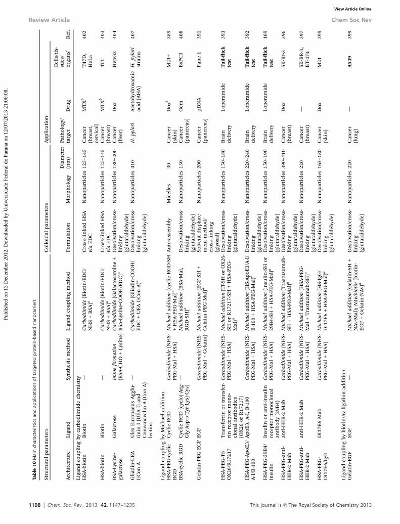
1198 Chem. Soc. Rev., 2013, 42, 1147--1235 This journal is c The Royal Society of Chemistry 2013
Tab
le1
0M
ain
char
acte
rist
ics
and
app
licat
ion
so
fta
rget
edp
rote
in-b
ased
nan
oca
rrie
rs
Stru
ctu
ral
para
met
ers
Col
loid
alpa
ram
eter
sA
ppli
cati
on
Ref
.A
rch
itec
ture
Liga
nd
Syn
thes
ism
eth
odLi
gan
dco
upl
ing
met
hod
Form
ula
tion
Mor
phol
ogy
Dia
met
er(n
m)
Path
olog
y/ta
rget
Dru
g
Cel
ls/t
is-
sues
/or
gan
sc
Liga
nd
cou
plin
gby
carb
odii
mid
ech
emis
try
HSA
-bio
tin
Bio
tin
—C
arbo
diim
ide
(Bio
tin
/ED
C/
NH
S+
BSA
)aC
ross
-lin
ked
HSA
via
ED
CN
anop
arti
cles
125–
145
Can
cer
(bre
ast,
cerv
ical
)
MT
Xb
T47
D,
HeL
a40
2
HSA
-bio
tin
Bio
tin
—C
arbo
diim
ide
(Bio
tin
/ED
C/
NH
S+
BSA
)aC
ross
-lin
ked
HSA
via
ED
CN
anop
arti
cles
125–
145
Can
cer
(bre
ast)
MT
Xb
4T1
403
BSA
-Lys
ine-
gala
ctos
eG
alac
tose
Imin
efo
rmat
ion
(BSA
-CH
O+
Lysi
ne)
Car
bodi
imid
e(G
alac
tosa
min
e+
BSA
-Lys
ine-
CO
OH
/ED
C)a
Des
olva
tion
/cro
ss-
lin
kin
g(g
luta
rald
ehyd
e)
Nan
opar
ticl
es18
0–20
0C
ance
r(l
iver
)D
oxH
epG
240
4
Gli
adin
-UE
AI/
Con
AU
lex
Eu
ropa
eus
Agg
lu-
tin
inI
(UE
AI)
and
Con
can
aval
inA
(Con
A)
lect
ins
—C
arbo
diim
ide
(Gli
adin
-CO
OH
/E
DC
+U
EA
I/C
onA
)aD
esol
vati
on/c
ross
-li
nki
ng
(glu
tara
ldeh
yde)
Nan
opar
ticl
es41
0H
.py
lori
Ace
toh
ydro
xam
icac
id(A
HA
)H
.py
lori
stra
ins
407
Liga
nd
cou
plin
gby
Mic
hae
lad
dit
ion
HSA
-PE
G-c
ycli
cR
GD
Cyc
lic
RG
DC
arbo
diim
ide
(NH
S-PE
G-M
al+
HSA
)M
icha
elad
diti
on(c
ycli
cR
GD
-SH
+H
SA-P
EG
-Mal
)aA
uto
-ass
embl
yM
icel
les
30C
ance
r(s
kin
)D
oxb
M21
+38
9
BSA
-cyc
lic
RG
DC
ycli
cR
GD
(cyc
lo(-
Arg
-G
ly-A
sp- D
-Tyr
-Lys
)-C
ys)
—M
icha
elad
diti
on(B
SA-M
al,
RG
D-S
H)a
Des
olva
tion
/cro
ss-
lin
kin
g(g
luta
rald
ehyd
e)
Nan
opar
ticl
es13
0C
ance
r(p
ancr
eas)
Gem
BxP
C3
408
Gel
atin
-PE
G-E
GF
EG
FC
arbo
diim
ide
(NH
S-PE
G-M
al+
Gel
atin
)M
icha
elad
diti
on(E
GF-
SH+
Gel
atin
-PE
G-M
al)a
Solv
ent
dis
plac
e-m
ent
met
hod
/cr
oss-
lin
kin
g(g
lyox
al)
Nan
opar
ticl
es20
0C
ance
r(p
ancr
eas)
pDN
APa
nc-
139
1
HSA
-PE
G-T
f/O
X26
/R17
217
Tra
nsf
erri
nor
tran
sfer
-ri
nre
cept
orm
ono-
clon
alan
tibo
die
s(O
X26
orR
1721
7)
Car
bodi
imid
e(N
HS-
PEG
-Mal
+H
SA)
Mic
hael
addi
tion
(Tf-
SHor
OX
26-
SHor
R17
217-
SH+
HSA
-PE
G-
Mal
)a
Des
olva
tion
/cro
ss-
lin
kin
g(g
luta
rald
ehyd
e)
Nan
opar
ticl
es15
0–18
0B
rain
del
iver
yLo
pera
mid
eT
ail-
flic
kte
st39
3
HSA
-PE
G-A
poE
3/A
-I/B
-100
Apo
E3,
A-I
,B
-100
Car
bodi
imid
e(N
HS-
PEG
-Mal
+H
SA)
Mic
hael
addi
tion
(HS-
Apo
E3/
A-I
/B
-100
+H
SA-P
EG
-Mal
)aD
esol
vati
on/c
ross
-li
nki
ng
(glu
tara
ldeh
yde)
Nan
opar
ticl
es22
0–24
0B
rain
del
iver
yLo
pera
mid
eT
ail-
flic
kte
st39
2
HSA
-PE
G-2
9B4/
insu
lin
Insu
lin
oran
ti-i
nsu
lin
rece
ptor
mon
oclo
nal
anti
bod
y(2
9B4)
Car
bodi
imid
e(N
HS-
PEG
-Mal
+H
SA)
Mic
hael
addi
tion
(in
suli
n-S
Hor
29B
4-SH
+H
SA-P
EG
-Mal
)aD
esol
vati
on/c
ross
-li
nki
ng
(glu
tara
ldeh
yde)
Nan
opar
ticl
es15
0–19
0B
rain
del
iver
yLo
pera
mid
eT
ail-
flic
kte
st16
9
HSA
-PE
G-a
nti
-H
ER
-2M
aban
ti-H
ER
-2M
abC
arbo
diim
ide
(NH
S-PE
G-M
al+
HSA
)M
icha
elad
diti
on(T
rast
uzu
mab
-SH
+H
SA-P
EG
-Mal
)aD
esol
vati
on/c
ross
-li
nki
ng
(glu
tara
ldeh
yde)
Nan
opar
ticl
es39
0–41
0C
ance
r(b
reas
t)D
oxSK
-Br-
339
6
HSA
-PE
G-a
nti
-H
ER
-2M
aban
ti-H
ER
-2M
abC
arbo
diim
ide
(NH
S-PE
G-M
al+
HSA
)M
icha
elad
diti
on(H
SA-P
EG
-M
al+
Tra
stu
zum
ab-S
H)a
Des
olva
tion
/cro
ss-
lin
kin
g(g
luta
rald
ehyd
e)
Nan
opar
ticl
es22
0C
ance
r(b
reas
t)—
SK-B
R-3
,B
T-4
7439
7
HSA
-PE
G-
DI1
7E6/
IgG
DI1
7E6
Mab
Car
bodi
imid
e(N
HS-
PEG
-Mal
+H
SA)
Mic
hael
addi
tion
(HS-
IgG
/D
I17E
6+
HSA
-PE
G-M
al)a
Des
olva
tion
/cro
ss-
lin
kin
g(g
luta
rald
ehyd
e)
Nan
opar
ticl
es16
5–18
0C
ance
r(s
kin
)D
oxM
2139
5
Liga
nd
cou
plin
gby
biot
in/A
vli
gati
onad
dit
ion
Gel
atin
-EG
FE
GF
—M
icha
elad
diti
on(G
elat
in-S
H+
NA
v-M
al);
NAv
/bio
tin
(bio
tin
-E
GF
+G
elat
in-N
Av)
a
Des
olva
tion
/cro
ss-
lin
kin
g(g
luta
rald
ehyd
e)
Nan
opar
ticl
es22
0C
ance
r(l
un
g)—
A54
939
9
Review Article Chem Soc Rev
Publ
ishe
d on
13
Dec
embe
r 20
12. D
ownl
oade
d by
Uni
vers
idad
e Fe
dera
l do
Para
na o
n 12
/07/
2013
21:
06:0
8.
View Article Online

This journal is c The Royal Society of Chemistry 2013 Chem. Soc. Rev., 2013, 42, 1147--1235 1199
Tab
le1
0(c
on
tin
ued
)
Stru
ctu
ral
para
met
ers
Col
loid
alpa
ram
eter
sA
ppli
cati
on
Ref
.A
rch
itec
ture
Liga
nd
Syn
thes
ism
eth
odLi
gan
dco
upl
ing
met
hod
Form
ula
tion
Mor
phol
ogy
Dia
met
er(n
m)
Path
olog
y/ta
rget
Dru
g
Cel
ls/t
is-
sues
/or
gan
sc
Gel
atin
-EG
FE
GF
—M
icha
elad
diti
on(G
elat
in-S
H+
NA
v-M
al);
NAv
/bio
tin
(bio
tin
-E
GF
+G
elat
in-N
Av)
a
Des
olva
tion
/cro
ss-
lin
kin
g(g
luta
rald
ehyd
e)
Nan
opar
ticl
es23
0C
ance
r(l
un
g)C
isPt
A54
9SC
IDm
odel
400
Gel
atin
-EG
FE
GF
—M
icha
elad
diti
on(G
elat
in-S
H+
NA
v-M
al);
NAv
/bio
tin
(bio
tin
-E
GF
+G
elat
in-N
Av)
a
Des
olva
tion
/cro
ss-
lin
kin
g(g
luta
rald
ehyd
e)
Nan
opar
ticl
es24
0C
ance
r(l
un
g)—
A54
9SC
IDm
odel
398
Gel
atin
-CD
3C
D3
Mab
—M
icha
elad
diti
on(G
elat
in-S
H+
NA
v-M
al);
NAv
/bio
tin
(bio
tin
-C
D3
+G
elat
in-N
Av)
a
Des
olva
tion
/cro
ss-
lin
kin
g(g
luta
rald
ehyd
e)
Nan
opar
ticl
es25
0–30
0T
-ly
mph
ocyt
es—
—40
1
HSA
-PE
G-A
poE
Apo
EC
arbo
diim
ide
(NH
S-PE
G-M
al+
HSA
)M
icha
elad
diti
on(N
Av-
SH+
HSA
-PE
G-M
al);
NAv
/bio
tin
(bio
tin
-A
poE
+H
SA-P
EG
-NA
v)a
Des
olva
tion
/cro
ss-
lin
kin
g(g
luta
rald
ehyd
e)
Nan
opar
ticl
es34
0B
rain
del
iver
yLo
pera
mid
eT
ail-
flic
kte
st39
0
Gel
atin
-PE
G-
tras
tuzu
mab
orH
AS-
PEG
-an
ti-
HE
R-2
Mab
anti
-HE
R-2
Mab
Car
bodi
imid
e(N
HS-
PEG
-Mal
+H
SAor
Gel
atin
)
Mic
hael
addi
tion
(Gel
atin
-SH
/H
SA-S
H+
NA
v-M
al);
NAv
/bio
tin
(tra
stu
zum
ab-b
ioti
n+
Gel
atin
-N
Av/
HSA
-NA
v)
Des
olva
tion
/cro
ss-
lin
kin
g(g
luta
r-al
deh
yde)
ord
oubl
ed
esol
vati
on
Nan
opar
ticl
es21
0–32
0C
ance
r(b
reas
t)—
MC
F7,
SK-
BR
-3,
BT
474
394
Liga
nd
cou
plin
gby
oth
erli
gati
onst
rate
gies
Gel
atin
-sil
oxan
e-T
atT
AT
Tw
o-st
epso
l–ge
lpr
oces
sD
isul
fide
brid
gefo
rmat
ion
(Tat
-SH
+G
elat
in-s
ilox
ane-
SH)a
See
syn
thes
ism
eth
odN
anop
arti
cles
210
Can
cer
(cer
vica
l)pD
NA
/pSV
b-ga
lH
eLa
406
Gel
atin
-sil
oxan
e-PE
G-S
ynB
Syn
BT
wo-
step
sol–
gel
pro-
cess
;ca
rbod
iim
ide
(HO
OC
-PE
G-N
H2/
ED
C/N
HS)
Dis
ulfi
debr
idge
form
atio
n(S
ynB
-SH
+G
elat
in-s
ilox
ane-
PEG
-PD
S)a
See
syn
thes
ism
eth
odN
anop
arti
cles
150–
200
Bra
ind
eliv
ery
—B
BB
mod
elB
rain
targ
etin
g
405
aC
oupl
ing
post
-nan
ocar
rier
form
atio
n.
bD
rug
cova
len
tly
lin
ked
toth
e(c
o)po
lym
er.
cIn
vivo
expe
rim
ents
are
wri
tten
inbo
ld.
Chem Soc Rev Review Article
Publ
ishe
d on
13
Dec
embe
r 20
12. D
ownl
oade
d by
Uni
vers
idad
e Fe
dera
l do
Para
na o
n 12
/07/
2013
21:
06:0
8.
View Article Online

1200 Chem. Soc. Rev., 2013, 42, 1147--1235 This journal is c The Royal Society of Chemistry 2013
such as MBS, sulfo-MBS etc.) and allowed linking at the surfaceof the protein (see Fig. 14 for the structures of the spacers). Thispathway was successfully applied to a broad variety of targetingpeptides (cyclic RGD),389 proteins (ApoE3, A-I, B-100, EGF,insulin, Tf)169,390–393 or antibodies (Trastuzumab, DI17E6,IgG, 29B4, OX26, R17217, see Fig. 36)169,393–397 for applicationin cancer and brain delivery (CNS-related disease).
Encapsulation of loperamide169,392,393 was reported as amodel drug to test the efficiency of the brain delivery whereaspDNA391 and Dox395,396 were employed for cancer therapy. Doxwas also covalently linked to the protein (Fig. 37).389
Biotin/avidin ligation. The NAv/biotin ligation strategy alsorepresents a method of choice for targeted protein NPs. Theglobal idea is to graft maleimide moieties at the surface ofprotein NPs and to use thiol-bearing ligands to form a thioetherbond. Proteins were turned into sulfhydryl-reactive NPs, eitherby thiolation with 2-iminothiolane or by reacting with a NHS-PEG-Mal. Note that the latter pathway also leads to PEGylatedNPs. NAv is activated with m-maleimidobenzoyl-N-hydroxy-succinimide (sulfo-MBS, Fig. 14), followed by its incubation withthe Mal-functionalized proteins to yield NAv-decorated proteinNPs. Then, the ligands were biotinylated with commerciallyavailable biotinylation reagents, such as sulfosuccinimidyl-6-[biotin-amido]hexanoate (sulfo-NHSLC-biotin, Fig. 14) or penta-fluorophenyl-ester biotin (PFP-biotin, Fig. 14) before their couplingto the NPs by simple incubation. This was efficiently applied toalbumin and gelatin NPs at the surface of which ApoE,390
EGF,398–400 CD3 MAb401 and trastuzumab394 were positioned.Drugs were either encapsulated/adsorbed (loperamide)390 orcovalently linked to the protein (CisPt) (Fig. 38).400
Carbodiimide coupling strategy. HSA was also functiona-lized with biotin via EDC/NHS chemistry to target biotinreceptors overexpressed at the surface of certain cancercells.402,403 This was readily achieved by a one-pot reactionbetween HSA, biotin and EDC/NHS. The same coupling chem-istry was also used to covalently link MTX as the anticancerdrug to HSA. In the same spirit, galactose was tethered at thesurface of doxorubicin-encapsulated albumin NPs (Fig. 39).404
In this example, glutaraldehyde was used to cross-link the NPsand to present aldehyde moieties at their surface for theirfurther conjugation to lysine. EDC was then used to performthe conjugation between galactosamine and the exposed car-boxylic acid of lysine residues.
Other ligation strategies. Interesting pathways have beenreported to link cell penetrating peptides, such as TAT or SynB,to gelatin NPs by means of a disulfide bridge.405,406 Gelatin-siloxane NPs were first prepared by a two-step sol–gel process.In the first route, the NPs were grafted with PEG via EDC/NHScoupling chemistry using surface amines of the NPs.405 Thenthe sulfhydryl-containing SynB peptide was tethered to the NPsby using the N-succinimidyl-3-(2-pyridyldithio) propionate(SPDP) heterobifunctional linker (Fig. 14), through the for-mation of a disulfide bridge. The second route reported thesurface modification of the gelatin-siloxane NPs by the Traut’sreagent (Fig. 14) in order to position thiol groups at theirsurface.406 The Cys-containing TAT peptide was then coupledto the NPs by forming the disulfide bridge.
3.4.5. POLY(ALKYL CYANOACRYLATE). Alkyl cyanoacrylates arewidely-known monomers, extremely appreciated for their veryhigh reactivity and the excellent adhesive properties of theresulting polymers. On one hand, the famous Superglues,which contains short alkyl chain cyanoacrylates, is commonlyemployed by the general public for repairing and do-it-yourselfactivities whereas longer alkyl chain cyanoacrylates have beendeveloped for biomedical purposes such as e.g. surgical glue forthe closure of skin wounds and embolic material for endo-vascular surgery. Their use for the construction of nano-particles has been discovered in the late seventies by Couvreuret al.409 and, since that time, a very important number ofpharmaceutical applications have been reported due to theease of preparation (in aqueous medium) and the biocompat-ibility of these nanoparticles. Importantly, they are currently ina phase III clinical trial for the treatment of the MDR resistanthepatocarcinoma.
Poly(alkyl cyanoacrylate) (PACA) can be synthesized accordingto three distinct types of polymerization: (i) anionic polymeriza-tion; (ii) zwitterionic polymerization and (iii) radical polymeriza-tion. However, due to their very high reactivity, anionic andzwitterionic polymerization mechanisms are predominant underconventional experimental conditions with respect to the radicalprocess.
The predominant degradation mechanism, which isbelieved to be the main degradation pathway in vivo, occursvia the hydrolysis of their side chain ester functions, producingthe corresponding alkyl alcohol and poly(cyanoacrylic acid) as
Fig. 30 Synthesis of poly(amino acid) (PAA) by the polymerization ofN-carboxyanhydride (NCA) initiated by amino compounds.
Fig. 31 Detailed chemical structure of the non-folate conjugated linear dendriticpolymer and illustration of its self-assembly into ‘‘patchy’’ micelles presentingclusters of folate of variable sizes on the surface of the micelle (reproduced withpermission from ref. 384. Copyright 2010, Wiley).
Review Article Chem Soc Rev
Publ
ishe
d on
13
Dec
embe
r 20
12. D
ownl
oade
d by
Uni
vers
idad
e Fe
dera
l do
Para
na o
n 12
/07/
2013
21:
06:0
8.
View Article Online

This journal is c The Royal Society of Chemistry 2013 Chem. Soc. Rev., 2013, 42, 1147--1235 1201
the degradation products, the latter being fully water-solubleand readily eliminated by kidney filtration.410 PACA are one ofthe fastest degrading polymers having degradation times
ranging from few hours to few days. However, a complete excretionof these materials would occur only for low-molecular weightpolymers, typically for a molecular weight below 10 000 g mol�1.
In the field of PACA, examples of successful functionaliza-tions to achieve active targeting are rather scarce because of thevery high reactivity of alkyl cyanoacrylate monomers and/or therapid degradability of PACA polymers/nanoparticles.411 How-ever, the tandem Knoevenagel condensation–Michael addition,which allowed preparing PEGylated PACA NPs (Fig. 40), alsorepresented a convenient strategy to position functional groupsat the extremity of the PEG chains for further coupling, eitheron the copolymer in organic medium or directly at the surfaceof the resulting NPs.
Couvreur and coworkers synthesized targeted PACA NPs bycoupling FA to P(HDCA-co-NH2PEGCA) NPs via an amidationreaction from its NHS-ester derivative.412,413 During the synthesis,a tBoc-protected amino PEG was used and the deprotection
Fig. 32 Synthetic scheme of the amphiphilic triblock copolymers and the preparation process of the SPIO/DOX-loaded vesicles with cross-linked inner hydrophilic PEGlayers (reproduced with permission from ref. 374. Copyright 2010, American Chemical Society).
Fig. 33 Schematic representation of PBLG-b-HA polymersomes, with a bilayerforming membrane with PBLG packed antiparallel (reproduced with permissionfrom ref. 378. Copyright 2012, American Chemical Society).
Chem Soc Rev Review Article
Publ
ishe
d on
13
Dec
embe
r 20
12. D
ownl
oade
d by
Uni
vers
idad
e Fe
dera
l do
Para
na o
n 12
/07/
2013
21:
06:0
8.
View Article Online

1202 Chem. Soc. Rev., 2013, 42, 1147--1235 This journal is c The Royal Society of Chemistry 2013
occurred prior to NP formation (Fig. 41). NPs exhibited an averagediameter of 80–90 nm. 15% of the PEG chain-ends were functio-nalized with FA and SPR accounted for a significant binding with
the folate-binding protein (FBP). The same targeted system was alsoemployed to encapsulate pDNA/PEI or pDNA/PLL for transfectionpurposes.414 In this case, bigger NPs of 260–390 nm were obtained.
Fig. 34 Synthesis of PBLG-b-PGG glycopeptide block copolymers by a combination of ring-opening polymerization of NCA and click chemistry (reproduced withpermission from ref. 386. Copyright 2012, Elsevier).
Fig. 35 Synthesis of functional poly(amino acid) based theranostic polymeric nanoparticles (reproduced with permission from ref. 387. Copyright 2011, AmericanChemical Society).
Review Article Chem Soc Rev
Publ
ishe
d on
13
Dec
embe
r 20
12. D
ownl
oade
d by
Uni
vers
idad
e Fe
dera
l do
Para
na o
n 12
/07/
2013
21:
06:0
8.
View Article Online

This journal is c The Royal Society of Chemistry 2013 Chem. Soc. Rev., 2013, 42, 1147--1235 1203
Periodate oxidation was also employed from P(HDCA-co-NH2PEGCA) NPs to link the Tf protein.415,416 W/O/W doubleemulsion was used to generate the amino-functionalized NPs
containing pDNA416 or pDNA/PLL.415 Average diameter rangedfrom 130–150 nm (pDNA) to 240–290 nm (pDNA/PLL).
Click chemistry,417 which has emerged as a powerful liga-tion strategy, was recently applied to PACA NPs.418 By means ofCuAAC, a versatile targeted nanoparticulate platform basedon PACA has been recently proposed and exhibited stealth,fluorescent and targeting abilities. A heterobifunctionalazidopoly(ethylene glycol) (N3PEG) was first derivatized withthe selected ligand by CuAAC using CuSO4/sodium ascorbate asthe catalytic system and turned into its cyanoacetate derivativeunder DCC-assisted chemistry (Fig. 42). Its terpolymerizationwith varying amount of MePEGCA (in order to tune the finalsurface density of ligand) and HDCA by tandem Knoevenagelcondensation–Michael addition led to the correspondingligand-containing amphiphilic copolymer. After concomitantself-assembly in aqueous solution with a rhodamine B-taggedPACA copolymer,419 functionalized NPs in the 85–150 nm rangedisplaying the ligand of interest were obtained. The versatilityof this methodology was illustrated by the coupling of a smalllibrary of biologically active ligands (biotin, selegiline, curcu-minoids) in order to target cancer (biotin) and Alzheimer’sdisease (selegiline, curcuminoids) via the Ab1-42 peptide bio-marker.420 To perform the CuAAc coupling, the ligands werefirst derivatized with an alkyne group except for selegiline,which natively contains such function. Interestingly, thanksto the surface availability of biotin, a monoclonal anti-Ab1-42
Fig. 36 Scanning electron microscopy (SEM) images showing HSA nanoparticleswith DI17E6 coupled on the surface (A, B = magnification of A in the redquadrangle) and nanoparticles without an antibody coupling (C) (reproducedwith permission from ref. 395. Copyright 2010, Elsevier).
Fig. 37 Synthetic pathway for targeted albumin-based micelles for delivery of amphipathic drugs (reprinted with permission from ref. 389. Copyright 2011, AmericanChemical Society).
Chem Soc Rev Review Article
Publ
ishe
d on
13
Dec
embe
r 20
12. D
ownl
oade
d by
Uni
vers
idad
e Fe
dera
l do
Para
na o
n 12
/07/
2013
21:
06:0
8.
View Article Online

1204 Chem. Soc. Rev., 2013, 42, 1147--1235 This journal is c The Royal Society of Chemistry 2013
Tab
le1
1M
ain
char
acte
rist
ics
and
app
licat
ion
so
fta
rget
edp
oly
(alk
ylcy
ano
acry
late
)-b
ased
nan
oca
rrie
rs
Stru
ctu
ral
para
met
ers
Col
loid
alpa
ram
eter
sA
ppli
cati
on
Ref
.A
rch
itec
ture
Liga
nd
Syn
thes
ism
eth
odC
oupl
ing
met
hod
Form
ula
tion
Mor
phol
ogy
Dia
met
er(n
m)
Path
olog
y/ta
rget
Dru
gC
ells
/tis
sues
/or
gan
s
Liga
nd
cou
plin
gby
carb
odii
mid
ech
emis
try
P(H
DC
A-c
o-FA
PEG
CA
)FA
Kno
even
agel
cond
ensa
tion
–M
icha
elad
diti
onC
arbo
diim
ide
(P(H
DC
A-c
o-H
2N
PEG
CA
)+
FA-N
HS)
bN
anop
reci
pita
tion
Nan
opar
ticl
es80
–90
Can
cer
——
412
P(H
DC
A-c
o-FA
PEG
CA
)FA
Kno
even
agel
cond
ensa
tion
–M
icha
elad
diti
onC
arbo
diim
ide
(P(H
DC
A-c
o-H
2N
PEG
CA
)+
FA-N
HS)
bN
anop
reci
pita
tion
Nan
opar
ticl
es80
–90
Can
cer
(ova
ry)
—K
B3-
141
3
P(H
DC
A-c
o-FA
PEG
CA
)FA
Kno
even
agel
cond
ensa
tion
–M
icha
elad
diti
onC
arbo
diim
ide
(P(H
DC
A-c
o-H
2N
PEG
CA
)+
FA-N
HS)
bN
anop
reci
pita
tion
Nan
opar
ticl
es26
0–39
0C
ance
r(o
vary
)pD
NA
/PLL
orpD
NA
/PE
I
KB
414
Liga
nd
cou
plin
gby
copp
er-c
atal
yzed
azid
e–al
kyn
ecy
cloa
dd
itio
n(C
uA
AC
)P(
HD
CA
-co-
biot
inPE
GC
A)
Bio
tin
Kno
even
agel
cond
ensa
tion
–M
icha
elad
diti
onC
lick
chem
istr
y(a
zid
oPE
G+
biot
in-a
lkyn
e)a
Nan
opre
cipi
tati
onor
emu
lsio
n/s
ol-
ven
tev
apor
atio
n
Nan
opar
ticl
es10
0C
ance
r(b
reas
t,lu
ng)
Ptx
MC
F7,
M10
942
2
P(H
DC
A-c
o-Se
lPE
GC
A)
Sele
gili
ne
(Sel
)K
noev
enag
elco
nden
sati
on–
Mic
hael
addi
tion
Cli
ckch
emis
try
(azi
doP
EG
+Se
l-alk
yne)
aN
anop
reci
pita
tion
Nan
opar
ticl
es90
–110
Alz
hei
mer
’sd
isea
se—
Ab 1
–42
420
P(H
DC
A-c
o-C
urP
EG
CA
)C
urc
um
inoi
ds
(Cu
r)K
noev
enag
elco
nden
sati
on–
Mic
hael
addi
tion
Cli
ckch
emis
try
(azi
doP
EG
+C
ur-
alky
ne)
aN
anop
reci
pita
tion
Nan
opar
ticl
es85
–105
Alz
hei
mer
’sd
isea
se—
Ab 1
–42
422
P(H
DC
A-c
o-bi
otin
PEG
CA
)an
ti-Ab 1
–42
MA
bK
noev
enag
elco
nden
sati
on–
Mic
hael
addi
tion
Cli
ckch
emis
try
(azi
doP
EG
+bi
otin
-alk
yne)
aan
dSA
v/bi
otin
(P(H
DC
A-c
o-bi
otin
PEG
CA
)+
Mab
-SA
v)b
Nan
opre
cipi
tati
onN
anop
arti
cles
150
Alz
hei
mer
’sd
isea
se—
Ab 1
–42
422
Liga
nd
cou
plin
gby
oth
erli
gan
dst
rate
gies
P(H
DC
A-c
o-M
ePE
GC
A)
PEG
Kno
even
agel
cond
ensa
tion
–M
icha
elad
diti
on—
Nan
opre
cipi
tati
onN
anop
arti
cles
90–1
10A
lzh
eim
er’s
dis
ease
—Ab 1
–42
423
P(H
DC
A-c
o-T
fPE
GC
A)
Tra
nsf
erri
n(T
f)K
noev
enag
elco
nden
sati
on–
Mic
hael
addi
tion
Imin
efo
rmat
ion
(P(H
DC
A-c
o-H
2N
PEG
CA
)+
Tf-
CH
O)b
Dou
ble
emu
lsio
n–
solv
ent
evap
orat
ion
Nan
opar
ticl
es13
0–15
0C
ance
r(l
euke
mia
)pD
NA
K56
242
4
P(H
DC
A-c
o-T
fPE
GC
A)
Tra
nsf
erri
n(T
f)K
noev
enag
elco
nden
sati
on–
Mic
hael
addi
tion
Imin
efo
rmat
ion
(P(H
DC
A-c
o-H
2N
PEG
CA
)+
Tf-
CH
O)b
Dou
ble
emu
lsio
n–
solv
ent
evap
orat
ion
Nan
opar
ticl
es24
0–29
0C
ance
r(l
euke
mia
)D
NA
/PLL
K56
241
5
aC
oupl
ing
prio
rto
nan
ocar
rier
form
atio
n.
bC
oupl
ing
post
-nan
ocar
rier
form
atio
n.
Review Article Chem Soc Rev
Publ
ishe
d on
13
Dec
embe
r 20
12. D
ownl
oade
d by
Uni
vers
idad
e Fe
dera
l do
Para
na o
n 12
/07/
2013
21:
06:0
8.
View Article Online

This journal is c The Royal Society of Chemistry 2013 Chem. Soc. Rev., 2013, 42, 1147--1235 1205
antibody was positioned by simple incubation at the surface ofthe NPs by means of the biotin/streptavidin ligation strategy.Note that the PEG chains exposed at the surface of the non-functionalized P(HDCA-co-MePEGCA) NPs also served as a ligandexhibiting a significant affinity for the Ab1-42 peptide.421,422
4. Biological applications of targetednanoparticles4.1. General considerations
The application of nanotechnologies in the biomedical fieldhas attracted a great deal of interest in the past decades due toits potential to revolutionize the treatment of severe diseasessuch as cancer, neurological and cardiovascular diseases. Inparticular, nanocarriers targeted to specific tissues offer theadvantages of improved drug therapeutic efficiency anddramatically reduced toxicity and unspecific side effects. Inthis perspective, in order to promote selective interactions,targeted nanocarriers have been designed taking inspirationfrom the specific characteristics of each disease. Due to thedramatically high number of deaths caused every year, mucheffort has been devoted to engineering actively targeted nano-carriers to improve the traditional treatment of cancer. There-fore, functionalized nanocarriers have been prepared usingligands able to specifically bind receptors with increasedexpression on malignant cells or molecules able to interferewith the signaling pathway unique to cancer cells.
Some in vitro proofs of concept of active targeting wereprovided using empty nanocarriers but mainly drug-loaded
Fig. 38 Schematic representation of gelatin NP formulations (GPs). (a) GPs wereprepared by desolvation method; (b) CDDP-loaded GPs (GP–Pt) werespontaneously formed via a ligand exchange reaction of Pt(II) from the chloride tothe carboxyl group in the GPs; (c) surface modification of GP–Pt was performedwith NeutrAvidin-FITC-biotinylated-EGF complex (reproduced with permissionfrom ref. 400. Copyright 2009, Elsevier).
Fig. 39 Schematic illustration for the preparation of Gal-Dox-albumin: (a) preparation of cross-linked Dox-albumin nanoparticles without free aldehyde groups onsurface; (b) conjugation of Gal onto the surface of Dox-albumin. The red small spheres represent Dox; the big spheres represent albumin; the yellow lines inside the bigspheres represent glutaraldehyde cross-linkage (reprinted with permission from ref. 404. Copyright 2011, Elsevier).
Chem Soc Rev Review Article
Publ
ishe
d on
13
Dec
embe
r 20
12. D
ownl
oade
d by
Uni
vers
idad
e Fe
dera
l do
Para
na o
n 12
/07/
2013
21:
06:0
8.
View Article Online

1206 Chem. Soc. Rev., 2013, 42, 1147--1235 This journal is c The Royal Society of Chemistry 2013
systems have been investigated in order to assess the therapeuticpotential of the ligand-targeted strategy. Considering that FA iscertainly one of the most represented targeting ligands, wedecided to present a more detailed description of in vitro experi-ments performed using FA-functionalized nanocarriers. Thefeasibility of this approach was evaluated by measuring theuptake of the nanocarriers and their cytotoxicity over specificcell lines, which well exemplifies the studies carried out withother targeting molecules. Indeed, although some details wereopportunely modified according to the used ligand, such in vitroexperimental protocol was generally described by all researchgroups.
Therefore, in this review, we preferred to highlight in vivo studiesby presenting the most attractive results obtained so far. Indeed, thein vivo pharmacological investigation is fundamental before thetranslation of the targeted nanomedicine concept into clinical trialsand, noteworthily, the increasing number of papers dealing with thein vivo evaluation of ligand-targeted nanocarriers is witnessed.
Ligand-targeted nanocarriers have found application notonly in the treatment of cancer but also in the managementof other severe diseases. A general overview of these therapeuticapplications will be proposed.
4.2. Cancer
4.2.1. VITAMIN TARGETED NANOCARRIERS
4.2.1.1. Folic acid targeted nanocarriers. A broad range ofcancers are characterized by a significant overexpression offolate receptors, up to 100–300 times higher than normaltissue.76,80,425 Therefore, FA has been widely employed as aligand for achieving targeted therapies of several carcinomas.Commonly used chemotherapeutic drugs (i.e. paclitaxel, doc-etaxel and doxorubicin, 5-FU and Oligomycin-A) were encapsu-lated in these drug delivery systems, resulting in improvedcytotoxicity against cancer cells. Interesting examples of bothphysical entrapment and covalent coupling of the drug to thecopolymers have been explored with the aim of increasing
Fig. 40 Synthesis of P(HDCA-co-MePEGCA) copolymer by tandem Knoevenagel condensation–Michael addition.
Fig. 41 Synthesis of P(HDCA-co-NH2PEGCA) copolymer for further NP surface functionalization.
Review Article Chem Soc Rev
Publ
ishe
d on
13
Dec
embe
r 20
12. D
ownl
oade
d by
Uni
vers
idad
e Fe
dera
l do
Para
na o
n 12
/07/
2013
21:
06:0
8.
View Article Online

This journal is c The Royal Society of Chemistry 2013 Chem. Soc. Rev., 2013, 42, 1147--1235 1207
the drug loading together with the therapeutic efficacy of thenanocarriers.245,246,295,329 Targeted nanocarriers were alsoexploited as carriers of plasmid DNA for gene ther-apy,221,238,239,335,342 The physical entrapment or the covalentconjugation of various fluorophores (e.g., FITC, Hylite 488,Alexa 488, Coumarin-6) allowed their accurate traceability andthe evaluation of specific in vitro and/or in vivo interactions.
The in vitro efficiency of folate-mediated targeting has beenevaluated using cell lines, which overexpress the folate recep-tor. Among these cell lines, the human epidermal carcinoma(KB), the human cervical carcinoma (Hela), the human breastcarcinoma (MCF7), the murine mammary carcinoma (4T1),human ovarian carcinoma (SKOV-3), human squamous carci-noma (SiHa), and the human gastric carcinoma (SGC-7901) arethe most well-known. These cells were compared, in terms ofuptake/targeting to folate receptor deficient cancer cells such ashuman alveolar carcinoma A549 and normal/healthy cells suchas murine fibroblast NIH/3T3, macrophages and humanendothelial EA.hy 926. Fluorescent nanocarriers allowed
monitoring and quantifying the specific folate-receptormediated binding and internalization by flow cytometry andconfocal microscopic analysis. Published results clearly demon-strated that the cellular uptake was enhanced by conjugation ofFA at the surface of the NPs compared to the unspecific inter-nalization of bare/naked nanocarriers. In addition, folate receptoroverexpressing cell lines showed the highest internalization of FA-functionalized nanocarriers. Competitive binding experiments,performed in the presence of an excess of free FA confirmed theoccurrence of a folate-mediated endocytosis mechanism.
The adequate targeting ligand density, which can be con-trolled by tuning the ratios of ligand-conjugated and ligand-lacking copolymer in the formulation, was also investigated.For instance, independently of the initial amount of PLGA-b-PEG-FA used (i.e., from 7 to 75 mol%), only 21% of folategroups were detected by Valencia and coworkers at the surfaceof their nanoparticles prepared using a blend of PLGA-b-PEGand PLGA-b-PEG-FA.225 This was probably due to the inter-actions between the low water soluble folate molecules and the
Fig. 42 Synthetic pathway to prepare fluorescent, PEGylated and biodegradable poly(alkyl cyanoacrylate) nanoparticles functionalized with biotin (VB7), selegiline(Sel) or curcuminoid derivatives (CurA and CurB).
Chem Soc Rev Review Article
Publ
ishe
d on
13
Dec
embe
r 20
12. D
ownl
oade
d by
Uni
vers
idad
e Fe
dera
l do
Para
na o
n 12
/07/
2013
21:
06:0
8.
View Article Online

1208 Chem. Soc. Rev., 2013, 42, 1147--1235 This journal is c The Royal Society of Chemistry 2013
PLGA during the formulation process, which caused the entrap-ment of a significant fraction of the FA into the core of the NPs.Consequently, fine tuning the initial stoichiometry of thedifferent copolymers is a crucial parameter to assure thepresence of a sufficient fraction of FA at the surface of thenanoparticles for achieving uptake on FA receptor-over-expressed cells.225 However, although the targeting capabilitycould be compromised when a too low amount of FA is presentat the surface of the nanocarriers, it was reported that an excessof FA could affect the aqueous stability of the resulting nano-carriers and cause formation of insoluble aggregates.242 Themanner by which FA molecules are presented to the cells (i.e.homogeneously or in spatially defined groupings on a nano-particle surface) and its influence on cellular uptake was alsoinvestigated. Both in vitro and in vivo studies were performed byPoon and coworkers,384 using linear dendritic PBLA(PED-b-PEG-FA)n polypeptide micelles with statistically similar amounts offolate in different spatial arrangements. In vivo results clearlyshowed that arrangement of FA molecules in clusters presentingan optimal size of, on average, three folate groups, enhanced theuptake of micelles owing to the resultant higher avidity andlonger residence times on the cell membrane (Fig. 43).
The in vitro cytotoxicity of drug-loaded nanocarriers wasevaluated in parallel to the assessment of their targetingfeatures by a routine MTT viability test. Folate-decorateddrug-loaded nanocarriers showed a higher toxicity comparedto untargeted NPs, due to the intracellular uptake enhancementmediated by folate receptors. Noteworthily, strong cytotoxicitywas also measured in multidrug resistant cell lines.226,314 WhenpDNA was encapsulated into polyester NPs, efficient gene deliverywas confirmed by successful transfection results.221,238,239 It is
therefore evident that the ability of these NPs to bind receptorswith high affinity offers the possibility of an efficient targeteddelivery of drugs to cancer cells, thus enhancing their thera-peutic efficacy.
The possibility to combine active targeting, therapeuticefficiency as well as imaging properties has been demonstratedby the encapsulation of Dox, quantum dots and iron oxidecrystals into the hydrophobic core of folate-functionalized PLGANPs.219 The specific targeting ability for cells overexpressing folatereceptors led to high NP uptake, which was further confirmed byT2-weighted MR images. Moreover, the targeting properties wereenhanced by application of an external magnetic field, causing theprogressive increase of the signal drop and a higher cytotoxicity.Fe3O4 was encapsulated also within the core of chitosan-basedNPs.346 The resulting magnetic properties allowed the guidance ofthe nanoparticles toward the tumor using an external magneticfield. Compared to control experiments, an approximately 50 timesgreater accumulation of magnetic nanoparticles was registered inS-180 sarcoma-bearing Kunming mice, 3 h after administration.
The in vivo validation of specific folate-mediated uptake ofdrug-loaded nanocarriers was provided by different groupsusing tumor xenograft animal models developed by transplant-ing folate overexpressing cancer cells. Tumor accumulation andtherapeutic efficacy of chemotherapeutic-loaded nanocarrierswere evaluated in KB,295,379 MCF7,73,379 Hela,334 A549,379 drugresistant A2780/AD,379 H-22,344 and SGC-7901 carcinoma301
models. In all these studies, intravenous administration offolate-decorated nanoparticulate systems resulted in a higherdrug accumulation at the tumor site and a slower rate of tumorgrowth compared to non-functionalized counterparts and tothe free drug used as controls. The enhanced permeability and
Fig. 43 In vivo evaluation of patchy micelles on nude mice bearing two different tumors (right flank: KB, left flank: A375). (a) Fluorescence 3D optical imaging oftumored nude mice 48 h after injection with different formulation micelles. Only fluorescence from tumors and a non-tumoral region of the mice (upper torso) isshown. (b) Normalized tumor fluorescence VivoTag680(VT680)/AngioSense 750(AS750)) of tumors (n = 4) showing the highest in vivo targeting with 20%F–60%mixmicelles. (c) Mean cell-associated micelle fluorescence (FI) (reproduced with permission from ref. 384. Copyright 2009, Wiley).
Review Article Chem Soc Rev
Publ
ishe
d on
13
Dec
embe
r 20
12. D
ownl
oade
d by
Uni
vers
idad
e Fe
dera
l do
Para
na o
n 12
/07/
2013
21:
06:0
8.
View Article Online

This journal is c The Royal Society of Chemistry 2013 Chem. Soc. Rev., 2013, 42, 1147--1235 1209
retention effect due to the large pores present in the tumorendothelial vessels (compared to healthy tissue) and thereduced tumor lymphatic drainage caused a passive accumula-tion of the nanocarriers at the tumor site. Then, the moreefficient internalization of functionalized systems via a folate-receptor mediated endocytosis led to an improved therapeuticresponse. An example of such successful results is provided inFig. 44. Therapeutic efficacy of FA-functionalized Ptx-loadedheparin nanoparticles was assessed in KB-3-1 xenograft-bearingmice.369 The treatment with free Ptx suppressed tumor growthonly initially, and after 3 weeks of treatment, the tumor nolonger responded and grew rapidly, likely due to the develop-ment of a drug resistance mechanism. Contrariwise, after thesame period of treatment with targeted nanocarriers, thisresurgence in tumor growth was not observed. Moreover,compared with control treatments (i.e. non-functionalizedNPs or free Ptx) FA-targeted NPs induced a significantlyhigher increase of acetylated tubulin levels, marker of a pro-apoptotic stage.
Interestingly, Werner and coworkers explored the potentialof folate-targeted nanoparticles, loaded with docetaxel (FT-NPDxtl), as radiosensitizers to improve the efficacy of the radio-therapy in the tumor and to reduce the toxicity in the healthytissues.222 Moreover, due to the importance of timing of radio-therapy, they were the first to determine the optimal intervalbetween chemo and radiotherapy. In vivo studies were performed
on mice bearing KB cells xenograft tumors, which were treatedwith FT-NP Dxtl and irradiation at 3, 12, 24 or 48 h postinjection. Due to the specific accumulation in the tumor,systemic administration of FT-NP Dxtl caused a significantinhibition of tumor progression compared to the controls (freedrug and non-targeted nanoparticles). The most importanttumor growth delay was observed in mice irradiated 12 h afternanoparticles administration. These results clearly demon-strated the potential of these nanoparticles as radiosensitizerand highlighted the importance of determining the optimaltime of irradiation to achieve successful treatments. The folate-targeting strategy was then enlarged leading to the develop-ment of so-called ‘‘ChemoRad’’ nanoparticles which offer thepossibility of concurrent use of chemo and radiotherapy.223 Ptxand yittrium-90 were used as chemo and radiotherapeuticisotope, respectively. The effectiveness of this dual systemwas investigated in an ovarian peritoneal metastasis modeldeveloped by intraperitoneal injection of folate overexpressingovarian cancer cells. Compared to mono treatments (chemotherapyor radiotherapy only), the best therapeutic outcome was achieved inmice treated with ChemoRad nanoparticles. Moreover, although notstatistically significant, there was a clear trend toward longer survivalin groups treated with folate-targeted nanoparticles (Fig. 45).
The design of nanocarriers combining FA receptor targetingand early endosomal pH sensitiveness was proposed by Kimand coworkers to overcome tumor multidrug resistance.226,243
Fig. 44 Antitumor effect of FA functionalized NPs (HFT-T) in an animal model. (a) The growth curve of KB-3-1 xenografts showed that all the treatment groupsincluding free Ptx, blank NPs (HT-t), and HFT-T significantly inhibited the growth of tumor as compared with the control (p o 0.0001 for each treatment). The tumorvolumes were similar in HT-T and free Ptx-treated groups (p o 0.608), but FA-decorated NPs more potently reduced tumor growth than free Ptx (p o 0.0001). (b) Arepresentative mouse from each group. (c) The body weight of the mice in all groups. (d) Representative images of H&E organ staining from control and HFT-T-treatedmice (magnification 200) (reproduced with permission from ref. 369. Copyright 2009, American Chemical Society).
Chem Soc Rev Review Article
Publ
ishe
d on
13
Dec
embe
r 20
12. D
ownl
oade
d by
Uni
vers
idad
e Fe
dera
l do
Para
na o
n 12
/07/
2013
21:
06:0
8.
View Article Online

1210 Chem. Soc. Rev., 2013, 42, 1147--1235 This journal is c The Royal Society of Chemistry 2013
Multidrug resistant ovarian tumor xenograft-bearing mice wereused to assess the anticancer activity of Dox-loaded pH-sensi-tive micelles made of a blend of P(His-co-Phe)-b-PEG andPLA-b-PEG-FA. These advanced micelles were able to induce asignificant inhibition of the tumor growth rate compared tocontrol groups, which received Dox-loaded pH-insensitivemicelles of free Dox. Despite the presence of the FA at theirsurface, which makes both pH-sensitive and pH-insensitivemicelles share the same endocytosis pathway, they caused adifferent therapeutic effect that might be explained consideringthe existence of different intracellular events such as drugrelease and endosomolysis. This observation was confirmedby non-invasive imaging experiments performed by using thedorsal window chamber model. The highest accumulation ofDox at the tumor site was observed 1 h after i.v. injection ofpH-sensitive micelles, while, in the case of pH-insensitive onesthe maximum was reached at 20 minutes, whereas at 1 h it wasalready in the decline phase. Therefore, pH-sensitive micellesprobably induced a high cytosolic accumulation of Dox, whichallowed overcoming the limited defense capacity of MDR cells.
A pH-responsive PLA-b-PEG-b-PHis targeted nanocarrierencapsulating Dox and bearing TAT and FA ligands was proposedby Lee and coworkers.379 This nanocarrier displayed the ability tohide TAT at its surface during circulation and to expose it at aslightly acidic tumor extracellular pH to facilitate the internalizationprocess. When tested on MCF-7, A549, KB and drug resistant A2780/AD xenograft tumor models, a significant regression of tumorgrowth was observed after three bolus injections of Dox-loadedmicelles (10 mg equivalent Dox per kg body weight per injection) at3-day interval, while minimum weight loss was observed.
4.2.1.2. Biotin-targeted nanocarriers. Biotin has often beenused to functionalize various targeting ligands for their furtherbinding with avidin, neutravidin or streptavidin. This ligationstrategy is useful when bulky ligands (protein, MAb etc.) areintended to be linked at the surface of NPs. In that case,biotinylated NPs can be either reacted with SAv/NAv/Av-ligandconjugates or with biotinylated ligands and SAv/NAv/Av areused as a sandwich moiety. However, the use of biotin as abiologically active ligand recently appeared as an elegant way totarget cancer cells and represents an original alternative to theextensive use of folic acid towards tumor overexpressed folatereceptors. Several authors investigated the use of biotin-func-tionalized biodegradable NPs to target breast cancer cells,which overexpress biotin receptors.224,422 Patil and coworkersco-encapsulated Ptx and P-gp-targeted siRNA into biotin-func-tionalized PLA-b-PEG NPs in order to overcome tumor drugresistance. Primary mammary carcinoma JC cells, which over-express the P-gp and exhibit a resistant phenotype both in vitroand in vivo, were used to assess the efficacy of the dual agentfunctionalized NPs. In vivo studies demonstrated that theincorporation of biotin resulted in an improved anticancereffectiveness compared to controls. This result was probablydue to a synergic effect between Ptx and P-gp-targeted siRNAsimultaneously delivered by a biotin-mediated mechanism totumor cells. Moreover, their different release profiles probablycaused the inactivation of the P-gp before complete release ofPtx, thus enhancing the therapeutic effect. Noteworthy, asynergy effect between chemo and gene therapy was observed;indeed, the same dose of Ptx was not effective in the absence ofgene silencing. Biotin was also positioned at the surface of HSA
Fig. 45 In vivo efficacy of Chemorad NPs. (A) Example of a murine ovarian intraperitoneal metastasis model. The mouse shown in the figure has ascites and diffuseperitoneal metastasis. (B) SKOV-3 cells implanted IP in Nu/Nu female mice, 3 weeks of incubation prior to treatment with NPs. FT-NP Ptxl 90Y appears to be the mosteffective treatment though statistically non-significant. (C) Survival analysis comparing folate targeted NP (FT-NP) treatment arms to combined NT-NP arms(reproduced with permission from ref. 223. Copyright 2011, Elsevier).
Review Article Chem Soc Rev
Publ
ishe
d on
13
Dec
embe
r 20
12. D
ownl
oade
d by
Uni
vers
idad
e Fe
dera
l do
Para
na o
n 12
/07/
2013
21:
06:0
8.
View Article Online

This journal is c The Royal Society of Chemistry 2013 Chem. Soc. Rev., 2013, 42, 1147--1235 1211
NPs conjugated to methotrexate (MTX) molecules in order totarget cancer cells.403 The anticancer efficacy of biotin targetedMTX-HSA NPs was evaluated in mice bearing 4T1 breastcarcinoma whereby a single dose of biotin-targeted MTX-HSANPs showed stronger in vivo antitumor activity than non-targeted MTX-HSA NPs and free MTX. After 7 days of treatment,average tumor volume in the biotin targeted MTX-HSA NPs-treated group decreased to B18% of the initial tumor volumewhen the number of attached biotin molecules on MTX-HSA-NPs was the highest. The average tumor volume in non-targetedMTX-HSA NPs-treated mice grew rapidly and reached 250% ofthe initial tumor volume. Biotin targeted MTX-HSA NPs causeda significant increase of the median survival time of tumor-bearing mice (47.5 � 0.7 days) compared with control groups(non-targeted MTX-HSA NPs or saline), which corresponded toan increase of their life span up to B220%.
4.2.2. LECTIN-TARGETED NANOCARRIERS. Lipids and proteins,which constitute the membrane of cells, are often glycosilatedand can be agglutinated by lectins. Different patterns of glycansare expressed by different cells. Therefore, lectin-functionalizednanocarriers can be used to selectively target specific cell types.Among the various lectins, the WGA from Triticum vulgare, ableto specifically recognize N-acetyl-glucosamine and sialic acid, isthe most-employed lectin for targeting purposes. WGA-targetedPLGA NPs have been proposed by Mo and coworkers205–207 asdrug delivery systems to enhance the therapeutic efficiency ofPtx and overcome side effects associated to its conventionalCremophores EL formulation.426 In vitro studies were performedusing cancer and normal pulmonary cell lines expressing differentglycosylation profiles to assess the selectivity of cell–nanoparticleinteractions. Compared to bare nanoparticles and conventional Ptxformulations, used as controls, a higher cytotoxicity and intra-cellular accumulation was observed after exposure to functiona-lized nanoparticles, which was clearly due to an efficientWGA-receptor mediated endocytosis. The in vivo anti-cancer evalua-tion was performed on mice bearing A549 xenograft tumors treatedwith a single intra-tumoral injection of targeted NPs at a Ptx dose of10 mg kg�1. A single injection inhibited the progression of tumorgrowth for 23 days post treatment and tumor doubling time wasestimated to be greater than 25 days compared to only 11 days forthe free drug-treated group. Conventional formulation had anegligible effect on the tumor growth, however the failure of thistreatment might be explained considering that the authors decidedto work at a low Ptx dose.427
4.2.3. SUGAR-TARGETED NANOCARRIERS. The functionalization ofnanoparticles with sugar moieties was described as a strategy toachieve an active targeting on hepatic cells, which largelyexpress the asialoglycoprotein receptors able to bind galactoseand N-acetylgalactosamine-terminated glycoproteins. Thus,galactosamine has been conjugated at the surface of g-PGA-b-PLA nanoparticles loaded with Ptx.250 The antitumor efficacy ofthe prepared nanoparticles was investigated in a HepG2 hepa-toma xenograft tumor model. Galactosamine-functionalizedNPs showed the highest therapeutic activity, with a significantinhibition of tumor growth compared to saline, free Ptx andnon-targeted NPs. This result was correlated to the large
accumulation of functionalized nanoparticles at the tumor site,clearly observed during the biodistribution studies. Remarkably,only the group treated with galactosamine-functionalized NPsdid not show any weight loss, thus confirming a specific target-ing of cancer cells without adverse effects for healthy cells.Galactosamine was also employed to decorate carboxypentyl-xyloglucan-galactose NPs, leading to similar results.371
HA was successfully employed as a ligand in doxorubicin-loaded PBLG-g-HA polymersomes, which suppressed growth ofbreast tumor in female Sprague-Dawley (SD) rats as opposed tothe control group.376 In addition, reduced level of serumenzymes (LDH and CPK) indicated reduced cardiotoxicity ofDox after loading in polymersomes. These results suggestedthat intracellular delivery of Dox-loaded PBLG-g-HA polymer-somes was dependent on the CD44 expression level in cells dueto the presence of HA on the surface of polymersomes, andcould be used as a self-targeting drug delivery cargo in over-expressed CD44 glycoprotein cells of breast cancer. In this view,it was shown that these polymersomes enhanced intracellularuptake of Dox or Dtx in a murine model of Ehrlich AscitesTumor (EAT) via CD44 receptor mediated endocytosis, resultingin prolonged tumor doubling time and in increasing the lifespan of mice (Fig. 46).375,378
4.2.4. PEPTIDE TARGETED NANOCARRIERS
4.2.4.1. RGD peptide. Several PLA/PLGA-based nanocarriersfunctionalized by RGD or RGD-containing peptides were designed,taking advantage from their ability to selectively target the anb3
integrin receptor.125,231,232,257,258,273,285,286,296,313,321,322,324 Anticancerdrugs (Ptx, mitoxantrone, Dox), vascular disrupting agents (combre-tastatin) as well as DNA were also encapsulated in these nanosys-tems. RGD-targeted theranostic PLA-b-PEG micelles, which combineimaging and therapeutic properties by encapsulation of both Doxand SPIO, were proposed by Nasongkla and coworkers.258 In vitrocytotoxicity and uptake studies were generally performed using thenormal HUVEC cell line to mimic the angiogenic tumor vasculaturecells and various malignant cell lines, all overexpressing the anb3
integrin receptor (B16F10 mouse melanoma cells; MDA-MB-231,Bacp-37, MCF7 human breast carcinoma cells; 4T1 murinemammary carcinoma; DU145 human prostate carcinoma cells;Hela human cervical carcinoma cells and A549 adenocarcinomacells). Models of co-culture (normal endothelial cells and cancercells) were also proposed to investigate selective uptake andtoxicity of drug loaded systems.231 In vivo administration offluorescent nanocarriers in tumor-bearing mice enabled assess-ment of the distribution of functionalized systems by non-invasiveimaging and quantification of the specific tumor accumulation.231
RGD-decorated nanocarriers induced a significant enhancement ofthe fluorescence intensity compared to non-targeted systems,which was clearly due to the combined effect of passive accumula-tion and ligand-mediated active targeting mechanism. However,results of dose–response studies showed that tumor accumulationdiscrepancies between targeted and non-targeted nanosystemswere reduced at high doses probably as a consequence of RGDreceptor saturation.257 Variable uptake in different cell lines mightbe explained considering the different expression of integrins onthe cell surface.428
Chem Soc Rev Review Article
Publ
ishe
d on
13
Dec
embe
r 20
12. D
ownl
oade
d by
Uni
vers
idad
e Fe
dera
l do
Para
na o
n 12
/07/
2013
21:
06:0
8.
View Article Online

1212 Chem. Soc. Rev., 2013, 42, 1147--1235 This journal is c The Royal Society of Chemistry 2013
An example of RGD-conjugated nanocarriers which exhib-ited a significant anticancer activity in vivo without showing thesystemic toxicity associated to conventional chemotherapieswas reported by Hu and coworkers.286 MDA-MB-435 breasttumor-bearing mice were treated once a week with Ptx-loadedRGD-targeted micelles of PLA-b-PEG-RGD or control formula-tions. Functionalized micelles demonstrated a dramatic effi-cacy, resulting in complete tumor regression in five of eightanimals at the end of the treatment and in a better survivalthan other treatments. Moreover, active targeting increased theapoptotic induction in tumor cells and prevented adverseeffects on healthy tissues which were instead observed afteradministration of Cremophores EL paclitaxel formulation.
A combined therapeutic approach, using two different RGD-targeted PLGA-b-PEG nanoparticles loaded with Ptx or the CDKinhibitor JNJ-7706621, was investigated in a fast growing trans-plantable liver tumor model.285 The two functionalized carrierswere sequentially administered and resulted in inhibition oftumor growth to a greater extent than either treatment. Non-functionalized NPs were instead ineffective in reducingthe tumor growth rate. A RGD-peptido mimetic was alsoinvestigated by the same group; however, although interestingresults were obtained in vitro, this ligand did not induce anyimprovement of Ptx efficacy in vivo.324
The RGD targeting strategy was also explored for nano-carriers based on PCL or PCL derivatives.319–322 The RGD4Cpeptide was conjugated to micelles made of the PEO-b-PCLcopolymer to which Dox was tethered via an amide bond[RGD4C-PEO-b-P(CL-Ami-Dox)] or a pH-sensitive hydrazonebond [RGD4C-PEO-b-P(CL-Hyd-Dox)].321 Targeting potentialand cytotoxicity of these nanocarriers were evaluated in bothwild type and MDR MDA-435/LCC6 cells. Significant increase inthe uptake of RGD-functionalized micelles compared to baresystems was observed in both cell lines. However, the presenceof a hydrazone or an amide bond led to a different intracellulardistribution of the released Dox, which was responsible for thedifferent cytotoxic response of sensitive and resistant cells.MDA-435/LCC6wt cells were more sensitive to RGD4C-PEO-b-P(CL-Hyd-Dox) micelles, which led to preferential nuclear accu-mulation of Dox. In contrast, RGD4C-PEO-b-P(CL-Ami-Dox)micelles were associated to mitochondrial drug localizationand were thus more toxic in MDA-435/LCC6MDR. The therapeu-tic potential of these micelles was evaluated in vivo in tumormodels developed by inoculation of wild type or MDR cells.Each group was then treated with the formulation that causedthe highest cytotoxic response in vitro. Targeted formulationsshowed the highest therapeutic efficacy with inhibition oftumor growth and longer life span compared to animals thatreceived the non-functionalized micelles. Noteworthily, theaccurate choice of the coupling strategy to conjugate thedoxorubicin to the polymer backbone led to the formulationof drug delivery systems effective even in MDR tumors.
Moreover, with the aim of finding successful strategies toovercome the multidrug resistance, a combined dual targeting/dual therapy approach was explored.322 Micelles were functio-nalized with both the RGD and TAT peptides as homing
Fig. 46 Tumor volume doubling time (DT) of EAT-bearing mice treated with freeDOX and PolyDOX at 5 mg kg�1 equivalent to free DOX (*p o 0.01 vs. DOX, *p o0.01 vs. control, **p > 0.05 vs. control). The data represent mean of six animals�SD. (B) Kaplan–Meier survival curve of mice treated with DOX and PolyDOX. (C)Percent increase in life span (ILS) of mice treated with DOX and PolyDOXcompared to control. PolyDOX-treated groups survived significantly longer ascompared to control and DOX (p o 0.001) (reproduced with permission fromref. 378. Copyright 2012, Elsevier).
Review Article Chem Soc Rev
Publ
ishe
d on
13
Dec
embe
r 20
12. D
ownl
oade
d by
Uni
vers
idad
e Fe
dera
l do
Para
na o
n 12
/07/
2013
21:
06:0
8.
View Article Online

This journal is c The Royal Society of Chemistry 2013 Chem. Soc. Rev., 2013, 42, 1147--1235 1213
devices. A siRNA against the P-gp was co-delivered with Dox,conjugated to the copolymer via the hydrazone bond. Thiscomplex nanosystem showed excellent results in vitro with highcytotoxicity in MDR resistant cells also. Although therapeuticefficacy was not demonstrated, the significant tumor accumu-lation observed in vivo represents a promising starting point toachieve this goal (Fig. 47).
The synthesis of triple-functionalized NPs based on PLGA-b-PLL-b-PEG for targeted and delivery of siRNA to tumors wasreported by Zhou and coworkers.125 These nanocarriers exhib-ited at the distal end of PEG chains iRGD (CRGDKGPDC) andtwo CPPs (i.e., mHph1 and mAP). When these NPs were loadedwith siRNA against PLK1, they provided knockdown of PLK1expression in A549 cells (91.0%), which was similar to theinhibition seen with Lipofectamine RNAiMAX (94.4%). In addi-tion, delivery siRNA against PLK1 significantly inhibited thein vitro proliferation of A549 cells. Intravenous injection ofthese nanoparticles significantly inhibited tumor growth andthe expression of the PLK1 gene in the treatment group was47% of the expression in the control group, even 7 days afterthe last treatment. Histologically, tumors from the control-treated group revealed a highly cellular mass with prominentnucleoli whereas tumors from animals treated with NPs loadedwith siRNA against PLK1 revealed a much less cellular masswith a lower nuclear/cytoplasmic ratio, and a marked increasein the number of apoptotic cells (Fig. 48).
Chitosan as polymeric scaffold was employed to designRGD-functionalized nanocarriers for gene silencing purposes.352
In vitro, these targeted nanoparticles labeled with Alexa555 wereable to selectively target anb3-positive ovarian cancer cell lines(SKOV3ip1) in a RGD concentration-dependent manner, while asignificant lower internalization (�94,25%) was observed withnon-RGD particles. In contrast, similar binding efficiency wasobserved in A2780ip2 cells (anb3-negative) for both functiona-lized and non-RGD nanoparticles. To evaluate the enhancedtargeted delivery in vivo, the authors quantified the siRNA-Alexa555 tumor accumulation 48 h after a single injection,observing a 3-fold higher accumulation of targeted nanoparticlescompared to non-functionalized counterparts. A co-localization,of anb3 integrin and NP fluorescence signals, was detected intumor slices stained with specific antibodies, confirming aspecific targeting. Furthermore, once loaded with a periostinspecific siRNA, the functionalized nanoparticles provided bettertumor growth inhibition in SKOV3ip1 tumor-bearing mice thanthe non-functionalized counterpart (Fig. 49).
4.2.4.2. Other peptides. The phage display strategy has beenexploited by various groups to screen peptides able to selec-tively bind a specific target. Short peptides were isolated andused to functionalize various types of nanocarriers. The LyP-1peptide is a 9-amino acid cyclic peptide able to selectively bindtumor cells and tumor-associated lymphatic vessels.131 Due tothese targeting abilities, LyP-1-targeted NPs have been exploited asnanocarriers for selective drug delivery to tumor and lymphaticmetastasis. Proof of concept of the feasibility of this approach wasprovided in vitro and in vivo in a BxPC-3 lymphatic metastasis
model.252 After administration of coumarin-6-loaded PLGA-b-PEG-LyP-1 NPs, an efficient active targeting, which led to a markedaccumulation in lymph nodes with metastasis, was observed bynon-invasive in vivo imaging. These results make the Lyp-1peptide extremely attractive to achieve a targeted delivery tolymphatic metastatic tumors.
A xenograft MDA-MB-231 human breast cancer model wasused to assess the therapeutic efficacy of two different nano-carriers: (i) Dox-loaded pH-sensitive PLA-b-PEG-AP/PAE-b-PEGmicelles functionalized with the AP peptide which specificallybind to the IL-4 receptor on breast cancer cells260 and (ii) Ptx-loaded PLA-b-PEG NPs functionalized with the K237 peptidespecifically targeted to the VEGFR of tumor neovessels.287 Inboth studies, an excellent therapeutic anticancer activity wasobserved after systemic administration due to an increased cellaccumulation via the receptor mediate mechanism. Noteworthily,in the latter case, the therapeutic effect was the consequence ofthe significant apoptosis of tumor neovasculature endothelialcells which led to large necrotic areas in the tumor due to thedeprivation of the blood supply.287
Research was also focused on a new peptidic sequence (Gly-Ile-Arg-Leu-Arg-Gly, GIRLRG) which can selectively bind toreceptors expressed only on cancer cells exposed to ionizingradiation therapy (XRT).133 These receptors represent an interestingtarget to achieve a controlled delivery of chemotherapeutic drugs toXRT-exposed cells and enhance the effectiveness of XRT. Thetargeting GIRLRG peptide was identified by phage display technol-ogy and used to functionalize PVL-based cross-linked nanoparticlesloaded with Ptx. Heterotopic breast and glioma mice tumor modelswere used for these in vivo studies. Mice were treated with 3 Gy XRTdaily for 3 days and Ptx-loaded functionalized NPs were i.v.administered at the second day. Due to the specific binding tothe GPR78 receptor, expressed only at the surface of irradiated cells,the GIRLRG peptide caused a specific accumulation of NPs in XRT-responding cells, which resulted in a greater reduction of the tumorgrowth rate compared to controls.
4.2.5. EGF-TARGETED NANOCARRIERS. Due to the overexpressionof EGFR in a wide range of solid tumors, EGF-conjugatedmicelles of PCL-b-PEG or PVl-b-PEG have been formu-lated.70,304–306 In these nanocarriers, EGF exerts the doublefunction of a specific targeting ligand as well as a therapeuticmolecule due to its capacity to activate apoptosis in cancer cellsexpressing high levels of EGFR.429,430 The possibility of usingdrug-loaded micelles for a combined therapy was assessedin vitro in MDA-MB-468 (high EGFR expression) and MCF7(low EGFR expression) breast cancer cells.304 A significantgrowth inhibitory effect was observed due to a synergic effectbetween EGF and the encapsulated anticancer drug, and to theligand-mediated active targeting. Noteworthily, these EGF-micelles represent one of the first examples of nanocarriersable to induce a strong bystander effect because of their specifictargeting capacity.431 Pharmacokinetic and biodistributionstudies were performed on breast cancer bearing mice andhighlighted the influence of the density and structural integrityof the tumor neovasculature on the accumulation of nano-carriers. The influence of the micelle size was also investigated,
Chem Soc Rev Review Article
Publ
ishe
d on
13
Dec
embe
r 20
12. D
ownl
oade
d by
Uni
vers
idad
e Fe
dera
l do
Para
na o
n 12
/07/
2013
21:
06:0
8.
View Article Online

1214 Chem. Soc. Rev., 2013, 42, 1147--1235 This journal is c The Royal Society of Chemistry 2013
Fig. 47 (i) Rational design of a multifunctional micellar nanomedicine for targeted co-delivery of siRNA and DOX to overcome multidrug resistance. (a) Chemicalstructure of functionalized copolymers with ligands at the end of the PEO block and conjugated moieties on the PCL block. (b) Assembly of multifunctional micelleswith DOX and siRNA in the micellar core and RGD and/or TAT on the micellar shell. (c) Schematic diagram showing the proposed model for the intracellular processingof targeted micelles in MDR cancer cells after receptor-mediated endocytosis, leading to cytoplasmic siRNA delivery followed by P-gp down-regulation on the cellularand nuclear membrane and endosomal DOX release, followed by DOX nuclear accumulation. (ii) Schematic illustration of polymer structures and assembled RGD-functionalized micelles containing NIRF imaging probes. The insets show images of one of those NIRF formulations. (iii) Athymic nude mice bearing MDA-MB-435/LCC6MDR1-resistant tumors injected with RGD- and NON-micelles-DOX-Cy5.5. (iv) Athymic nude mice bearing MDA-MB-435/LCC6MDR1-resistant tumors injectedwith RGD- and NON-micelles/Dy677-siRNA. MDA-MB-435/LCC6MDR1-resistant cells were transplanted subcutaneously into the right rear flanks of the mice, andtumor was approximately 0.1 cm3 17 days after implant. The mice were imaged at 24 h after injection. The intensity of fluorescence is expressed by different colors,with pink reflecting the lowest intensity (background) and red reflecting the highest intensity. The tumor is indicated by the thick arrow (reproduced with permissionfrom ref. 322. Copyright 2011, American Chemical Society).
Review Article Chem Soc Rev
Publ
ishe
d on
13
Dec
embe
r 20
12. D
ownl
oade
d by
Uni
vers
idad
e Fe
dera
l do
Para
na o
n 12
/07/
2013
21:
06:0
8.
View Article Online

This journal is c The Royal Society of Chemistry 2013 Chem. Soc. Rev., 2013, 42, 1147--1235 1215
demonstrating that smaller micelles were easily uptaken,although a certain delay was observed when micelles werefunctionalized due to the so-called ‘‘binding site barriereffect’’.305,306 Indeed, according to this effect, internalizationof ligand-functionalized nanocarriers requires the successfulbinding to their target, which therefore halt their penetration insolid tumors compared with free molecules.432
4.2.6. TRANSFERRIN-TARGETED NANOCARRIERS. Transferrin is a glyco-protein which plays a key role in several biological functions ofthe organism. Among them, Tf shows the capacity to bind Fe3+
and interact with its corresponding receptors to induce thedelivery of iron to the brain by a receptor-mediated mechanism.Therefore, Tf-functionalized nanocarriers have been exploitedmainly to overcome the BBB and to achieve an efficient deliveryto the CNS. However, Tf receptors are overexpressed also insolid tumors such as breast and prostate cancers. With the aimto reduce side effects associated to the conventional Cremo-phores EL formulation, Ptx has been encapsulated into PLGANPs decorated with Tf.289 The effectiveness of a single-doseintratumoral delivery of this formulation was evaluated in vivoin pancreatic xenograft tumor-bearing mice. A significanttumor inhibition and longer survival was achieved with Ptxloaded-Tf targeted NPs than with control NPs and Cremophores
EL formulation, which was correlated to an increased NP uptakevia the Tf receptor-mediated mechanism.
Significant results were also obtained using transferrin-targeted nanocarriers based on CDs for the delivery of anti-cancer siRNA. The ability of this nanoparticulate system topredominantly target tumor cells was first identified in vitro,where the co-administration with free Tf clearly reduced theuptake of the nanoparticles in HeLa cells.361 The in vivo phar-macokinetics of these particles was studied using 64Cu labelingfor micro-PET imaging. The results highlighted that thepresence of Tf at the surface of the nanocarrier had a negligibleimpact on the biodistribution and tumor accumulation kinetic,likely because of the EPR effect. However, bioluminescenceanalysis showed that the functionalization enhanced the siRNAintracellular localization. These results confirmed that target-ing moieties did not noticeably affect the NP accumulation intumor environment but contributed to the increase of theinternalization in tumor cells.360,363 The activity of this non-viral nanocarrier system was assessed using different siRNAs totarget specific tumor mutations. Anti-EWS-FLI1 siRNA wasemployed to combat Ewing’s sarcoma, leading to impressiveresults. Three consecutive daily injections of targeted NPsresulted in a decreased tumor signal lasting for 2 to 3 days,
Fig. 49 Therapeutic efficacy of PLXDC1 siRNA/RGD-CH-NP, PLXDC1 siRNA/CH-NP or CH-NP in A2780 tumor-bearing mice: (a) and (b) antitumor effect; (c) apoptosis intumor endothelial cells (data taken from ref. 352).
Fig. 48 (a) Antitumor effects of nanoparticles loaded with siRNA against PLK1. (b) Expression of the PLK1 gene in residual tumors. (c) H&E staining of the control tumor(left) and the siPLK1 treated tumor (right), 40� magnification. (d) TUNEL staining of the control tumor (left) and the siPLK1 treated tumor (right), 20� magnification(reproduced with permission from ref. 125. Copyright 2012, Elsevier).
Chem Soc Rev Review Article
Publ
ishe
d on
13
Dec
embe
r 20
12. D
ownl
oade
d by
Uni
vers
idad
e Fe
dera
l do
Para
na o
n 12
/07/
2013
21:
06:0
8.
View Article Online

1216 Chem. Soc. Rev., 2013, 42, 1147--1235 This journal is c The Royal Society of Chemistry 2013
related to a 60% decrease of EWS-FLI1 expression by the tumortissue while no effect was observed with scramble siRNA. Mostnotably, a long-term treatment regimen in which formulationswere given twice a week, beginning the same day as injection oftumor cells (TC71-LUC), was evaluated. The targeted NPsdramatically inhibited the engraftment of tumor cells with20% of the mice showing no tumor growth compared to90–100% in control groups. Further analysis also confirmedthe absence of immune response or major organs damage inthe treated animals.366
Safety, systemic doses and pharmacokinetics of thisCD-based nanocarrier, charged with anti-M2 subunit of ribo-nucleotide reductase siRNA (siR2B + 5), were widely investi-gated in non-human primates. When administered tocynomolgus monkeys at doses of 3 and 9 mg siRNA kg�1, theNPs were well tolerated, while at 27 mg siRNA kg�1, a signifi-cant toxicity was observed in the kidneys and in the liver.Analysis of complement factors did not reveal any change thatcould be clearly assigned to NPs injections doses even if a mildimmune response was detected at the highest dose. Biodistri-bution and pharmacokinetic parameters were also investigatedand identified.365 These results were considered very encoura-ging and led to a phase I clinical trial in patients with solidtumors (see Section 4.7 for more details).362
4.2.7. MAB TARGETED NANOCARRIERS. Upregulation of theepidermal growth factor receptors (EGFR) and the humanepidermal growth factor receptor 2 (HER2) is associated tothe development of tumors. Specific monoclonal antibodiesable to target these tumor-associated receptors have beenwidely used for functionalization of biodegradable polyester-based drug delivery systems.209,211,233,240,247,269–271,274,294,297,308
Noteworthily, these Mabs not only provide an active targetingbut can also exert a therapeutic activity by inhibiting theligand–receptor interaction and consequently blocking theassociated signaling pathway.
The PE38KDEL toxin was encapsulated in PLGA NPs deco-rated with anti-HER2 Mab (PE-NPs-HER) and its therapeuticefficacy was investigated in a HER2-overexpressing breast cancermodel and compared to a control immunotoxine synthetized byconjugation of anti-HER2 Mab to PE38KDEL (PE-HER).209
Encapsulation of PE38KDEL in immuno NPs was associated toa reduced non-specific toxicity, compared with free PE38KDEL.Internalization of functionalized NPs via a receptor-mediatedmechanism led to the release of the drug in the intracellularcompartment, which prevented its interaction with specificreceptors for protein toxins in normal tissues. In protectiveexperiments, tumor development was inhibited in a dose depen-dent manner, without significant difference, after administra-tion of both anti-HER2-PLGA NPs and PE-HER. Nevertheless, thepotent antitumor activity of antibody-functionalized NPs com-pared to the control toxin was clearly demonstrated undertherapeutic experiments.
A similar approach was described in vitro by using chitosanpolymeric backbone decorated with anti-HER2 MAb encapsu-lating Gemcitabine (Gem) or Dox. An increased internalizationof functionalized nanoparticles was observed in MiaPaca2,
PANC-1 and ovarian carcinoma cell line SKOV-3, comparedwith the HER2 deficient MCF7cell line.347,351
As an alternative to the conventional methods to evaluatethe tumor progression, the non-invasive bioluminescence ima-ging was selected to assess in vivo the effectiveness of Ptx-palmitate-loaded anti-HER2 functionalized PLA-b-PEG NPs inan orthotopic model of prostate cancer developed by injectionof PC-3.38/luc cells. A significant decrease in tumor growth ratewas observed after administration of targeted formulationcompared to controls as results of a tissue specific accumula-tion mediated by the Mab (Fig. 50).274
In vivo ligand-mediated targeted delivery of fluorescentrhodamine loaded PLA-PLL-EGFRmAb NPs in a SMMC-7721hepatocellular carcinoma xenograft mouse model was investi-gated by non-invasive imaging.240 In contrast with controls(free rhodamine and non-functionalized NPs), fluorescenceintensities of tumor in groups treated with PLA-PLL-EGFRmAbNPs was stronger and lasted for a longer time, thus confirmingan active targeting of the tumor via the receptor–ligandrecognition.
An optimized nanoparticulate system based on Heparin,encapsulating CisPt and decorated with a single chain antibodyagainst EGFR, which contains the binding region able torecognize the receptor but lacks the Fc region, was reported.368
This approach led to several advantages compared to the use ofthe whole Ab: (i) a decrease in the molecular weight of thewhole nanoassemblies, (ii) a decreased immunogenicity and(iii) a low accumulation in normal organs. Compared to freeCisPt, the designed Heparin nanoparticles showed a prolongedblood circulation time, improved pharmacokinetics and significantenhancement of the anti-tumor activity, without leading to weight
Fig. 50 Detection and quantification of tumor size in living mice by a luciferaseassay using a CCD camera following 3 consecutive treatment injections ofdifferent pcpl formulations 54 days post cell inoculation. Paclitaxel palmitateloaded immuno NPs inhibit the tumor growth much more than the pcpl solutionand pcpl NPs (p o 0.05). N = 8, values are mean� SE. Mice pictures were taken atday 95 (reproduced with permission from ref. 274. Copyright 2008, Elsevier).
Review Article Chem Soc Rev
Publ
ishe
d on
13
Dec
embe
r 20
12. D
ownl
oade
d by
Uni
vers
idad
e Fe
dera
l do
Para
na o
n 12
/07/
2013
21:
06:0
8.
View Article Online

This journal is c The Royal Society of Chemistry 2013 Chem. Soc. Rev., 2013, 42, 1147--1235 1217
loss or damage to the kidney and the spleen in nude mice bearingthe H292 lung mucoepidermoid tumor (Fig. 51).
4.2.8. APTAMER-TARGETED NANOCARRIERS. Aptamers are DNA orRNA oligonucleotides that, through intramolecular inter-actions, fold into a unique conformation able to bind targetligands with specificity and affinity comparable to thoseobserved for antibodies.184
The A10 RNA aptamer that recognizes the extracellulardomain of the prostate specific membrane antigen (PSMA)was widely exploited by Langer and coworkers to design PEGy-lated PLGA-b-PEG NP-aptamer bioconjugates.185,186,235,236,298
The anticancer activity of drug loaded nanocarriers was eval-uated in vivo using a LNCaP mouse model of pancreatic cancerand compared with traditional chemotherapeutics (i.e., Ptx, CisPt,Dtx). Intratumoral administration of Apt NPs-bioconjugates wassignificantly more efficient in tumor reduction than non-targetedNPs and controls due to efficient cell internalization after bindingthe PSMA protein onto the cell surface (Fig. 52). Interestingly, asignificant reduction of systemic toxicity was also observed. Themost efficient cell uptake both in vitro and in vivo was correlatedto an optimal density of aptamers and PEG molecules at thesurface of NPs, which modulated targeting and stealth properties,respectively.
A DNA aptamer (AS1411) which binds to nucleolin, amembrane protein highly expressed in cancer cells, was insteadinvestigated by Guo and coworkers to mediate specific drugdelivery to gliomas.237 Pharmacokinetics and biodistributionanalysis of aptamer-functionalized Ptx-loaded PLA-b-PEG NPs
clearly demonstrated an improved accumulation in C6 gliomacells due to specific aptamer–nucleolin interactions. Significanttumor inhibition and longer survival times were observed insubcutaneous and orthotopic glioma tumor models after intra-venous administration of functionalized NPs (Fig. 53).
4.3. CNS delivery
The BBB is formed at the level of the endothelial cells of thecerebral capillaries and essentially composes the major inter-face between the blood and the brain.433,434 This barrier is themost important factor that limits drug delivery to the centralnervous system. Indeed, even small therapeutic molecules aregenerally not able to cross the BBB. In recent years, manyattempts were focused on the development of variousapproaches to overcome the limitations inherent to BBB pas-sage. Among them, BBB-targeted surface-functionalized nano-carriers appeared to be an extremely attractive and promisingstrategy.
4.3.1. BRAIN CANCER
4.3.1.1. Peptide-functionalized nanocarriers. Candotoxin is asnake neurotoxin, which binds with high affinity the nicotineacetylcholine receptors (nAChRs). nAChRs are widely expressedon the capillary endothelium of the brain and are involved in apeptide-mediated delivery to the CNS of various molecules.Therefore, short candotoxin-derived peptides (CDX) have beenexploited as targeting ligands to promote the transport to thebrain of PEGylated PLA-b-PEG micelles.147 The therapeuticpotential of Ptx-loaded functionalized micelles was assessedusing intracranial glioblastoma-bearing nude mice. NAChRs-mediated transport across the BBB was responsible for theefficiency of these micelles which led to a significant longersurvival time compared to non-functionalized ones. Resultsclearly highlighted the higher potential of the peptide-targetedstrategy in the management of brain tumors as well as otherCNS diseases.
Fig. 51 Effects of free CisPt (DDP), DDP loaded heparin NPs (HDDP) andScFvEGFR-decorated/DDP loaded heparin nanoparticles (EHDDP) on tumorgrowth and body weight. Tumor growth rate (A) and body weight change (%) (B)of nude mice treated with DDP, HDDP, and EHDDP nanoparticles (Pt 2.5 mg kg�1,5 injections, 3 day intervals, 6 mice per group). The data are shown as mean (SD,*p o 0.05, **p o 0.01) (reproduced with permission from ref. 368. Copyright2009, American Chemical Society).
Fig. 52 (a) Comparative efficacy study in LNCaP s.c. xenograft nude mouse modelof PCa. of a single intratumoral injection (day 0) of (i) saline (black); (ii) pegylatedPLGA NP without drug (NP, brown); (iii) emulsified Dtxl (Dtxl, green), 40 mg kg�1;(iv) Dtxl-encapsulated NPs (Dtxl-NP, red), 40 mg kg�1; or (v) Dtxl-encapsulatedNP-Apt bioconjugates (Dtxl-NP-Apt, blue), 40 mg kg�1 evaluated over 109 days.*, Data points for the Dtxl-NP-Apt group that were statistically significantcompared with all other groups (p o 0.05). (b) Representative mouse at the endpoint for each group is shown (Left) alongside images of excised tumors (Right).Black arrows point to the position of the implanted tumor on each mouse(reproduced with permission from ref. 186. Copyright 2006, PNAS).
Chem Soc Rev Review Article
Publ
ishe
d on
13
Dec
embe
r 20
12. D
ownl
oade
d by
Uni
vers
idad
e Fe
dera
l do
Para
na o
n 12
/07/
2013
21:
06:0
8.
View Article Online

1218 Chem. Soc. Rev., 2013, 42, 1147--1235 This journal is c The Royal Society of Chemistry 2013
With the aim of finding a new strategy for efficient deliveryof drugs to the CNS, in vivo phage display was proposed toisolate peptides able to target the brain with high specificity
and thus potentially promote the passage across the BBB ofdrug delivery nanoconstructs. A 12 amino acids sequence,denoted Pep-TGN, was isolated by Li et al.150 and covalentlyconjugated at the surface of PLGA-b-PEG NPs. As observed byNIR imaging, pep-TGP-functionalized NPs were able to crossthe BBB with a significant accumulation in the brain comparedto non-functionalized NPs, which mainly accumulated in theliver and the spleen. Higher uptake in the brain was achievedby increasing the Pep-TGP density at the surface of the NPs.Although extremely interesting results were obtained, mucheffort is needed to establish the exact mechanism by which theBBB passage is achieved.
By decorating the surface of PCL-b-PEG NPs both with theTGN peptide and the AS1411 DNA aptamer, Gao and coworkersengineered a double functionalized colloidal system, able tofacilitate the NP transport in the brain and to selectively targetgliomas cells.303 This aptamer binds with high affinity thenucleolin protein highly expressed at the surface of varioustumor cells including gliomas. In vivo NIR imaging allowedmonitoring distribution of fluorescent DiR-loaded NPs andshowed that the highest accumulation in the glioma wasobserved with bi-functionalized NPs, thus confirming that theTGN peptide could facilitate the transport of the NPs from theblood to the brain and the AS1411 aptamer could recognizeglioma cells and enrich NPs in the glioma. Mono-functionalizedNPs caused instead a diffuse brain distribution of NPs withoutselectivity. Efficacy studies were performed by i.v. administrationof Dtx-loaded NPs. Due to the specific glioma accumulation,bi-functionalized NPs resulted in higher survival time comparedto non- or mono-functionalized NPs.
The low-density lipoprotein receptor-related protein (LRP) ishighly expressed both at the BBB, which is involved in thetranscytosis of several molecules, and at the surface of humangliomas cells. Therefore, the Angiopep-2, a small peptide ableto bind the LRP, is extremely attractive for the design of dual-targeted drug delivery systems. This strategy has been appliedto the development of Ptx-loaded Angiopep-targeted PCL-b-PEGNPs to achieve an improved passage of the BBB and an efficientaccumulation in gliomas cells.309,310 This dual targeting wasinvestigated in vitro and in vivo, whereby a marked anti-proliferative effect against U87 MG glioma cells was observedusing a transwell co-culture model. Distribution studies clearlyshowed the penetration of angiopep-targeted NPs in the braintissue via a receptor-mediated endocytosis and a specific accu-mulation both in glioma bed and infiltrating margins. Con-versely, only a moderate accumulation was noticed using non-functionalized NPs which penetrated only by EPR effect.
4.3.1.2. Transferrin-targeted nanocarriers. An increasedexpression of Tf receptors has been found in various malignantbrain tumors. This feature has been exploited to achieve aspecific delivery to the CNS by designing Tf-functionalizedPLA-248 and PCL-based nanocarriers,311 encapsulating Dtxand Dox, respectively. C6 glioma cells were used to providean in vitro proof of concept of the feasibility of such systems astargeted drug carriers. Surface conjugation of Tf led to a higher
Fig. 53 Anti-tumor efficacy of Taxols, PTX-NP and Ap-PTX-NP on nude micebearing glioma xenograft. (a) Tumor xenografts alignment of each group takenout from the sacrificed mice, (b) tumor volume and (c) tumor weight at the studyend point. Significant differences found between the Ap-PTX-NP/PTX-NP and theTaxols groups, and marked as *p o 0.05, ***p o 0.001 respectively. Significantdifferences found between the Ap-PTX-NP and the PTX-NP groups, and labeledwith #p o 0.05. (d) Kaplan–Meier survival log-rank analysis of Wistar rats bearingintracranial C6 glioma (n = 6). The survived time of the animals received Ap-PTX-NP was significantly longer than that of those received PTX-NP (p o 0.05), Taxols
(p o 0.01) and saline (p o 0.001) (reproduced with permission from ref. 237.Copyright 2011, Elsevier).
Review Article Chem Soc Rev
Publ
ishe
d on
13
Dec
embe
r 20
12. D
ownl
oade
d by
Uni
vers
idad
e Fe
dera
l do
Para
na o
n 12
/07/
2013
21:
06:0
8.
View Article Online

This journal is c The Royal Society of Chemistry 2013 Chem. Soc. Rev., 2013, 42, 1147--1235 1219
cell accumulation and enhanced cytotoxicity compared to non-functionalized systems. Biodistribution studies confirmed thepossibility to target the brain through a receptor-mediatedendocytosis, thus achieving CNS accumulation of functiona-lized nanocarriers for an extended period of time. Noteworthily,NPs formulated with the TGSP, although not functionalized,were able to accumulate to a certain extent in the brain,probably as a consequence of the P-gp transporter inhibitionmediated by the TGPS.248
Encapsulation of Dox in PCL-b-PEG-Tf polymersomesresulted in a significant increase of mice survival time and areduction of tumor volume in an orthotopic model ofglioma.311 Immunohistological analysis of brain tumors clearlyshowed a marked apoptotic response in mice treated withdoxorubicin loaded Tf-targeted polymersomes, while only afew apoptotic cells were detected in control groups.
4.3.1.3. Lactoferrin-targeted nanocarriers. Similarly to Tf, lac-toferrin (Lf) is another iron-binding protein which has beeninvestigated as targeting molecule to overcome the BBB and topromote the delivery of anticancer drugs to the brain.307
Pharmacokinetic and biodistribution of Lf-functionalizedPCL-b-PEG polymersomes encapsulating both Dox and thepotent MDR inhibitor tetrandrine were performed in an ortho-topic glioma model in rats. NIR fluorescence images showed ahigher accumulation of targeted DiR-loaded polymersomes inbrain tumors compared to non-functionalized counterpartswhich confirmed the ability of Lf to facilitate the passage ofLf-conjugated systems across the BBB. Administration of Lf-targeted polymersomes loaded with the two therapeutic agentsresulted in decrease of tumor growth rate and extended long-term survival compared to control treatments (Fig. 54) Combi-nation of both active targeting and tetrandrine-mediatedinhibition of MDR dramatically increased the therapeuticefficacy of Dox.
4.3.2. NEURODEGENERATIVE DISEASES
4.3.2.1. Chitosan-functionalized nanocarriers. Another possi-bility to deliver drugs to the brain relies on the adsorptive-mediated transcytosis (AMT) through the cerebral endotheliumbased on specific charge interactions.435 This pathwayallows the efficient internalization of polycationic moleculesafter binding to the negatively charged cell membrane. Being
positively charged under physiological conditions, the tri-methylated chitosan (TMC), a permanent quaternized chitosanderivative,436 might promote an active transport via the AMTmechanism and thus allow the bran internalization of TMC-modified NPs. This approach has been investigated to achievean efficient delivery to the brain of the coenzyme Q10-loadedPLGA-TMC NPs.103 Behavioral experiments carried out on anAlzheimer’s disease (AD) transgenic animal model enabledassessment of the neuroprotective effect of these formulationsafter i.v. injection. The Morris water maze task demonstratedan improvement in the memory impairment after administra-tion of coenzyme Q10-loaded PLGA-TMC NPs with a rise inlearning ability and significant reduction of the escape latencycompared to control animals. Analysis of brain parenchyma inAD mice clearly showed the presence of senile plaques. How-ever, a progressive reduction of the number and the size ofthese plaques was observed after treatment with coenzyme Q10-loaded PLGA-TMC NPs. The concentration of Ab was reducedand the deposition of Ab fibrils was inhibited. This work clearlydemonstrated that TMC-modified NPs crossed the BBB and itconstitutes the first proof of the ability of the coenzyme Q10 todissolve senile plaques in vivo. Nevertheless all these results arein conflict with the recent failure of highly promising phase IIIclinical trial showing that a dramatic reduction of amyloidpeptides in the brain (obtained using monoclonal antibody,Solanezumabs, Eli Lilly) is not associated with recovery ofcognitive or functional state, thus underlining the complexityof the disease and the difficulty of animals–human translation.
4.3.2.2. Lectin-functionalized NPs. Intranasal administrationhas been proposed as an alternative strategy to cross the BBBand to achieve an efficient delivery of drugs to the CNS. Due tothe specific capacity to bind to the glycosylated nasal mucosa,lectin-functionalized nanoparticles were formulated to enhancethe nose to brain delivery of biodegradable NPs (PLA-b-PEG andPLGA-b-PEG). Different lectins were employed: (i) WGA,264,265
(ii) odorranalectin (OD),255,256 and (iii) solanum tuberosumlectin (SLT).267 OD is the smallest peptide with lectin-likeproperties and presents low immunogenicity while STS isattractive for its stability over a wide range of pHs (4–10) andtemperatures (above 50 1C). Extensive studies were performedto investigate the pathways followed by these NPs after
Fig. 54 (a) Brain fluorescence imaging of glioma model rats at a series of time points after i.v. injection of Lf-PO-DiR (the upper row) or PO-DiR (the lower row) at thedose of 0.5 mg kg�1 of DiR (n = 4). (b) Percentage of survival (Kaplan–Meier plot) of glioma bearing rats after intravenous injection of 1.5 mg kg�1 Dox on days 2, 5, 8,and 11 with different formulations (n = 11) (adapted with permission from ref. 307. Copyright 2010, American Chemical Society).
Chem Soc Rev Review Article
Publ
ishe
d on
13
Dec
embe
r 20
12. D
ownl
oade
d by
Uni
vers
idad
e Fe
dera
l do
Para
na o
n 12
/07/
2013
21:
06:0
8.
View Article Online

1220 Chem. Soc. Rev., 2013, 42, 1147--1235 This journal is c The Royal Society of Chemistry 2013
administration and to assess their potential toxicity. Significantbrain accumulation was obtained after intranasal administra-tion compared to intravenous injection. WGA-functionalizedNPs were transported to the brain via an extracellular mecha-nism along the olfactory and trigeminal nerves in 2 h followingnasal administration.266,267 All these NPs showed very lowtoxicity and represent a biocompatible platform to achieve acontrolled delivery of drugs to the brain. Proof of concept of thetherapeutic potential of this strategy has been provided byformulating drug loaded lectin-functionalized NPs.256,265 Theneuroprotective effect of the vasoactive intestinal peptide (VIP)and the corticotrophin releasing factor urocortin was investi-gated on a Parkinson disease and a learning impairmentanimal model, respectively. Intranasal administration of theurocortin-loaded OD-targeted NPs was responsible for a sig-nificant therapeutic efficacy in a hemiparkinsonian rat modelas demonstrated by a dramatically reduced rotational beha-vior.256 Similarly, VIP-loaded WGA targeted NPs exerted aneuroprotective effect with a significant improvement in learn-ing in the Morris maze task, as a result of an increasedcholinergic activity.265
4.3.2.3. Lactoferrin-targeted nanocarriers. Lf has been investi-gated as targeting molecule not only for the transport ofchemotherapeutics to the CNS, but also of molecules whichcan exert a neuroprotective effect in various neurodegenerativediseases.261–263 Biodistribution studies, performed using cou-marin-6 loaded PLA-b-PEG-Lf NPs, demonstrated an extensivedistribution in the cortex, the striatum substantia nigra, thethird ventricle and the periventricular regions. In these areas,the fluorescence associated to non-functionalized NPs wasnegligible. The possibility of using these Lf-functionalizedNPs to deliver therapeutic molecules to the brain was alsoinvestigated in animal models of neurodegenerative diseases.A neuroprotective agent, urocortin, was encapsulated in Lf-targeted NPs, and their therapeutic efficacy was evaluated in aParkinson model induced by unilateral intra-striatal infusion of6-HODA in rats. Administration of urocortin-loaded Lf-targetedNPs caused a significant reduction of the apomorphine-induced rotational behavior, thus confirming a potential anti-parkinsonian activity. Intravenously injected urocortin couldnot cross the BBB, but the encapsulation in Lf-functionalizedNPs led to a specific brain accumulation and an attenuation ofthe brain lesions induced by the 6-HODA. Similarly, S14G–humanin, a neuroprotective peptide able to inhibit Alzheimer’sdisease-associated apoptosis and amiloid-b related toxicity, wasencapsulated in Lf-targeted PLA-b-PEG polymersomes. Thetherapeutic potential of this actively targeted drug deliverysystem was evaluated by immuno-histochemistry in rats treatedwith amyloid-b25–35 to induce a model of AD. Intravenousadministration of S14G–humanin-loaded Lf-targeted polymer-somes caused an increase of the activity of the cholinesteraseand remarkable inhibition of Bax expression and caspase-3-related neuronal apoptosis. All these results suggest the possi-bility to achieve an active targeting using Lf functionalizednanocarriers.263
4.3.2.4. Mab-targeted nanocarriers. Another strategy to targetthe Tf receptors located at the BBB and to promote the passageacross the BBB consists in using specific anti-Tf Mab. Amongthem, the OX26, previously used to decorate the surface of lipidvesicular carriers and chitosan nanoparticles, has been chosento functionalize PCL-b-PEG polymersomes and transport effi-ciently to the brain the neuroprotective peptide NC-1900.312 Arat model of learning and memory deficit was successfullydeveloped by i.p. injection of scopolamine. Then, the Morriswater maze task was used to assess the therapeutic potential ofNC-1900-loaded OX-26 polymersomes. Even at the very low doseof 1 ng kg�1 of NC-1900, functionalized nanocarriers inducedan improvement of the spatial memory impairment with asignificant shortening of the latency time, while the s.c. admin-istration of the free drug at the same dose was totally ineffec-tive. A progressive dose-dependent improvement was alsoobserved, reaching a plateau at 10 ng kg�1. Results obtainedwere promising and offered the possibility to enhance thetransport and the bioavailability of various therapeutic peptidesinto the brain.
4.3.3. MISCELLANEOUS APPLICATIONS
4.3.3.1. FA-targeted nanocarriers for treatment of rheumatoidarthritis. FA-decorated chitosan NPs have been investigated asnon-viral carrier for gene delivery of IL-Ra (interleukin 1 recep-tor antagonist), a natural blocker of the inflammatory cytokineIL-1, which plays a crucial role in the progressive cartilagedestruction and bone damage that characterize rheumatoidarthritis. Gene therapy offers the possibility to overcome thelimitation of the conventional therapy (i.e. recombinantIL-1Ra), which requires high-yield daily subcutaneous injec-tions to maintain the therapeutic serum levels. The activetargeting approach was selected according to the fact that,interestingly, articular macrophages isolated from rats withadjuvant-induced arthritis overexpress folate and exhibitsignificantly higher binding capacity for folate conjugates thanmacrophages obtained from healthy rats.437 In vitro, the func-tionalized carrier did not demonstrate increased transfectionefficacy compared with non-functionalized ones.341 However, inan adjuvant induced arthritis (AIA) model in rats, treatmentwith IL-1Ra-loaded folate-functionalized chitosan NPs signifi-cantly inhibited elevated levels of IL-1b, which is known to be apotent mediator of bone resorption, by acting as an osteoclast-activating factor.438 Therefore, blocking or lowering the activityof proinflammatory IL-1b should protect against inflammatorybone resorption. This result was confirmed by the investigationof three different bone metabolism biomarkers, namely alka-line phosphatase (ALP), tartrate-resistant acid phosphatase(TRAP) and osteocalcin (OC). Indeed, functionalized NPs wereable to reduce the activity of ALP and the number of TRAP-positive osteoclasts and to increase the osteoblastic levels ofOC, confirming the partial disease recovery. Moreover, theparticles induced a slight decrease of prostaglandin (PGE2),indicating a reduction of inflammatory conditions.
4.3.3.2. Cell penetrating peptide-functionalized nanocarriers forHIV management. Functionalization of nanocarriers with cell
Review Article Chem Soc Rev
Publ
ishe
d on
13
Dec
embe
r 20
12. D
ownl
oade
d by
Uni
vers
idad
e Fe
dera
l do
Para
na o
n 12
/07/
2013
21:
06:0
8.
View Article Online

This journal is c The Royal Society of Chemistry 2013 Chem. Soc. Rev., 2013, 42, 1147--1235 1221
penetrating peptides has been proposed as a way to cross theBBB as an alternative to receptor-mediated transport.439 Amongthem, the TAT peptide is one of the widely used in the design offunctionalized drug delivery nanoassemblies.123 The capacity ofthis peptide to cross the BBB has been investigated to achievean efficient delivery to the brain of ritonavir-loaded NPs and toincrease the bioavailability of the anti-HIV drug.290 In vivostudies were performed on healthy mice and aimed at assessingthe localization of NPs and the distribution of ritonavir in thebrain as well as any potential adverse effect of the functiona-lized NPs on the BBB integrity. Results clearly demonstrated anefficient uptake of TAT-targeted NPs and a sustained release ofritonavir, reaching concentration that corresponds to therequired therapeutic levels. Ritonavir first accumulated in theventricles and then drug biodistribution changed with expo-sure time. Noteworthily, no modification of the BBB perme-ability was observed. Even though the use of CPPs represents asmart method to promote the crossing of the plasmamembrane, the lack of cell/tissue specificity remains a signifi-cant limitation.
4.3.3.3. Functionalized nanocarriers for management of painand depression. Considering that some naturally occurringpeptides (e.g., opioid peptides) are able to cross the BBB byreceptor-mediated transport, the use of specific peptides fortargeted delivery to the brain has been investigated by severalgroups. Different glycosylated simil-opioid heptapeptides (g7)were tethered at the surface of PLGA NPs to confer them theability to cross the BBB.200,201,215 To perform pharmacologicaland biodistribution studies, loperamide and rhodamine-123,both unable to cross the BBB when injected intravenously, wereencapsulated into these nanocarriers. The hot-plate test wasused to measure the nociceptive threshold. The insurgence ofthe opioid effect as early as 30–60 min after NP injection clearlydemonstrated the rapid penetration of the NPs into the brain.Moreover, a strong analgesic effect was assured for 5 h, thusdemonstrating a sustained drug release. Dual surface functio-nalization with g7 peptide and sialic acid residues,215 able tobind specific brain receptors, caused an even quicker analgesiceffect (with 15 min post-injection) and a long lasting constantsedative effect was observed up to 15 h and then slowlydecreased. Confocal microscopy analysis showed, at least qua-litatively, the ability of targeted NPs to accumulate within thebrain parenchyma and a prolonged residence until 15 days wasobserved for double targeted NPs.215 The in vitro proof ofconcept concerning the possibility of using g7-functionalizedNPs to deliver Zn2+ to the brain, whose altered concentrationseemed to be associated to neuropsychological disorders suchas depression, was also provided.173
Functionalized NPs based on proteins such as HSA orgelatin were also selected as potential nanocarriers for BBBcrossing and CNS delivery of active drugs such as lopera-mide.169,390,392,393 Their functionalization was achieved withapolipoproteins (e.g., ApoE, A-I, B-100),169,390,392,393 insulin oranti-insulin receptor monoclonal antibody (29B4),169 transfer-rin or anti-transferrin receptor monoclonal antibodies (OX26 or
R17217).393 In particular, HSA-PEG NPs decorated with apoli-poproteins induced antinociceptive effects in the tail-flick testin ICR mice after i.v. injection. In contrast, nanoparticles linkedto apolipoprotein E variants that do not recognize lipoproteinreceptors failed to induce these effects.390 It was also shownthat the delivery of drugs to the brain by means of HSA NPs, inaddition to the Apo E and B pathway, may also be improved bythe interaction of apolipoprotein A-I covalently attached tothese nanoparticles with the scavenger receptor SR-BI locatedat the BBB.392 Similar results were obtained with insulin, 29B4,Tf, OX26 or R17217 ligands.169,393
4.3.3.4. Mab-targeted nanocarriers for treatment of cerebralischemia. During the last few years, diverse therapeutic peptidesable to specifically interfere with cerebral ischemia-derived celldeath have been discovered. Caspase-3 plays an important roleas mediator of cell death in acute and chronic neurologicaldisorders. The Z-DEVD-FMK peptide, a specific inhibitor, isreported to significantly reduce vulnerability to the neuronalcell death following cerebral ischemia. Although very active, thepeptide has to be administered intracerebroventricularly due tothe lack of BBB-crossing ability and its limited (several min-utes) plasma stability. To bypass all these restrictions, thegroup of Couvreur designed a nanocarrier system based onchitosan-g-PEG, encapsulating Z-DEVD-FMK and decoratedwith Tfr-specific monoclonal Ab (OX26, CD71) to promote thepassage of the BBB.354 Fluorescence and TEM microscopy onbrain slices demonstrated the penetration of functionalizednanocarriers to the brain parenchyma 2 h after intravenousadministration in healthy mice (Fig. 55). The neuroprotectiveeffect of the designed system was studied in a mouse focalcerebral ischemia model where the targeted nanocarrierssignificantly reduced the infarct volume (43 � 4 mm3) com-pared with the group receiving the peptide-free nanospheres(50 � 4 mm3). An even stronger effect was obtained by increas-ing the treatment dose, confirming a dose-dependent activity.Most notably, a significant recovery in neurological deficitsscore was identified 24 h after the reperfusion in animalstreated with targeted NPs, while no effect was detected incontrol mice.353
4.4. Other applications
Despite the preferential application of targeted-nanomedicineto diagnosis and treatment of cancer and CNS diseases, a fewstudies describe the benefits of active targeting strategy direc-ted toward other pathologies.
4.4.1. MANNAN-TARGETED NANOCARRIERS FOR VACCINATION. Mannanis a natural polysaccharide able to bind with high affinity themannose receptors localized on antigen-presenting cells suchas dendritic cells and macrophages. Targeting antigens tomannose receptors has been demonstrated to be an interestingstrategy to enhance antigen uptake, processing and presenta-tion, which resulted in improved antigen-specific T-cellsresponse.98 Mannan has therefore been proposed to functiona-lize the surface of antigen-loaded PLGA NPs, in order toenhance their effectiveness as vaccines. Proof of concept studies
Chem Soc Rev Review Article
Publ
ishe
d on
13
Dec
embe
r 20
12. D
ownl
oade
d by
Uni
vers
idad
e Fe
dera
l do
Para
na o
n 12
/07/
2013
21:
06:0
8.
View Article Online

1222 Chem. Soc. Rev., 2013, 42, 1147--1235 This journal is c The Royal Society of Chemistry 2013
has been obtained both in vitro and in vivo using the ovalbumin asmodel antigen. Vaccination was carried out in wild type C57BL/6mice which received two subcutaneous injections of functionalizedNPs or controls. These immunization experiments demonstratedthe possibility to use MN-decorated nanoparticles to enhance bothCD4+ and CD8+ T cell responses. Although the response of CD4+ Tcells was predominant, opportune modulation of NPs physico-chemical properties could enhance their ability to stimulate CD8+
associated immune responses.213
4.4.2. PEPTIDE-TARGETED NANOCARRIERS FOR DIABETES MANAGEMENT.A proof of concept for the development of islet targeting NPs forthe immunomodulatory therapy of autoimmune type 1 diabeteswas reported.202 To achieve this goal, PLGA-b-PEG NPs weredecorated with a cyclic peptide sequence able to target specifi-cally the microvessels of the pancreatic islets and loaded withGenistein, a tyrosin kinase inhibitor agent, which impairs
leucocyte adhesion to activated endothelial cells. In vivo inter-nalization studies, performed both in static and dynamic con-ditions (using a microfluidic system), showed the high affinityof peptide-functionalized NPs towards the islet capillaryendothelial cells with up to 3-fold greater uptake compared tothe skin capillary endothelial cells. Moreover, the significantinhibition of leucocyte adhesion to TNF-activated endotheliumafter incubation with genistein-loaded NPs was achieved at a200-fold lower dose than with the free drug. A clear superiortherapeutic effect was obtained by using these site-specificdelivery systems, which might have implications not only fordiabetes therapy but also for diagnostic screening of pre-diabetic subjects through incorporation of imaging agents thatcan permit real-time monitoring of pancreatic islets.
4.4.3. PEPTIDE-TARGETED NANOCARRIERS FOR THROMBOLYTIC THERAPIES.Targeted delivery of thrombolytic agents via peptide-functionalized
Fig. 55 (a) Nanospheres are rapidly transported to brain parenchyma after systemic administration. The graph illustrates the change in fluorescence recorded in thebrain over the course of 3 h after injection of Nile red-loaded to TfRMAb-conjugated (K) or unconjugated (’) nanospheres. The difference between the two lines (redline) reflects the fluorescence coming from the nanospheres within the parenchyma and illustrates the time course of nanosphere penetrance to the brain. (b) Nile redconcentration in brain post-vascular tissue increased only when TfRMAb-conjugated nanospheres were administered. The graph illustrates spectrophotometricmeasurements at 549 nm from brain homogenates obtained 1 h after injection of TfRMAb-conjugated or unconjugated nanospheres or from sham-operated mice.Only values above the horizontal red line, below which values correspond to the tissue background readings, were taken into consideration. *p o 0.05. (c) FITC-conjugated anti-rat IgG antibody (green) labeled the nanospheres bearing TfRMAb, clearly demonstrating that the nanospheres were dispersed within theextracellular space outside the vessel lumens. Vessels were visualized by nonspecific labeling obtained with a high concentration of the antibody. Some FITC-conjugated nanospheres exhibited green as well as red fluorescence because they had not released all the Nile red loaded within an hour (right). A lowerconcentration of the antibody was used to stain these sections; hence, vessels were not labeled (reproduced with permission from ref. 353. Copyright 2009, Society ofNeuroscience).
Review Article Chem Soc Rev
Publ
ishe
d on
13
Dec
embe
r 20
12. D
ownl
oade
d by
Uni
vers
idad
e Fe
dera
l do
Para
na o
n 12
/07/
2013
21:
06:0
8.
View Article Online

This journal is c The Royal Society of Chemistry 2013 Chem. Soc. Rev., 2013, 42, 1147--1235 1223
NPs has been proposed as an alternative strategy to overcomeside effects associated with the antibody-mediated thromboly-sis.254 Due to the fundamental role of the interaction betweencoagulation and tissue factors in the formation of intravascularthrombi, the EGF1 domain of the coagulation factor FVIIa waschosen as a targeting ligand due to its specific affinity to thetissue factor. Fusion protein EGF1-EGFP, expressed to achieveboth targeting and in vitro imaging purposes, was conjugated atthe surface of PLA-b-PEG NPs. The NIR Dir dye was encapsu-lated to monitor non-invasively the distribution of EGF1-EGFP-NPs in a model of cerebral thrombosis photochemicallyinduced. Highest fluorescence was observed in the irradiatedregions due to the specific interaction between the EGF1 andthe expressed tissue factor. Although promising, the realthrombolytic potential in vivo still needs to be investigated(Fig. 56).
4.4.4. PEPTIDE-FUNCTIONALIZED NANOCARRIERS FOR INJURED VASCULA-
TURE TARGETING. An interesting work proposed by Farokhzad andcoworkers aimed at developing original PLA-based nanocarriersable to target the vessel basement membrane.134 These nano-systems might be useful for targeted drug delivery in patho-logical conditions characterized by injured vasculature,
neo-angiogenesis and increased vascular permeability, suchas cardiovascular and oncologic diseases. Heptapeptidesspecific to collagen IV, the most important component of thebasal membrane, were screened by phage display biopanningand the peptide sequence YHWYGYTPQNVI (GE11 peptide)was selected to functionalize the surface of NPs named‘‘nanoburrs’’. In this core–shell hybrid lipid–polymer system,Ptx was covalently conjugated to PLA in order to provide abetter-controlled sustained release. The affinity of peptide-targeted NPs was evaluated ex vivo and in vivo in different ratmodels of injured vasculature. Both intra-arterial and intra-venous administration revealed the accumulation of the nano-burrs at the injured sites (Fig. 57). The controlled andprolonged drug release over 12 days observed in vitro couldexplain the obtained in vivo pharmacological efficacy.
4.4.5. PEPTIDE-FUNCTIONALIZED NANOCARRIERS FOR ATHEROSCLEROTIC
LESION TARGETING. Cardiovascular diseases represent the leadingcause of death in developed countries and atherosclerosis is amajor contributor of myocardial and cerebral infarction. Theformation and evolution of atherosclerotic plaques play pivotalrole in the pathogenesis. Catheter-based intravascular ultra-sound, CT and MRI imaging system was demonstrated to beeffective for the study/analysis of the plaques. However, therecent advances of nanobiotechnology gave the chance for thedevelopment of novel specific probes for plaque imaging. A newatherosclerotic lesion-imaging probe was designed by conjugat-ing a plaque homing peptide with chitosan-based nano-particles.336 This smart probe demonstrated in vitro theability to recognize activated bovine aortic endothelial cells(BAECs) but most notably showed a higher binding affinity tothe atherosclerotic aorta of an Ldlr�/� mouse than to the aortaof normal animals (Fig. 58). The reported system, coupled withdifferent classes of contrast agents, could present an interest-ing tool for the development of an innovative imaging approachfor the visualization of atherosclerotic plaques.
4.4.6. PEPTIDE-FUNCTIONALIZED NANOCARRIERS FOR TREATMENT OF
HEARING LOSS. Due to the lack of effective therapeutic strategiesfor the treatment of sensorineural hearing loss, newapproaches must be developed to achieve a controlled andtargeted delivery of drugs to the inner ear. Tet-1 peptide-functionalized PCL-b-PEG polymersomes were proposed byZhang and coworkers as novel nanocarriers to target thecochlear nerve and the spiral ganglion cells.326 This peptidewas chosen for its capacity to specifically bind the neuronalGT1b receptors. Proof of concept was provided in vivo bytranstympanic injection or cochleostomy. Only in the lattercase, polymersomes accumulated in spiral ganglion cells andneurofilaments. Although no difference between functionalizedand bare polymersomes was observed in the ganglion cells, theformer showed axonal accumulation. Accumulation in neuro-nal soma was not detected; however, the specific targeting toaxons could be opportunely exploited for controlled deliveryof drugs.
4.4.7. MAB-FUNCTIONALIZED NANOCARRIERS FOR INFLAMED ENDOTHE-
LIUM TARGETING. Sites of pathological inflammation are character-ized by the overexpression of endothelial cell adhesion
Fig. 56 Distribution of Dir-loaded EGFP-EGF1-NP and EGFP-NP In vivo. I. (A)Model rats treated by normal saline (2 ml kg�1); (B) Control rats treated byDir-loaded EGFP-EGF1-NP (80 mg kg�1); (C) Model rats treated by Dir-loadedEGFP-EGF1-NP (80 mg kg�1); (D) Model rats treated by Dir-loaded EGFP-NP(80 mg kg�1); E, F, G, and H are the close-ups of the head part of figures A, B, C,and D. The white arrows show the regions illuminated by glass-fiber light and thelight bar indicates fluorescence intensity, from low intensity (violet) to highintensity (red). II. Organ imaging results corresponding to A, B, C and D in I(reproduced with permission from ref. 254. Copyright 2010, Elsevier).
Chem Soc Rev Review Article
Publ
ishe
d on
13
Dec
embe
r 20
12. D
ownl
oade
d by
Uni
vers
idad
e Fe
dera
l do
Para
na o
n 12
/07/
2013
21:
06:0
8.
View Article Online

1224 Chem. Soc. Rev., 2013, 42, 1147--1235 This journal is c The Royal Society of Chemistry 2013
molecules (ECAMs), which mediate the recruitment of leukocytes.Taking inspiration from the leukocyte-endothelial cell biochemis-try, adhesive PEGylated PLA NPs, able to selectively adhere to theinflamed endothelium, were prepared by using as targeting ligandsdifferent Mabs specific to the most expressed ECAMs (e.g. E-selec-tin, P-selectin, vascular cell adhesion molecule 1 and intercellularadhesion molecule 1).281 Specific adhesion of functionalized NPs tothe inflamed endothelium was observed under in vitro flow condi-tions (that simulate fluid dynamic conditions present in vivo), aswell as during in vivo studies, performed on cytokine or trauma-induced activated endothelium, which demonstrated the possibilityto achieve both firm adhesion and rolling as a function of theselected ligand.
4.4.8. MAB-TARGETED NANOCARRIERS FOR IMMUNOMODULATION.Recognition of an antigen causes activation of naıve CD4+ Tlymphocytes and their differentiation into effector cells (Teff) orregulatory cells (Treg). In this process, the composition of themicroenvironment and in particular the type of cytokynes playsa key role.440 The modulation of T cell development seems to becrucial for the therapy of immune-mediated diseases. In thiscontext, Park and coworkers evaluated the possibility of specificand controlled delivery of the leukemia inhibitory factor (LIF)and IL-6 by their encapsulation in anti-CD4+-targeted PLGANPs.284 In vitro preliminary studies revealed the ability offunctionalized LIF-loaded NPs to inhibit the IL-6-mediatedmaturation of the CD4+ cells into the aggressive Th17
Fig. 57 (a) Schematic representation of nanoburrs synthesis by nanoprecipitation and self-assembly. Ptxl–PLA in acetone was added dropwise to a heated lipidsolution, vortexed vigorously, and allowed to self-assemble for 2 h to form NPs. The NPs were peptide-functionalized using maleimide–thiol chemistry. Nanoburrs havea drug-eluting polymeric core, a lipid monolayer, a PEG layer, and peptide ligands to adhere to the exposed basement membrane during vascular injury. (b) Ex vivodelivery in an abdominal aorta injury model. Samples were delivered into the aorta segment for 5 min in situ. Non-adsorbed samples were flushed out by salineinfusion for 15 min. (c) Fluorescence images overlaid on photographs of balloon-injured aortas incubated with nanoburrs, compared with scrambled-peptide and non-targeted NPs. (d) Carotid injury model. A catheter was inserted via the external carotid into the common carotid and advanced into the aortic arch. Samples weredelivered at 1 mL min�1 for 1 min and allowed to circulate for 1 h before the animals were killed. (e) Fluorescence images overlaid on photographs of carotid arteriesincubated with nanoburrs, compared with scrambled-peptide and non-targeted NPs (reproduced with permission from ref. 134. Copyright 2010, PNAS).
Review Article Chem Soc Rev
Publ
ishe
d on
13
Dec
embe
r 20
12. D
ownl
oade
d by
Uni
vers
idad
e Fe
dera
l do
Para
na o
n 12
/07/
2013
21:
06:0
8.
View Article Online

This journal is c The Royal Society of Chemistry 2013 Chem. Soc. Rev., 2013, 42, 1147--1235 1225
regulatory phenotype. Moreover, these NPs induced prolongedsurvival in a mouse model of hearth allograft and increased thenumber of CD4+ Foxp3-expressing cells in a non-human pri-mate in vitro model. The demonstrated tolerogenic effect mightbe further explored to achieve the development of cytokine-based therapies.
4.5. Oral drug delivery
4.5.1. WGA-TARGETED NANOCARRIERS FOR IMMUNOMODULATION.Although functionalized drug delivery systems have beenmainly conceived for intravenous administration, surface func-tionalization can be exploited also to increase the biodispon-ibility of drugs after oral administration. Thymopentin (TP5) isa synthetic pentapeptide generally used as an immunomodu-lating agent in the treatment of autoimmune diseases.441,442
However, due to its high water solubility, poor membranepermeability and a t1/2 of 30 s, repeated i.v. injections arerequired to reach therapeutic concentrations. Thus, use ofWGA-functionalized PLGA NPs was proposed as an alternative
approach for the oral delivery of thymopentin.203,204 Due to thecapacity of WGA to specifically bind the intestinal mucosa, thisligand offers the possibility to design a promising bioadhesivedrug delivery system. The retention of the WGA biorecognitiveactivity, after covalent conjugation to the PLGA, was assessedin vitro by haemagglutination test. Ex vivo studies, performed toevaluate the interaction of functionalized NPs with the differentstructures of the intestine, revealed a significant enhancedadhesion to the intestinal segments with or without Peyer’spatches compared to control NPs without WGA conjugation. Asignificant accumulation in the small intestine, due to theimproved bio-adhesion and lectin-induced cellular uptake,was confirmed also by in vivo biodistribution analysis. Note-worthily, the enhanced absorption led to a remarkable immu-nomodulating effect, which was associated to changes of thecount of peripheral blood lymphocytes (CD4+ and CD8+). WGAconjugation clearly improved the oral adsorption of thymopen-tin and could be therefore exploited to improve uptake andbioavailability of other poorly absorbable drugs such as pep-tides and proteins. Interestingly, the bioadhesion capacity wasonly marginally influenced by the WGA density at the surface ofthe NPs. A minimal WGA amount was required to bind tointestinal mucosa but higher WGA density did not induce aproportional uptake enhancement.
4.5.2. PEPTIDE-TARGETED NANOCARRIERS FOR VACCINE DELIVERY. Oraladministration of targeted nanoparticles was also proposed asnovel strategy to deliver antigens to the M cells located in theintestinal mucosa and thus enhance the effectiveness of orallydelivered vaccines.138,323 According to the expression of b1integrins on M cells, the GRGDS peptide was used as a targetingligand to decorate the surface of PCL-based NPs loaded withovalbumin as model antigen. Immunization experiment wasperformed on mice and IgG titers were determined 10 weeksafter the first oral administration of GRGDS-targeted NPs.Unfortunately, despite the interesting theoric approach, resultswere not satisfactory. Indeed, although a slight increase of thenumber of mice producing IgG after administration of thetargeted formulation was observed, the mean concentrationof serum IgG was not modified.
An M cell-homing peptide, isolated by phage display wasinstead conjugated at the surface of chitosan NPs by Yoo andcoworkers.138 Specific targeting to Peyer’s patches region,where the M cells are abundant, was monitored in vivo afterinjection in a closed ideal loop.
4.5.3. VITAMIN B12-FUNCTIONALIZED NANOCARRIERS FOR INSULIN
DELIVERY. The oral delivery of peptide/protein drugs still repre-sents a major challenge due to their poor intrinsic permeabilityacross the intestinal epithelium and the rapid degradation inthe gastrointestinal tract. Insulin is being used for decades forthe treatment of diabetes but the molecule does not immedi-ately reach the liver post-subcutaneous administration. Duringthe last few decades, several delivery mechanisms have beenproposed for effective oral delivery of insulin including lipo-somes and polymeric nanocapsules.443 Although the reportedsystems provided interesting results, none of these led to acommercially viable product. Chalasani and coworkers
Fig. 58 In vivo imaging of atherosclerotic lesion targeting by AP-tagged Chitosan-Cy5.5 nanoparticles. (a) The representative NIR fluorescence image of theexposed aorta in an Ldlr�/� mouse (1) and in a normal mouse (2). AP-taggedChitosan-Cy5.5 nanoparticles (5 mg kg�1) were injected via the lateral tail vein.After 6 h, the exposed aortas were imaged using the eXplore Optix system. (b) OilRed O lipid staining of aortas in an Ldlr�/� mouse (1) and in a normal mouse (2).(c) NIR fluorescence microscopy of aortas in an Ldlr�/� mouse (1) and in a normalmouse (2) injected with AP-tagged Chitosan-Cy5.5 nanoparticles (reproducedwith permission from ref. 336. Copyright 2008, Elsevier).
Chem Soc Rev Review Article
Publ
ishe
d on
13
Dec
embe
r 20
12. D
ownl
oade
d by
Uni
vers
idad
e Fe
dera
l do
Para
na o
n 12
/07/
2013
21:
06:0
8.
View Article Online

1226 Chem. Soc. Rev., 2013, 42, 1147--1235 This journal is c The Royal Society of Chemistry 2013
designed a dextran-based nanocarrier system decorated withvitamin B12,372 a molecule with confirmed ability to promotethe intestine up-take of pharmaceuticals.444 The efficacy of theobtained nanoparticles for the delivery of insulin was tested ona streptozotocin-induced and congenital diabetic mice model(NOD/J). The functionalized carriers demonstrated significanthypoglycemic action for a prolonged period (54 h) with doses(10 and 20 IU kg�1) comparable to those clinically employed aswell as an interesting dose-response effect that may offer theflexibility to adjust the doses for different conditions. Interest-ingly, the insulin delivery was found to be predominantly VB1-mediated, as the co-administration of VB12 in excess signifi-cantly reduced the hypoglycemic effect. The reported resultsdemonstrated the efficacy of VB12 as a targeting moiety forintestinal delivery; however, the supremacy of these deliverysystems compared to those clinically employed for insulindelivery needs to be proven.
4.6. Pulmonary delivery
Administration of drug-loaded nanocarriers via the pulmonaryroute represents an alternative to the parenteral one due to thehigh bioavailability of nanoparticulate systems in the lungs,their high retention in situ and their low capture by alveolarmacrophages, providing their diameter is around 250 nm.445
Interesting results have been obtained after aerosol adminis-tration of EGF-functionalized gelatin NPs398–400 in an animalmodel of pulmonary metastasis induced by i.v. injection ofA549 cells (Fig. 59).
Conjugation of FITC at the NPs surface enabled us to followtheir biodistribution and to clearly demonstrate their capacity
of specific targeting and accumulation in the lungs (Fig. 60).398
Therapeutic potential of CisPt-loaded EGF-functionalized NPswas evaluated after aerosol administration using the sametumor model. This treatment showed that inhaled nano-particles could target EGFR-overexpressing cells to achieve highCisPt dosage in cancerous lungs.400 Furthermore, CisPt-loadedEGF-functionalized NPs were given through intra-tumoralinjections to SCID mice in a subcutaneous model. This treat-ment showed a stronger anti-tumor activity and was less toxicthan free CisPt. Indeed, NPs-treated mice showed slight bodyweight loss, whereas free drug treatment at the same dosecaused a body weight loss of 20–30%.400
4.7. Clinical applications of targeted-nanocarriers
Despite the numerous promising results described previously,translations of designed targeted-nanocarriers from preclinicalin vivo results to the clinical practice are rather scarce. Never-theless, a successful example of clinical application of targetednanoparticles has been recently reported by Langer, Farokzhadand coworkers.188 Firstly, a systematic investigative physico-chemical study conducted to an optimized formulation of Dtx-loaded biodegradable PLA-b-PEG/PLGA-b-PEG NPs targetedtowards PSMA via the use of the ACUPA molecule as a homingdevice. Then, pharmacokinetics, biodistribution and tolerabil-ity studies were investigated in multiple animal species, andseveral xenograft tumor models were used to evaluate theirtherapeutic potential. The promising results obtained in pre-clinical studies allowed starting a phase I human clinical trialin which patients with advanced or metastatic cancers weretreated every three weeks with Dtx-loaded ACUPA-functiona-lized NPs administered by intravenous infusion. The prelimin-ary data were highly promising and showed pharmacokineticprofiles similar to those observed during the preclinical trialsand, in some patients, a reduction of tumor volume at doses ofDtx lower than those conventionally used in clinical therapeuticprotocols.
Fig. 59 The in vivo fluorescence images of tumor-induced mice following aerosoldelivery 24 h later by treatment with different nanoparticles’ solution: PBS-treated group (a), GP-Av-treated group (b), and GP-Av-bEGF conjugate-treatedgroup (c). FITC green fluorescence spectra were obtained from live micexenografted with the human lung adenocarcinoma cells (A549) (reproducedwith permission from ref. 399. Copyright 2008, Elsevier).
Fig. 60 Fluorescent image of lungs. The sites of color spot on the tissue wereinitiated by green fluorescence from FITC on the gelatin nanoparticles (GP-Av-bEGF). The cancerous lung had much higher fluorescent spots on it: (a) the resultof 0.5 h after inhalation and (b) the image obtained 24 h later. (N: normal SCIDmice; T: tumor inoculation by A549 cells in SCID mice) (reproduced withpermission from ref. 398. Copyright 2007, Elsevier).
Review Article Chem Soc Rev
Publ
ishe
d on
13
Dec
embe
r 20
12. D
ownl
oade
d by
Uni
vers
idad
e Fe
dera
l do
Para
na o
n 12
/07/
2013
21:
06:0
8.
View Article Online

This journal is c The Royal Society of Chemistry 2013 Chem. Soc. Rev., 2013, 42, 1147--1235 1227
Recent advances have also been witnessed in the field of genetherapy using siRNA. Several clinical trials are now ongoing usingsiRNA for the treatment of severe diseases.446 An attractive con-cept was proposed by the group of Davis for the use Tf-decoratedCDs nanoparticles as siRNA tumor-specific carrier (CALAA-01).After positive outcomes in wide preclinical studies, the authorsmoved to human evaluations (Clinical trial NCT00689065,Calando Pharmaceuticals). In patients with metastatic melanoma,refractory to standard therapies were administered with targetednanoparticles (18, 24 or 30 mg m�2 siRNA) on days 1, 3, 8 and 10in a 21-day cycle by a 30 min intravenous infusion. Tumorbiopsies from post-treatment melanoma showed the presence ofintracellularly localized NPs in amounts that correlated with dosesof the administered nanocarriers. Most notably, a reduction wasfound in both the specific messenger RNA (M2 subunit ofribonucleotide reductase (RRM2)) and the protein (RRM2) levelswhen compared to pre-dosing tissues (Fig. 61).362 A short Phase Ibstudy started in August 2011 was undertaken to find whether thetherapeutic window of CALAA-01 could be further increasedsimply by changing the dosing schedule. In addition, these resultsdemonstrated the feasibility of using both RNAi-based thera-peutics and targeted NPs, and opened the door for future break-through therapeutics that could fight cancer and other diseases atthe genetic level.
5. Concluding remarks
Due to the high flexibility offered by macromolecular synthesismethods, the almost infinite diversity of polymers in terms ofnature, properties and composition, and their ease of function-alization, a broad range of functionalized nanoparticulatesystems have been witnessed over the past decades. Complexmacromolecular architectures and efficient coupling methodsconjugated to the discovery of various biologically activeligands gave access to a wide collection of targeted systems.Their successful applications in many different pathologies(although cancer was the most representative) confirmed their
high potential and hold great promise concerning the develop-ment of future nanomedicines.
However, despite the wide enthusiasm associated with thepresentation of every new and always more sophisticatedtargeted nanocarrier, much work still needs to be done beforefunctionalized nanoparticulate systems could be effectivelyintroduced into the medical practice. This can be mainlyexplained by several obstacles encountered before clinical trialsare foreseen: (i) the difficulty to perform efficient scale-up of thenanocarrier formulation while maintaining all the parametersthat would govern its in vivo fate and its therapeutic efficiency(e.g., purity of the compounds and building blocks, colloidalstability, drug loading, ligand spatial organization, biologicalactivity) and (ii) the commonly employed murine models thatperhaps do not perfectly reflect the pathological conditions inhumans. Consequently, an urgent need to tackle these pro-blems would be to develop both some more robust andreproducible, scalable preparation methodologies to readilyfulfill the need of clinical trials and some animal models thatwould give more reliable results closer to the clinic. Further-more, as well demonstrated by Langer, Farokzhad and cow-orkers,188 the assessment of the efficacy of functionalizednanocarriers in patients requires that safety, biodistributionand pharmacokinetic/pharmacodynamic parameters must beinvestigated in multiple animal species. The cost of suchexperimental protocols could represent a limit for translationof the laboratory-based nanotechnologies to clinical trials.
Abbreviations
6-HODA 6 Hydroxy dopamineACUPA S,S-2-[5-Amino-1-carboxypentyl]-ureido]-
pentanedioic acidACVA 4,40-Azobis (4-cyanovaleric acid)AD Alzheimer’s diseaseADR AdriamycinAEM N-(2-Aminoethyl) maleimide
Fig. 61 RRM2 mRNA and protein expression in tumor tissue. (a) qRT–PCR analysis of RRM2 mRNA levels in samples from patients A and B before and after dosing.RRM2 mRNA levels are normalized to TATA box binding protein (TBP) mRNA levels. Results are presented as the percentage of the pre-dosing RRM2/TBP mRNA levelsfor each patient. (b) qRT–PCR and western blot analysis of RRM2 protein expression from patient samples C2pre and C2post. The bar graph shows the average volumeof western blot bands from two independent experiments; one representative blot is pictured. Asterisk denotes archived samples; dagger symbol denotes samplesobtained during the trial (reproduced with permission from ref. 362. Copyright (2010) Nature Publishing Group).
Chem Soc Rev Review Article
Publ
ishe
d on
13
Dec
embe
r 20
12. D
ownl
oade
d by
Uni
vers
idad
e Fe
dera
l do
Para
na o
n 12
/07/
2013
21:
06:0
8.
View Article Online

1228 Chem. Soc. Rev., 2013, 42, 1147--1235 This journal is c The Royal Society of Chemistry 2013
AMT Adsorptive mediated transcytosisAsp Aspartic acidATRP Atom transfer radical polymerizationAv AvidinBMP-2 Bone morphogenetic protein-2BSA Bovine serum albuminCD CyclodextrinsCDI N0,N0-carbonyldiimidazoleCE Capillary electrophoresisCEA Carcinoembryonic antigenCisPt CisplatinCNS Central nervous systemCPADB (4-Cyanopentanoic acid) dithiobenzoateCPP Cell penetrating peptideCPK Creatine phosphokinaseCPT CamptothecinDC Dendritic cellsDCC N,N0-DicyclohexylcarbodiimideDiR 1,10-Dioctadecyl-3,3,3 0,30-tetramethyl indotricar-
bocyanine iodideDMBA 7,12-Dimethylbenz[a]anthraceneDNA Deoxyribonucleic acidDox DoxorubicinDSS Disuccinimidyl subernateDtx DocetaxeleCl e-CaprolactoneEDC 1-Ethyl-3-(-3-dimethylaminopropyl) carbodiimide
hydrochlorideEGF Epidermal growth factorEGFR Epidermal growth factor receptorELPbc Elastin-like polypeptide diblock copolymerEMA Ethyl methacrylateEMCS N-e-Maleimidocaproyl-oxysuccinimide esterENX EnoxacinFA Folic acidFACS Fluorescence-activated cell sortingFBP Folate-binding proteinFTSC Fluorescein-5-thiosemicarbazideGBM GlioblastomaGem GemcitabineH. pylori. Helicobacter pyloriH40 Hyperbranched polyester Boltorn H40HA HyaluronanHER2 Human epidermal growth factor receptor 2HPAE Hyperbranched poly(amine-ester)HSA Human serum albumineI.p. IntraperitonealI.v. IntravenousICAM-1 Intercellular cell-adhesion molecule-1ICR Imprinting control regionLDH Lactate dehydrogenaseLf LactoferrinLRP Low-density lipoprotein receptor-related proteinMAb Monoclonal antibodyMal MaleimideMAO-B Monoamine oxidase-B
MBS m-Maleimidobenzoyl-N-hydroxysuccinimide esterMCL 4-Maleate-e-caprolactoneMDR Multidrug resistanceMMC Mitomycin CMPBH 4-(4-N-maleimidophenyl)butyric acid hydrazid�HClMTX MethotrexatenAChR Nicotine acetylcholine receptorsNAv NeutravidinNHS N-HydroxysuccinimideNP NanoparticleNSCL cancer Non small cells lung cancerOD OdorranalectinOli-A Oligomycin-AOMCCA Octadecyl-4-(maleimido methyl) cyclohexane-
carboxylic acidOQLCS Octadecyl-quaternised lysine modified chitosanP(His-co-Phe) Poly(l-histidine-co-phenylalanine)PAE Polyb(amino-ester)PAsp Poly(aspartic acid)PBLA Poly(benzyl-l-aspartic acid)PBLG Poly(benzyl-l-glutamate)PCB Poly(L-cysteinenisamide)PCBS Poly(L-cysteinebisamide-g-sulfadiazine)PCL Poly(e-caprolactone)Pcpl Paclitaxel palmitatePD PharmacodistributionpDNA Plasmid deoxyribonucleic acidPED Polyester dendronPEEP Poly(ethyl ethylene phosphate)PEG Poly(ethylene glycol)PEGCA Poly(ethylene glycol cyanoacrylate)PEI PolyethyleneiminePG PropargylglycinePHDCA Poly(hexadecyl cyanoacrylate)PHis PolyhistidinePK PharmacokineticsPLA PolylactidePLGA Poly(lactide-co-glycolide)PLL Poly-L-lysinePMPI p-MaleimidophenylisocyanatePSD Poly(methacryloyl sulfadimethoxine)PSMA Prostate specific membrane antigenPTD Protein transduction domainPtx PaclitaxelQC QuinacrineROP Ring-opening polymerizationSAv StreptavidinSCK Shell cross linkedSel SelegilineSiRNA Small interfering RNASPDP N-succinimidyl-3-(2-pyridyldithio) propionateSPR Surface plasmon resonanceSulfo-MBS m-Maleimidobenzoyl-N-hydroxysulfosuccinimide
esterSMCC Succinimidyl-4-(N-maleimidomethyl)cyclohexane-
1-carboxylate
Review Article Chem Soc Rev
Publ
ishe
d on
13
Dec
embe
r 20
12. D
ownl
oade
d by
Uni
vers
idad
e Fe
dera
l do
Para
na o
n 12
/07/
2013
21:
06:0
8.
View Article Online

This journal is c The Royal Society of Chemistry 2013 Chem. Soc. Rev., 2013, 42, 1147--1235 1229
Sulfo-SMCC Sulfosuccinimidyl-4-(N-maleimidomethyl)cyclo-hexane-1-carboxylate
TAT Trans activating transcriptorTCEP Tris(2-carboxyethyl)phosphineTCHD Transcyclohexane-1,2-diolTf TransferrinTFA Trifluoro acetic acidTGPS D-a-Tocopheryl polyethylene glycol succinateThT Thioflavin-T assayTLT Transplantable liver tumor modelTMC Trimethylated chitosanToF-SIMMS Time-of-flight secondary ion mass spectrometryUEA-I Ulex europaeus agglutinin IVEGF Vascular endothelium growth factorWGA Wheat germ agglutininXPS X-ray photoelectron spectroscopy
Acknowledgements
The authors acknowledge the financial support of CNRS andthe French Ministry of Research.
References1 O. C. Farokhzad and R. Langer, ACS Nano, 2009, 3, 16.2 D. Brambilla, B. Le Droumaguet, J. Nicolas, S. H. Hashemi, L.-P. Wu,
S. M. Moghimi, P. Couvreur and K. Andrieux, Nanomedicine, 2011,7, 521.
3 I. Brigger, C. Dubernet and P. Couvreur, Adv. Drug Delivery Rev.,2002, 54, 631.
4 W. R. Sanhai, J. H. Sakamoto, R. Canady and M. Ferrari, Nat.Nanotechnol., 2008, 3, 242.
5 S. E. Erb, Drug Delivery Syst., 2009, 24, 63.6 K. Riehemann, S. W. Schneider, T. A. Luger, B. Godin, M. Ferrari
and H. Fuchs, Angew. Chem., Int. Ed., 2009, 48, 872.7 M. L. Hans and A. M. Lowman, Curr. Opin. Solid State Mater. Sci.,
2002, 6, 319.8 J. Panyam and V. Labhasetwar, Adv. Drug Delivery Rev., 2003, 55, 329.9 L. Brannon-Peppas, Int. J. Pharm., 1995, 116, 1.
10 R. Sinha, G. J. Kim, S. Nie and D. M. Shin, Mol. Cancer Ther., 2006,5, 1909.
11 H. Cabral, N. Nishiyama and K. Kataoka, Acc. Chem. Res., 2011, 44, 999.12 K. S. Soppimath, T. M. Aminabhavi, A. R. Kulkarni and
W. E. Rudzinski, J. Controlled Release, 2001, 70, 1.13 P. Couvreur and C. Vauthier, Pharm. Res., 2006, 23, 1417.14 G. B. Sukhorukov, A. L. Rogach, B. Zebli, T. Liedl, A. G. Skirtach,
K. Kohler, A. A. Antipov, N. Gaponik, A. S. Susha, M. Winterhalterand W. J. Parak, Small, 2005, 1, 194.
15 F. Zhang, E. Lees, F. Amin, P. Rivera_Gil, F. Yang, P. Mulvaney andW. J. Parak, Small, 2011, 7, 3113.
16 S. Laurent, D. Forge, M. Port, A. Roch, C. Robic, L. Vander Elst andR. N. Muller, Chem. Rev., 2008, 108, 2064.
17 S. S. Kelkar and T. M. Reineke, Bioconjugate Chem., 2011, 22, 1879.18 R. Mout, D. F. Moyano, S. Rana and V. M. Rotello, Chem. Soc. Rev.,
2012, 41, 2539.19 K. Miyata, N. Nishiyama and K. Kataoka, Chem. Soc. Rev., 2012,
41, 2562.20 N. Lee and T. Hyeon, Chem. Soc. Rev., 2012, 41, 2575.21 M. Perfezou, A. Turner and A. Merkoci, Chem. Soc. Rev., 2012, 41, 2606.22 J. L. Vivero-Escoto, R. C. Huxford-Phillips and W. Lin, Chem. Soc.
Rev., 2012, 41, 2673.23 E. C. Dreaden, A. M. Alkilany, X. Huang, C. J. Murphy and M. A.
El-Sayed, Chem. Soc. Rev., 2012, 41, 2740.24 J. Khandare, M. Calderon, N. M. Dagia and R. Haag, Chem. Soc.
Rev., 2012, 41, 2824.25 M. Elsabahy and K. L. Wooley, Chem. Soc. Rev., 2012, 41, 2545.26 S. Mizrahy and D. Peer, Chem. Soc. Rev., 2012, 41, 2623.
27 N. Kamaly, Z. Xiao, P. M. Valencia, A. F. Radovic-Moreno andO. C. Farokhzad, Chem. Soc. Rev., 2012, 41, 2971.
28 D.-E. Lee, H. Koo, I.-C. Sun, J. H. Ryu, K. Kim and I. C. Kwon, Chem.Soc. Rev., 2012, 41, 2656.
29 W. T. Al-Jamal and K. Kostarelos, Acc. Chem. Res., 2011, 44, 1094.30 R. H. Muller and C. M. Keck, J. Biotechnol., 2004, 113, 151.31 D. Peer, J. M. Karp, S. Hong, O. C. Farokhzad, R. Margalit and
R. Langer, Nat. Nanotechnol., 2007, 2, 751.32 M. L. Adams, A. Lavasanifar and G. S. Kwon, J. Pharm. Sci., 2003,
92, 1343.33 C. Oerlemans, W. Bult, M. Bos, G. Storm, J. Nijsen and
W. Hennink, Pharm. Res., 2010, 27, 2569.34 V. Torchilin, Pharm. Res., 2007, 24, 1.35 U. Kedar, P. Phutane, S. Shidhaye and V. Kadam, Nanomedicine,
2010, 6, 714.36 H. Hillaireau and P. Couvreur, in Polymers in Drug Delivery,
ed. I. F. Uchegbu and A. G. Schatzlein, Taylor & Francis, New York,2006, pp. 101.
37 K. Letchford and H. Burt, Eur. J. Pharm. Biopharm., 2007, 65, 259.38 J. Liu, Y. Xiao and C. Allen, J. Pharm. Sci., 2004, 93, 132.39 C. LoPresti, H. Lomas, M. Massignani, T. Smart and G. Battaglia,
J. Mater. Chem., 2009, 19, 3576.40 B. M. Discher, Y.-Y. Won, D. S. Ege, J. C.-M. Lee, F. S. Bates,
D. E. Discher and D. A. Hammer, Science, 1999, 284, 1143.41 K. Kita-Tokarczyk, J. Grumelard, T. Haefele and W. Meier, Polymer,
2005, 46, 3540.42 G. Battaglia and A. J. Ryan, J. Am. Chem. Soc., 2005, 127, 8757.43 T. P. Lodge, B. Pudil and K. J. Hanley, Macromolecules, 2002,
35, 4707.44 R. Gurny, N. A. Peppas, D. D. Harrington and G. S. Banker, Drug
Dev. Ind. Pharm., 1981, 7, 1.45 A. Sanchez, J. L. Vila-Jato and M. J. Alonso, Int. J. Pharm., 1993,
99, 263.46 J.-P. Plard and D. Bazile, Colloids Surf., B, 1999, 16, 173.47 J.-C. Leroux, E. Allemann, F. De Jaeghere, E. Doelker and R. Gurny,
J. Controlled Release, 1996, 39, 339.48 H. Fessi, F. Puisieux, J. P. Devissaguet, N. Ammoury and S. Benita,
Int. J. Pharm., 1989, 55, R1.49 S. Stainmesse, H. Fessi, J. P. Devissaguet, F. Puisieux and C. Theis,
US, 1989-374246, 1992, p. 8.50 O. Thioune, H. Fessi, J. P. Devissaguet and F. Puisieux, Int. J.
Pharm., 1997, 146, 233.51 J.-W. Nah, Y.-W. Paek, Y.-I. Jeong, D.-W. Kim, C.-S. Cho, S.-H. Kim
and M.-Y. Kim, Arch. Pharmacal Res., 1998, 21, 418.52 P. Calvo, C. Remunan-Lopez, J. L. Vila-Jato and M. J. Alonso, J. Appl.
Polym. Sci., 1997, 63, 125.53 Z. L. Tyrrell, Y. Shen and M. Radosz, Prog. Polym. Sci., 2010,
35, 1128.54 M. Yokoyama, P. Opanasopit, T. Okano, K. Kawano and Y. Maitani,
J. Drug Targeting, 2004, 12, 373.55 S. Elhasi, R. Astaneh and A. Lavasanifar, Eur. J. Pharm. Biopharm.,
2007, 65, 406.56 C. E. Mora-Huertas, H. Fessi and A. Elaissari, Int. J. Pharm., 2010,
385, 113.57 J. J. Marty, R. C. Oppenheim and P. Speiser, Pharm. Acta Helv.,
1978, 53, 17.58 K. Langer, S. Balthasar, V. Vogel, N. Dinauer, H. von Briesen and
D. Schubert, Int. J. Pharm., 2003, 257, 169.59 C. Weber, C. Coester, J. Kreuter and K. Langer, Int. J. Pharm., 2000,
194, 91.60 K. Strebhardt and A. Ullrich, Nat. Rev. Cancer, 2008, 8, 473.61 J. Shi, Z. Xiao, N. Kamaly and O. C. Farokhzad, Acc. Chem. Res.,
2011, 44, 1123.62 M. R. Dreher, W. Liu, C. R. Michelich, M. W. Dewhirst, F. Yuan and
A. Chilkoti, J. Natl. Cancer Inst., 2006, 98, 335.63 H. Maeda, J. Wu, T. Sawa, Y. Matsumura and K. Hori, J. Controlled
Release, 2000, 65, 271.64 A. K. Iyer, G. Khaled, J. Fang and H. Maeda, Drug Discovery Today,
2006, 11, 812.65 K. Greish, J. Drug Targeting, 2007, 15, 457.66 A. Gabizon, R. Catane, B. Uziely, B. Kaufman, T. Safra, R. Cohen,
F. Martin, A. Huang and Y. Barenholz, Cancer Res., 1994, 54, 987.67 T.-Y. Kim, D.-W. Kim, J.-Y. Chung, S. G. Shin, S.-C. Kim, D. S. Heo,
N. K. Kim and Y.-J. Bang, Clin. Cancer Res., 2004, 10, 3708.
Chem Soc Rev Review Article
Publ
ishe
d on
13
Dec
embe
r 20
12. D
ownl
oade
d by
Uni
vers
idad
e Fe
dera
l do
Para
na o
n 12
/07/
2013
21:
06:0
8.
View Article Online

1230 Chem. Soc. Rev., 2013, 42, 1147--1235 This journal is c The Royal Society of Chemistry 2013
68 T. Ishida and H. Kiwada, Int. J. Pharm., 2008, 354, 56.69 E. Gullotti and Y. Yeo, Mol. Pharmaceutics, 2009, 6, 1041.70 F. Zeng, H. Lee and C. Allen, Bioconjugate Chem., 2006, 17, 399.71 M. E. Gindy, S. Ji, T. R. Hoye, A. Z. Panagiotopoulos and
R. K. Prud’homme, Biomacromolecules, 2008, 9, 2705.72 E. Jule, Y. Nagasaki and K. Kataoka, Bioconjugate Chem., 2003,
14, 177.73 Y. B. Patil, U. S. Toti, A. Khdair, L. Ma and J. Panyam, Biomaterials,
2009, 30, 859.74 D. Sutton, N. Nasongkla, E. Blanco and J. Gao, Pharm. Res., 2007,
24, 1029.75 C. Zhan, B. Gu, C. Xie, J. Li, Y. Liu and W. Lu, J. Controlled Release,
2010, 143, 136.76 P. S. Low, W. A. Henne and D. D. Doorneweerd, Acc. Chem. Res.,
2008, 41, 120.77 Y. Lu, E. Sega, C. P. Leamon and P. S. Low, Adv. Drug Delivery Rev.,
2004, 56, 1161.78 G. Russell-Jones, K. McTavish, J. McEwan, J. Rice and D. Nowotnik,
J. Inorg. Biochem., 2004, 98, 1625.79 D. Goren, A. T. Horowitz, D. Tzemach, M. Tarshish, S. Zalipsky and
A. Gabizon, Clin. Cancer Res., 2000, 6, 1949.80 J. F. Ross, P. K. Chaudhuri and M. Ratnam, Cancer, 1994, 73,
2432.81 W. Yang, Y. Cheng, T. Xu, X. Wang and L. P. Wen, Eur. J. Med.
Chem., 2009, 44, 862.82 G. J. Russell-Jones, S. W. Westwood and A. D. Habberfield,
Bioconjugate Chem., 1995, 6, 459.83 G. J. Kelloff, J. A. Crowell, E. T. Hawk, V. E. Steele, R. A. Lubet,
C. W. Boone, J. M. Covey, L. A. Doody, G. S. Omenn, P. Greenwald,W. K. Hong, D. R. Parkinson, D. Bagheri, G. T. Baxter, M. Blunden,M. K. Doeltz, K. M. Eisenhauer, K. Johnson, G. G. Knapp,D. G. Longfellow, W. F. Malone, S. G. Nayfield, H. E. Seifried,L. M. Swall and C. C. Sigman, J. Cell. Biochem., 1996, 63, 54.
84 Y. Porat, A. Abramowitz and E. Gazit, Chem. Biol. Drug Des., 2006,67, 27.
85 K. Ono, K. Hasegawa, H. Naiki and M. Yamada, J. Neurosci. Res.,2004, 75, 742.
86 F. Yang, G. P. Lim, A. N. Begum, O. J. Ubeda, M. R. Simmons,S. S. Ambegaokar, P. P. Chen, R. Kayed, C. G. Glabe, S. A. Frautschyand G. M. Cole, J. Biol. Chem., 2005, 280, 5892.
87 J. Tetrud and J. Langston, Science, 1989, 245, 519.88 M. Sano, C. Ernesto, R. G. Thomas, M. R. Klauber, K. Schafer,
M. Grundman, P. Woodbury, J. Growdon, C. W. Cotman,E. Pfeiffer, L. S. Schneider and L. J. Thal, N. Engl. J. Med., 1997,336, 1216.
89 C. Ehrhardt, C. Kneuer and U. Bakowsky, Adv. Drug Delivery Rev.,2004, 56, 527.
90 E. E. Simanek, G. J. McGarvey, J. A. Jablonowski and C. H. Wong,Chem. Rev., 1998, 98, 833.
91 H. Fleisch, Metab. Bone Dis. Relat. Res., 1981, 3, 279.92 T. M. Allen, Nat. Rev. Cancer, 2002, 2, 750.93 A. A. Reinke and J. E. Gestwicki, Chem. Biol. Drug Des., 2007,
70, 206.94 M. Goto, H. Yura, C.-W. Chang, A. Kobayashi, T. Shinoda,
A. Maeda, S. Kojima, K. Kobayashi and T. Akaike, J. ControlledRelease, 1994, 28, 223.
95 J. H. Han, Y. K. Oh, D. S. Kim and C. K. Kim, Int. J. Pharm., 1999,188, 39.
96 Y.-I. Jeong, S.-J. Seo, I.-K. Park, H.-C. Lee, I.-C. Kang, T. Akaike andC.-S. Cho, Int. J. Pharm., 2005, 296, 151.
97 R. A. Dwek, Chem. Rev., 1996, 96, 683.98 T. Keler, V. Ramakrishna and M. W. Fanger, Expert Opin. Biol.
Ther., 2004, 4, 1953.99 K. L. White, T. Rades, R. H. Furneaux, P. C. Tyler and S. Hook,
J. Pharm. Pharmacol., 2006, 58, 729.100 E. A. Turley, P. W. Noble and L. Y. Bourguignon, J. Biol. Chem.,
2002, 277, 4589.101 R. E. Eliaz and F. C. Szoka, Cancer Res., 2001, 61, 2592.102 M. G. Ismair, C. Stanca, H. R. Ha, E. L. Renner, P. J. Meier and
G. A. Kullak-Ublick, Hepatol. Res., 2003, 26, 343.103 Z. H. Wang, Z. Y. Wang, C. S. Sun, C. Y. Wang, T. Y. Jiang and
S. L. Wang, Biomaterials, 2010, 31, 908.104 S. Kelm, R. Brossmer, R. Isecke, H. J. Gross, K. Strenge and
R. Schauer, Eur. J. Biochem., 1998, 255, 663.
105 C. Lemarchand, R. Gref, C. Passirani, E. Garcion, B. Petri, R. Muller,D. Costantini and P. Couvreur, Biomaterials, 2006, 27, 108.
106 R. C. Ladner, A. K. Sato, J. Gorzelany and M. de Souza, DrugDiscovery Today, 2004, 9, 525.
107 H. Koo, M. S. Huh, I.-C. Sun, S. H. Yuk, K. Choi, K. Kim andI. C. Kwon, Acc. Chem. Res., 2011, 44, 1018.
108 M. de Jong, W. A. P. Breeman, D. J. Kwekkeboom, R. Valkema andE. P. Krenning, Acc. Chem. Res., 2009, 42, 873.
109 M. A. Clark, B. H. Hirst and M. A. Jepson, Adv. Drug Delivery Rev.,2000, 43, 207.
110 Y. Nagata and M. M. Burger, J. Biol. Chem., 1974, 249, 3116.111 J. Li, H. Wu, J. Hong, X. Xu, H. Yang, B. Wu, Y. Wang, J. Zhu, R. Lai,
X. Jiang, D. Lin, M. C. Prescott and H. H. Rees, PLoS One, 2008,3, e2381.
112 I. Damjanov, Lab. Invest., 1987, 57, 5.113 C. Bies, C. M. Lehr and J. F. Woodley, Adv. Drug Delivery Rev., 2004,
56, 425.114 A. E. Clausen, C. E. Kast and A. Bernkop-Schnurch, Pharm. Res.,
2002, 19, 602.115 E. Ruoslahti, Annu. Rev. Cell Dev. Biol., 1996, 12, 697.116 C. C. Kumar, Curr. Drug Targets, 2003, 4, 123.117 P. C. Brooks, R. A. Clark and D. A. Cheresh, Science, 1994, 264, 569.118 R. Haubner, R. Gratias, B. Diefenbach, S. L. Goodman, A. Jonczyk
and H. Kessler, J. Am. Chem. Soc., 1996, 118, 7461.119 R. Haubner, W. Schmitt, G. Hoelzemann, S. L. Goodman,
A. Jonczyk and H. Kessler, J. Am. Chem. Soc., 1996, 118, 7881.120 L. Hetian, A. Ping, S. Shumei, L. Xiaoying, H. Luowen, W. Jian,
M. Lin, L. Meisheng, Y. Junshan and S. Chengchao, J. Biol. Chem.,2002, 277, 43137.
121 K. M. Stewart, K. L. Horton and S. O. Kelley, Org. Biomol. Chem.,2008, 6, 2242.
122 K. S. Rao and V. Labhasetwar, J. Biomed. Nanotechnol., 2006, 2, 173.123 V. P. Torchilin, Adv. Drug Delivery Rev., 2008, 60, 548.124 J.-M. Choi, M.-H. Ahn, W.-J. Chae, Y.-G. Jung, J.-C. Park,
H.-M. Song, Y.-E. Kim, J.-A. Shin, C.-S. Park, J.-W. Park,T.-K. Park, J.-H. Lee, B.-F. Seo, K.-D. Kim, E.-S. Kim, D.-H. Lee,S.-K. Lee and S.-K. Lee, Nat. Med., 2006, 12, 574.
125 J. Zhou, T. R. Patel, M. Fu, J. P. Bertram and W. M. Saltzman,Biomaterials, 2012, 33, 583.
126 V. P. Torchilin, Adv. Drug Delivery Rev., 2012, DOI: 10.1016/j.addr.2012.09.031.
127 E. Koren, A. Apte, R. R. Sawant, J. Grunwald and V. P. Torchilin,Drug Delivery, 2011, 18, 377.
128 G. Drin, S. Cottin, E. Blanc, A. R. Rees and J. Temsamani, J. Biol.Chem., 2003, 278, 31192.
129 R. Pasqualini and E. Ruoslahti, Nature, 1996, 380, 364.130 G. P. Smith and V. A. Petrenko, Chem. Rev., 1997, 97, 391.131 P. Laakkonen, K. Porkka, J. A. Hoffman and E. Ruoslahti, Nat.
Med., 2002, 8, 751.132 T. Oyama, K. F. Sykes, K. N. Samli, J. D. Minna, S. A. Johnston and
K. C. Brown, Cancer Lett., 2003, 202, 219.133 R. J. Passarella, D. E. Spratt, A. E. van der Ende, J. G. Phillips,
H. M. Wu, V. Sathiyakumar, L. Zhou, D. E. Hallahan, E. Harth andR. Diaz, Cancer Res., 2010, 70, 4550.
134 J. M. Chan, L. Zhang, R. Tong, D. Ghosh, W. Gao, G. Liao,K. P. Yuet, D. Gray, J.-W. Rhee, J. Cheng, G. Golomb, P. Libby,R. Langer and O. C. Farokhzad, Proc. Natl. Acad. Sci. U. S. A., 2010,107, 2213.
135 C. Eigenbrot, Curr. Protein Pept. Sci., 2002, 3, 287.136 H. Y. Hong, H. Y. Lee, W. Kwak, J. Yoo, M. H. Na, I. S. So,
T. H. Kwon, H. S. Park, S. Huh, G. T. Oh, I. C. Kwon, I. S. Kimand B. H. Lee, J. Cell. Mol. Med., 2008, 12, 2003.
137 V. J. Yao, M. G. Ozawa, M. Trepel, W. Arap, D. M. McDonald andR. Pasqualini, Am. J. Pathol., 2005, 166, 625.
138 M.-K. Yoo, S.-K. Kang, J.-H. Choi, I.-K. Park, H.-S. Na, H.-C. Lee,E.-B. Kim, N.-K. Lee, J.-W. Nah, Y.-J. Choi and C.-S. Cho, Biomaterials,2010, 31, 7738.
139 J. Mendelsohn, Endocr.-Relat.Cancer, 2001, 8, 3.140 Z. Li, R. Zhao, X. Wu, Y. Sun, M. Yao, J. Li, Y. Xu and J. Gu, FASEB J.,
2005, 19, 1978.141 H. Yusuf-Makagiansar and T. J. Siahaan, Pharm. Res., 2001, 18, 329.142 T. R. Daniels, E. Bernabeu, J. A. Rodrıguez, S. Patel, M. Kozman,
D. A. Chiappetta, E. Holler, J. Y. Ljubimova, G. Helguera andM. L. Penichet, Biochim. Biophys. Acta, –Gen. Subj., 2012, 1820, 291.
Review Article Chem Soc Rev
Publ
ishe
d on
13
Dec
embe
r 20
12. D
ownl
oade
d by
Uni
vers
idad
e Fe
dera
l do
Para
na o
n 12
/07/
2013
21:
06:0
8.
View Article Online

This journal is c The Royal Society of Chemistry 2013 Chem. Soc. Rev., 2013, 42, 1147--1235 1231
143 E. Wagner, D. Curiel and M. Cotten, Adv. Drug Delivery Rev., 1994,14, 113.
144 C. Fillebeen, L. Descamps, M. P. Dehouck, L. Fenart, M. Benaissa,G. Spik, R. Cecchelli and A. Pierce, J. Biol. Chem., 1999, 274, 7011.
145 R. Polt and M. M. Palian, Drug Future, 2001, 26, 561.146 A. F. Casy and R. T. Parfitt, J. Clin. Pharmacol., 1988, 28, 191.147 C. Zhan, B. Li, L. Hu, X. Wei, L. Feng, W. Fu and W. Lu, Angew.
Chem., Int. Ed., 2011, 50, 5482.148 M. Demeule, A. Regina, C. Che, J. Poirier, T. Nguyen,
R. Gabathuler, J. P. Castaigne and R. Beliveau, J. Pharmacol. Exp.Ther., 2008, 324, 1064.
149 M. Demeule, J. C. Currie, Y. Bertrand, C. Che, T. Nguyen, A. Regina,R. Gabathuler, J. P. Castaigne and R. Beliveau, J. Neurochem., 2008,106, 1534.
150 J. Li, L. Feng, L. Fan, Y. Zha, L. Guo, Q. Zhang, J. Chen, Z. Pang,Y. Wang, X. Jiang, V. C. Yang and L. Wen, Biomaterials, 2011,32, 4943.
151 J. Kreuter, D. Shamenkov, V. Petrov, P. Ramge, K. Cychutek,C. Koch-Brandt and R. Alyautdin, J. Drug Targeting, 2002, 10, 317.
152 M. J. Coloma, H. J. Lee, A. Kurihara, E. M. Landaw, R. J. Boado,S. L. Morrison and W. M. Pardridge, Pharm. Res., 2000, 17, 266.
153 S. C. Benn, I. Ay, E. Bastia, R. J. Chian, S. A. Celia, R. B. Pepinsky,P. S. Fishman, R. H. Brown, Jr. and J. W. Francis, J. Neurochem.,2005, 95, 1118.
154 E. V. Surovtseva, A. H. Johnston, W. Zhang, Y. Zhang, A. Kim,M. Murakoshi, H. Wada, T. A. Newman, J. Zou and I. Pyykko, Int. J.Pharm., 2012, 424, 121.
155 J. K. Liu, Q. Teng, M. Garrity-Moses, T. Federici, D. Tanase,M. J. Imperiale and N. M. Boulis, Neurobiol. Dis., 2005, 19, 407.
156 S. Roy, A. H. Johnston, T. A. Newman, R. Glueckert, J. Dudas,M. Bitsche, E. Corbacella, G. Rieger, A. Martini and A. Schrott-Fischer, Int. J. Pharm., 2010, 390, 214.
157 A. V. Kabanov, V. P. Chekhonin, V. Y. Alakhov, E. V. Batrakova,A. S. Lebedev, N. S. Melik-Nubarov, S. A. Arzhakov, A. V. Levashov,G. V. Morozov, E. S. Severin and V. A. Kabanov, FEBS Lett., 1989,258, 343.
158 V. P. Torchilin, A. N. Lukyanov, Z. Gao and B. Papahadjopoulos-Sternberg, Proc. Natl. Acad. Sci. U. S. A., 2003, 100, 6039.
159 M. Buschle, M. Cotten, H. Kirlappos, K. Mechtler, G. Schaffner,W. Zauner, M. L. Birnstiel and E. Wagner, Hum. Gene Ther., 1995,6, 753.
160 R. Nahta and F. J. Esteva, Cancer Lett., 2006, 232, 123.161 Y. Zhou, D. C. Drummond, H. Zou, M. E. Hayes, G. P. Adams,
D. B. Kirpotin and J. D. Marks, J. Mol. Biol., 2007, 371, 934.162 F. Mitjans, D. Sander, J. Adan, A. Sutter, J. M. Martinez, C. S. Jaggle,
J. M. Moyano, H. G. Kreysch, J. Piulats and S. L. Goodman, J. CellSci., 1995, 108, 2825.
163 B. C. Trauth, C. Klas, A. M. Peters, S. Matzku, P. Moller, W. Falk,K. M. Debatin and P. H. Krammer, Science, 1989, 245, 301.
164 L. Beketic-Oreskovic, B. Sarcevic, B. Malenica and D. Novak,Neoplasma, 1993, 40, 69.
165 R. L. J. Kadouche, USA, 2006.166 J. Boye, T. Elter and A. Engert, Ann. Oncol., 2003, 14, 520.167 F. V. Ona, N. Zamcheck, P. Dhar, T. Moore and H. Z. Kupchik,
Cancer, 1973, 31, 324.168 A. Serrano-Pozo, C. M. William, I. Ferrer, E. Uro-Coste,
M. B. Delisle, C. A. Maurage, C. Hock, R. M. Nitsch, E. Masliah,J. H. Growdon, M. P. Frosch and B. T. Hyman, Brain, 2010,133, 1312.
169 K. Ulbrich, T. Knobloch and J. Kreuter, J. Drug Targeting, 2011,19, 125.
170 Z. M. Qian, H. Li, H. Sun and K. Ho, Pharmacol. Rev., 2002, 54, 561.171 J. Lesley, R. Schulte and J. Woods, Exp. Cell Res., 1989, 182, 215.172 S. M. Ranuncolo, V. Ladeda, S. Specterman, M. Varela, J. Lastiri,
A. Morandi, E. Matos, E. Bal de Kier Joffe, L. Puricelli andM. G. Pallotta, J. Surg. Oncol., 2002, 79, 30.
173 A. M. Grabrucker, C. C. Garner, T. M. Boeckers, L. Bondioli,B. Ruozi, F. Forni, M. A. Vandelli and G. Tosi, PLoS One, 2011,6, e17851.
174 G. Horneff, G. R. Burmester, F. Emmrich and J. R. Kalden, ArthritisRheum., 1991, 34, 129.
175 M. Everts, A. J. Schraa, L. F. M. H. de Leij, D. K. F. Meijer andG. Molema, in Drug Targeting, Wiley-VCH Verlag GmbH, 2001,pp. 171.
176 E. Horak, T. Heitner, M. K. Robinson, H. H. Simmons, J. Garrison,M. Russeva, P. Furmanova, J. Lou, Y. Zhou, Q. A. Yuan,L. M. Weiner, G. P. Adams and J. D. Marks, Cancer Biother.Radiopharm., 2005, 20, 603.
177 M. Peipp, H. Kupers, D. Saul, B. Schlierf, J. Greil, S. J. Zunino,M. Gramatzki and G. H. Fey, Cancer Res., 2002, 62, 2848.
178 P. Carter, Nat. Rev. Cancer, 2001, 1, 118.179 A. A. Gabizon, H. Shmeeda and S. Zalipsky, J. Liposome Res., 2006,
16, 175.180 A. Orlova, M. Magnusson, T. L. Eriksson, M. Nilsson, B. Larsson,
I. Hoiden-Guthenberg, C. Widstrom, J. Carlsson, V. Tolmachev,S. Stahl and F. Y. Nilsson, Cancer Res., 2006, 66, 4339.
181 Z. Xiao, E. Levy-Nissenbaum, F. Alexis, A. Luptak, B. A. Teply,J. M. Chan, J. Shi, E. Digga, J. Cheng, R. Langer andO. C. Farokhzad, ACS Nano, 2012, 6, 696.
182 S. E. Lupold, B. J. Hicke, Y. Lin and D. S. Coffey, Cancer Res., 2002,62, 4029.
183 W. Hwang do, H. Y. Ko, J. H. Lee, H. Kang, S. H. Ryu, I. C. Song,D. S. Lee and S. Kim, J. Nucl. Med., 2010, 51, 98.
184 A. D. Ellington and J. W. Szostak, Nature, 1990, 346, 818.185 O. C. Farokhzad, S. Jon, A. Khademhosseini, T.-N. T. Tran,
D. A. LaVan and R. Langer, Cancer Res., 2004, 64, 7668.186 O. C. Farokhzad, J. Cheng, B. A. Teply, I. Sherifi, S. Jon,
P. W. Kantoff, J. P. Richie and R. Langer, Proc. Natl. Acad. Sci.U. S. A., 2006, 103, 6315.
187 K. P. Maresca, S. M. Hillier, F. J. Femia, D. Keith, C. Barone,J. L. Joyal, C. N. Zimmerman, A. P. Kozikowski, J. A. Barrett,W. C. Eckelman and J. W. Babich, J. Med. Chem., 2009, 52, 347.
188 J. Hrkach, D. Von Hoff, M. Mukkaram Ali, E. Andrianova, J. Auer,T. Campbell, D. De Witt, M. Figa, M. Figueiredo, A. Horhota,S. Low, K. McDonnell, E. Peeke, B. Retnarajan, A. Sabnis,E. Schnipper, J. J. Song, Y. H. Song, J. Summa, D. Tompsett,G. Troiano, T. Van Geen Hoven, J. Wright, P. LoRusso,P. W. Kantoff, N. H. Bander, C. Sweeney, O. C. Farokhzad,R. Langer and S. Zale, Sci. Transl. Med., 2012, 4, 128ra39.
189 F. Re, C. Airoldi, C. Zona, M. Masserini, B. La Ferla, N. Quattrocchiand F. Nicotra, Curr. Med. Chem., 2010, 17, 2990.
190 L. Nobs, F. Buchegger, R. Gurny and E. Allemann, J. Pharm. Sci.,2004, 93, 1980.
191 R. P. Haugland, The Handbook A Guide to Fluorescent Probes andLabeling Technologies, 2005.
192 P. Crosasso, P. Brusa, F. Dosio, S. Arpicco, D. Pacchioni, F. Schuberand L. Cattel, J. Pharm. Sci., 1997, 86, 832.
193 W. R. Algar, D. E. Prasuhn, M. H. Stewart, T. L. Jennings,J. B. Blanco-Canosa, P. E. Dawson and I. L. Medintz, BioconjugateChem., 2011, 22, 825.
194 A. Warnecke and F. Kratz, Bioconjugate Chem., 2003, 14, 377.195 J.-F. Lutz, Angew. Chem., Int. Ed., 2007, 46, 1018.196 V. Hong, S. I. Presolski, C. Ma and M. G. Finn, Angew. Chem., Int.
Ed., 2009, 48, 9879.197 M. Moro, M. Pelagi, G. Fulci, G. Paganelli, P. Dellabona,
G. Casorati, A. G. Siccardi and A. Corti, Cancer Res., 1997, 57,1922.
198 S. Y. Mao, Methods Mol. Biol., 2010, 588, 49.199 T. Yoshikawa and W. M. Pardridge, J. Pharmacol. Exp. Ther., 1992,
263, 897.200 L. Costantino, F. Gandolfi, G. Tosi, F. Rivasi, M. A. Vandelli and
F. Forni, J. Controlled Release, 2005, 108, 84.201 G. Tosi, A. V. Vergoni, B. Ruozi, L. Bondioli, L. Badiali, F. Rivasi,
L. Costantino, F. Forni and M. A. Vandelli, J. Controlled Release,2010, 145, 49.
202 K. Ghosh, M. Kanapathipillai, N. Korin, J. R. McCarthy andD. E. Ingber, Nano Lett., 2011, 12, 203.
203 Y. Yin, D. Chen, M. Qiao, Z. Lu and H. Hu, J. Controlled Release,2006, 116, 337.
204 Y. Yin, D. Chen, M. Qiao, X. Wei and H. Hu, J. Controlled Release,2007, 123, 27.
205 Y. Mo and L.-Y. Lim, J. Pharm. Sci., 2004, 93, 20.206 Y. Mo and L.-Y. Lim, J. Controlled Release., 2005, 108, 244.207 Y. Mo and L.-Y. Lim, J. Controlled Release, 2005, 107, 30.208 P. A. McCarron, W. M. Marouf, D. J. Quinn, F. Fay, R. E. Burden,
S. A. Olwill and C. J. Scott, Bioconjugate Chem., 2008, 19, 1561.209 H. Chen, J. Gao, Y. Lu, G. Kou, H. Zhang, L. Fan, Z. Sun, Y. Guo and
Y. Zhong, J. Controlled Release, 2008, 128, 209.
Chem Soc Rev Review Article
Publ
ishe
d on
13
Dec
embe
r 20
12. D
ownl
oade
d by
Uni
vers
idad
e Fe
dera
l do
Para
na o
n 12
/07/
2013
21:
06:0
8.
View Article Online

1232 Chem. Soc. Rev., 2013, 42, 1147--1235 This journal is c The Royal Society of Chemistry 2013
210 P. Kocbek, N. Obermajer, M. Cegnar, J. Kos and J. Kristl,J. Controlled Release, 2007, 120, 18.
211 S. Acharya, F. Dilnawaz and S. K. Sahoo, Biomaterials, 2009,30, 5737.
212 W. Hyung, H. Ko, J. Park, E. Lim, S. B. Park, Y.-J. Park, H. G. Yoon,J. S. Suh, S. Haam and Y.-M. Huh, Biotechnol. Bioeng., 2008, 99, 442.
213 S. Hamdy, A. Haddadi, A. Shayeganpour, J. Samuel andA. Lavasanifar, Pharm. Res., 2011, 28, 2288.
214 S.-W. Choi and J.-H. Kim, J. Controlled Release, 2007, 122, 24.215 G. Tosi, L. Costantino, F. Rivasi, B. Ruozi, E. Leo, A. V. Vergoni,
R. Tacchi, A. Bertolini, M. A. Vandelli and F. Forni, J. ControlledRelease, 2007, 122, 1.
216 S. H. Kim, J. H. Jeong, K. W. Chun and T. G. Park, Langmuir, 2005,21, 8852.
217 F. Esmaeili, M. H. Ghahremani, S. N. Ostad, F. Atyabi,M. Seyedabadi, M. R. Malekshahi, M. Amini and R. Dinarvand,J. Drug Targeting, 2008, 16, 415.
218 Y. Liu, K. Li, J. Pan, B. Liu and S.-S. Feng, Biomaterials, 2010,31, 330.
219 J. Kim, J. E. Lee, S. H. Lee, J. H. Yu, J. H. Lee, T. G. Park andT. Hyeon, Adv. Mater., 2008, 20, 478.
220 H. Zhao and L. Y. L. Yung, Int. J. Pharm., 2008, 349, 256.221 Y. Nie, Z. Zhang, L. Li, K. Luo, H. Ding and Z. Gu, J. Mater. Sci.:
Mater. Med., 2009, 20, 1849.222 M. E. Werner, J. A. Copp, S. Karve, N. D. Cummings, R. Sukumar,
C. Li, M. E. Napier, R. C. Chen, A. D. Cox and A. Z. Wang, ACS Nano,2011, 5, 8990.
223 M. E. Werner, S. Karve, R. Sukumar, N. D. Cummings, J. A. Copp,R. C. Chen, T. Zhang and A. Z. Wang, Biomaterials, 2011, 32, 8548.
224 Y. B. Patil, S. K. Swaminathan, T. Sadhukha, L. Ma and J. Panyam,Biomaterials, 2010, 31, 358.
225 P. M. Valencia, M. H. Hanewich-Hollatz, W. Gao, F. Karim,R. Langer, R. Karnik and O. C. Farokhzad, Biomaterials, 2011,32, 6226.
226 D. Kim, E. S. Lee, K. T. Oh, Z. G. Gao and Y. H. Bae, Small, 2008,4, 2043.
227 E. S. Lee, K. Na and Y. H. Bae, J. Controlled Release, 2003, 91, 103.228 C. Chittasupho, S.-X. Xie, A. Baoum, T. Yakovleva, T. J. Siahaan and
C. J. Berkland, Eur. J. Pharm. Sci., 2009, 37, 141.229 N. Zhang, C. Chittasupho, C. Duangrat, T. J. Siahaan and
C. Berkland, Bioconjugate Chem., 2007, 19, 145.230 V. Sanna, G. Pintus, A. M. Roggio, S. Punzoni, A. M. Posadino,
A. Arca, S. Marceddu, P. Bandiera, S. Uzzau and M. Sechi, J. Med.Chem., 2011, 54, 1321.
231 Z. Wang and P. C. Ho, Small, 2010, 6, 2576.232 Z. Wang, W.-K. Chui and P. C. Ho, Pharm. Res., 2009, 26, 1162.233 J. S. Lee, T. Groothuis, C. Cusan, D. Mink and J. Feijen, Biomater-
ials, 2011, 32, 9144.234 J. Cheng, B. A. Teply, I. Sherifi, J. Sung, G. Luther, F. X. Gu, E. Levy-
Nissenbaum, A. F. Radovic-Moreno, R. Langer andO. C. Farokhzad, Biomaterials, 2007, 28, 869.
235 F. Gu, L. Zhang, B. A. Teply, N. Mann, A. Wang, A. F. Radovic-Moreno, R. Langer and O. C. Farokhzad, Proc. Natl. Acad. Sci. U. S. A.,2008, 105, 2586.
236 S. Dhar, N. Kolishetti, S. J. Lippard and O. C. Farokhzad, Proc. Natl.Acad. Sci. U. S. A., 2011, 108, 1850.
237 J. Guo, X. Gao, L. Su, H. Xia, G. Gu, Z. Pang, X. Jiang, L. Yao, J. Chenand H. Chen, Biomaterials, 2011, 32, 8010.
238 H. Wang, P. Zhao, W. Su, S. Wang, Z. Liao, R. Niu and J. Chang,Biomaterials, 2010, 31, 8741.
239 C.-H. Wang and G.-H. Hsiue, Bioconjugate Chem., 2005, 16, 391.240 P. Liu, Z. Li, M. Zhu, Y. Sun, Y. Li, H. Wang and Y. Duan, J. Mater.
Sci.: Mater. Med., 2010, 21, 551.241 W. Cao, J. Zhou, Y. Wang and L. Zhu, Biomacromolecules, 2010,
11, 3680.242 W. Cao, J. Zhou, A. Mann, Y. Wang and L. Zhu, Biomacromolecules,
2011, 12, 2697.243 D. Kim, Z. G. Gao, E. S. Lee and Y. H. Bae, Mol. Pharmaceutics, 2009,
6, 1353.244 J. Pan and S.-S. Feng, Biomaterials, 2008, 29, 2663.245 J. Wang, W. Liu, Q. Tu, J. Wang, N. Song, Y. Zhang, N. Nie and
J. Wang, Biomacromolecules, 2010, 12, 228.246 Z. Zhang, S. Huey Lee and S.-S. Feng, Biomaterials, 2007, 28, 1889.247 J. Zhao, Y. Mi, Y. Liu and S. S. Feng, Biomaterials, 2012, 33, 1948.
248 C. W. Gan and S.-S. Feng, Biomaterials, 2010, 31, 7748.249 H.-F. Liang, S.-C. Chen, M.-C. Chen, P.-W. Lee, C.-T. Chen and
H.-W. Sung, Bioconjugate Chem., 2006, 17, 291.250 H.-F. Liang, C.-T. Chen, S.-C. Chen, A. R. Kulkarni, Y.-L. Chiu,
M.-C. Chen and H.-W. Sung, Biomaterials, 2006, 27, 2051.251 L. Milane, Z. Duan and M. Amiji, Mol. Pharmaceutics, 2010, 8, 185.252 G. Luo, X. Yu, C. Jin, F. Yang, D. Fu, J. Long, J. Xu, C. Zhan and
W. Lu, Int. J. Pharm., 2010, 385, 150.253 J. S. Guthi, S.-G. Yang, G. Huang, S. Li, C. Khemtong,
C. W. Kessinger, M. Peyton, J. D. Minna, K. C. Brown and J. Gao,Mol. Pharmaceutics, 2009, 7, 32.
254 H. Mei, W. Shi, Z. Pang, H. Wang, W. Lu, X. Jiang, J. Deng, T. Guoand Y. Hu, Biomaterials, 2010, 31, 5619.
255 Z. Wen, Z. Yan, R. He, Z. Pang, L. Guo, Y. Qian, X. Jiang andL. Fang, Drug Delivery, 2011, 18, 555.
256 Z. Wen, Z. Yan, K. Hu, Z. Pang, X. Cheng, L. Guo, Q. Zhang,X. Jiang, L. Fang and R. Lai, J. Controlled Release, 2011, 151, 131.
257 U. S. Toti, B. R. Guru, A. E. Grill and J. Panyam, Mol. Pharmaceutics,2010, 7, 1108.
258 N. Nasongkla, E. Bey, J. Ren, H. Ai, C. Khemtong, J. S. Guthi,S.-F. Chin, A. D. Sherry, D. A. Boothman and J. Gao, Nano Lett.,2006, 6, 2427.
259 F. Alexis, P. Basto, E. Levy-Nissenbaum, A. F. Radovic-Moreno,L. Zhang, E. Pridgen, A. Z. Wang, S. L. Marein, K. Westerhof,L. K. Molnar and O. C. Farokhzad, ChemMedChem, 2008, 3, 1839.
260 X. L. Wu, J. H. Kim, H. Koo, S. M. Bae, H. Shin, M. S. Kim,B.-H. Lee, R.-W. Park, I.-S. Kim, K. Choi, I. C. Kwon, K. Kim andD. S. Lee, Bioconjugate Chem., 2010, 21, 208.
261 K. Hu, J. Li, Y. Shen, W. Lu, X. Gao, Q. Zhang and X. Jiang,J. Controlled Release, 2009, 134, 55.
262 K. L. Hu, Y. B. Shi, W. M. Jiang, J. Y. Han, S. X. Huang andX. G. Jiang, Int. J. Pharm., 2011, 415, 273.
263 Y. Yu, Z. Pang, W. Lu, Q. Yin, H. Gao and X. Jiang, Pharm. Res.,2012, 29, 83.
264 X. Gao, W. Tao, W. Lu, Q. Zhang, Y. Zhang, X. Jiang and S. Fu,Biomaterials, 2006, 27, 3482.
265 X. Gao, B. Wu, Q. Zhang, J. Chen, J. Zhu, W. Zhang, Z. Rong,H. Chen and X. Jiang, J. Controlled Release, 2007, 121, 156.
266 Q. Liu, Y. Shen, J. Chen, X. Gao, C. Feng, L. Wang, Q. Zhang andX. Jiang, Pharm. Res., 2012, 29, 546.
267 J. Chen, C. Zhang, Q. Liu, X. Shao, C. Feng, Y. Shen, Q. Zhang andX. Jiang, J. Drug Targeting, 2012, 20, 174.
268 C. K. Huang, C. L. Lo, H. H. Chen and G. H. Hsiue, Adv. Funct.Mater., 2007, 17, 2291.
269 L. Nobs, F. Buchegger, R. Gurny and E. Allemann, BioconjugateChem., 2005, 17, 139.
270 A. Cirstoiu-Hapca, L. Bossy-Nobs, F. Buchegger, R. Gurny andF. Delie, Int. J. Pharm., 2007, 331, 190.
271 A. Cirstoiu-Hapca, F. Buchegger, L. Bossy, M. Kosinski, R. Gurnyand F. Delie, Eur. J. Pharm. Sci., 2009, 38, 230.
272 Y. Zheng, B. Yu, W. Weecharangsan, L. Piao, M. Darby, Y. Mao,R. Koynova, X. Yang, H. Li, S. Xu, L. J. Lee, Y. Sugimoto,R. W. Brueggemeier and R. J. Lee, Int. J. Pharm., 2010, 390, 234.
273 Q. Xu, Y. Liu, S. Su, W. Li, C. Chen and Y. Wu, Biomaterials, 2012,33, 1627.
274 N. Debotton, M. Parnes, J. Kadouche and S. Benita, J. ControlledRelease, 2008, 127, 219.
275 X. Yang, J. J. Grailer, I. J. Rowland, A. Javadi, S. A. Hurley,D. A. Steeber and S. Gong, Biomaterials, 2010, 31, 9065.
276 V. A. Sethuraman and Y. H. Bae, J. Controlled Release, 2007, 118, 216.277 V. Sethuraman, M. Lee and Y. Bae, Pharm. Res., 2008, 25, 657.278 C.-M. J. Hu, S. Kaushal, H. S. T. Cao, S. Aryal, M. Sartor, S. Esener,
M. Bouvet and L. Zhang, Mol. Pharmaceutics, 2010, 7, 914.279 A. O. Saeed, J. P. Magnusson, E. Moradi, M. Soliman, W. Wang,
S. Stolnik, K. J. Thurecht, S. M. Howdle and C. Alexander, Biocon-jugate Chem., 2011, 22, 156.
280 J. Lu, M. Shi and M. S. Shoichet, Bioconjugate Chem., 2008, 20, 87.281 H. S. Sakhalkar, M. K. Dalal, A. K. Salem, R. Ansari, J. Fu,
M. F. Kiani, D. T. Kurjiaka, J. Hanes, K. M. Shakesheff andD. J. Goetz, Proc. Natl. Acad. Sci. U. S. A., 2003, 100, 15895.
282 S. A. Townsend, G. D. Evrony, F. X. Gu, M. P. Schulz, R. H. Brown, Jrand R. Langer, Biomaterials, 2007, 28, 5176.
283 J. Park, T. Mattessich, S. M. Jay, A. Agawu, W. M. Saltzman andT. M. Fahmy, J. Controlled Release, 2011, 156, 109.
Review Article Chem Soc Rev
Publ
ishe
d on
13
Dec
embe
r 20
12. D
ownl
oade
d by
Uni
vers
idad
e Fe
dera
l do
Para
na o
n 12
/07/
2013
21:
06:0
8.
View Article Online

This journal is c The Royal Society of Chemistry 2013 Chem. Soc. Rev., 2013, 42, 1147--1235 1233
284 J. Park, W. Gao, R. Whiston, T. B. Strom, S. Metcalfe andT. M. Fahmy, Mol. Pharmaceutics, 2010, 8, 143.
285 F. Danhier, B. Ucakar, N. Magotteaux, M. E. Brewster and V. Preat,Int. J. Pharm., 2010, 392, 20.
286 Z. Hu, F. Luo, Y. Pan, C. Hou, L. Ren, J. Chen, J. Wang andY. Zhang, J. Biomed. Mater. Res., Part A, 2008, 85A, 797.
287 D.-H. Yu, Q. Lu, J. Xie, C. Fang and H.-Z. Chen, Biomaterials, 2010,31, 2278.
288 Y. Nagasaki, K. Yasugi, Y. Yamamoto, A. Harada and K. Kataoka,Biomacromolecules, 2001, 2, 1067.
289 S. K. Sahoo, W. Ma and V. Labhasetwar, Int. J. Cancer, 2004,112, 335.
290 K. S. Rao, M. K. Reddy, J. L. Horning and V. Labhasetwar,Biomaterials, 2008, 29, 4429.
291 X. Banquy, G. g. Leclair, J.-M. Rabanel, A. Argaw, J.-F. o. Bouchard,P. Hildgen and S. Giasson, Bioconjugate Chem., 2008, 19, 2030.
292 T. Hammady, J.-M. Rabanel, R. S. Dhanikula, G. Leclair andP. Hildgen, Eur. J. Pharm. Biopharm., 2009, 72, 418.
293 K. Yasugi, T. Nakamura, Y. Nagasaki, M. Kato and K. Kataoka,Macromolecules, 1999, 32, 8024.
294 M. Shi, K. Ho, A. Keating and M. S. Shoichet, Adv. Funct. Mater.,2009, 19, 1689.
295 H. S. Yoo and T. G. Park, J. Controlled Release, 2004, 96, 273.296 P. Liu, X. Qi, Y. Sun, H. Wang, Y. Li and Y. Duan, J. Nanosci.
Nanotechnol., 2011, 11, 10760.297 Y. Liu, K. Li, B. Liu and S.-S. Feng, Biomaterials, 2010, 31, 9145.298 S. Dhar, F. X. Gu, R. Langer, O. C. Farokhzad and S. J. Lippard,
Proc. Natl. Acad. Sci. U. S. A., 2008, 105, 17356.299 V. R. Sinha, K. Bansal, R. Kaushik, R. Kumria and A. Trehan, Int. J.
Pharm., 2004, 278, 1.300 S. Ponsart, J. Coudane and M. Vert, Biomacromolecules, 2000,
1, 275.301 Y. Zhang, J. Li, M. Lang, X. Tang, L. Li and X. Shen, J. Colloid
Interface Sci., 2011, 354, 202.302 J. Liu, Q. Zhang, E. E. Remsen and K. L. Wooley, Biomacro-
molecules, 2001, 2, 362.303 H. Gao, J. Qian, S. Cao, Z. Yang, Z. Pang, S. Pan, L. Fan, Z. Xi,
X. Jiang and Q. Zhang, Biomaterials, 2012, 33, 5115.304 H. Lee, M. Hu, R. M. Reilly and C. Allen, Mol. Pharmaceutics, 2007,
4, 769.305 H. Lee, H. Fonge, B. Hoang, R. M. Reilly and C. Allen, Mol.
Pharmaceutics, 2010, 7, 1195.306 H. Lee, B. Hoang, H. Fonge, R. Reilly and C. Allen, Pharm. Res.,
2010, 27, 2343.307 Z. Pang, L. Feng, R. Hua, J. Chen, H. Gao, S. Pan, X. Jiang and
P. Zhang, Mol. Pharmaceutics, 2010, 7, 1995.308 T. Noh, Y. H. Kook, C. Park, H. Youn, H. Kim, E. T. Oh, E. K. Choi,
H. J. Park and C. Kim, J. Polym. Sci., Part A: Polym. Chem., 2008,46, 7321.
309 H. Xin, X. Jiang, J. Gu, X. Sha, L. Chen, K. Law, Y. Chen, X. Wang,Y. Jiang and X. Fang, Biomaterials, 2011, 32, 4293.
310 H. Xin, X. Sha, X. Jiang, L. Chen, K. Law, J. Gu, Y. Chen, X. Wangand X. Fang, Biomaterials, 2012, 33, 1673.
311 Z. Pang, H. Gao, Y. Yu, L. Guo, J. Chen, S. Pan, J. Ren, Z. Wen andX. Jiang, Bioconjugate Chem., 2011, 22, 1171.
312 Z. Pang, W. Lu, H. Gao, K. Hu, J. Chen, C. Zhang, X. Gao, X. Jiangand C. Zhu, J. Controlled Release, 2008, 128, 120.
313 N. Nasongkla, X. Shuai, H. Ai, B. D. Weinberg, J. Pink,D. A. Boothman and J. Gao, Angew. Chem., Int. Ed., 2004, 43, 6323.
314 X. Yang, W. Deng, L. Fu, E. Blanco, J. Gao, D. Quan and X. Shuai,J. Biomed. Mater. Res., Part A, 2008, 86A, 48.
315 J. Yang, E.-J. Cho, S. Seo, J.-W. Lee, H.-G. Yoon, J.-S. Suh, Y.-M. Huhand S. Haam, J. Biomed. Mater. Res., Part A, 2008, 84A, 273.
316 Y.-C. Wang, X.-Q. Liu, T.-M. Sun, M.-H. Xiong and J. Wang,J. Controlled Release, 2008, 128, 32.
317 E. K. Park, S. B. Lee and Y. M. Lee, Biomaterials, 2005, 26, 1053.318 E. K. Park, S. Y. Kim, S. B. Lee and Y. M. Lee, J. Controlled Release,
2005, 109, 158.319 X.-B. Xiong, A. Mahmud, H. Uludag and A. Lavasanifar, Biomacro-
molecules, 2007, 8, 874.320 X.-B. Xiong, A. Mahmud, H. Uludag and A. Lavasanifar, Pharm.
Res., 2008, 25, 2555.321 X.-B. Xiong, Z. Ma, R. Lai and A. Lavasanifar, Biomaterials, 2010,
31, 757.
322 X.-B. Xiong and A. Lavasanifar, ACS Nano, 2011, 5, 5202.323 M. Garinot, V. Fievez, V. Pourcelle, F. Stoffelbach, A. des Rieux,
L. Plapied, I. Theate, H. Freichels, C. Jerome, J. Marchand-Brynaert, Y.-J. Schneider and V. Preat, J. Controlled Release, 2007,120, 195.
324 F. Danhier, B. Vroman, N. Lecouturier, N. Crokart, V. Pourcelle,H. Freichels, C. Jerome, J. Marchand-Brynaert, O. Feron andV. Preat, J. Controlled Release, 2009, 140, 166.
325 J. Rieger, H. l. n. Freichels, A. Imberty, J.-L. Putaux, T. Delair,C. Jeroome and R. Auzely-Velty, Biomacromolecules, 2009, 10, 651.
326 Y. Zhang, W. Zhang, A. H. Johnston, T. A. Newman, I. Pyykko andJ. Zou, Int. J. Nanomed., 2012, 7, 1015.
327 R. Gref, P. Couvreur, G. Barratt and E. Mysiakine, Biomaterials,2003, 24, 4529.
328 T. Liu, Y. Qian, X. Hu, Z. Ge and S. Liu, J. Mater. Chem., 2012,22, 5020.
329 X. Yang, J. J. Grailer, S. Pilla, D. A. Steeber and S. Gong, Bioconju-gate Chem., 2010, 21, 496.
330 M. Prabaharan, J. J. Grailer, S. Pilla, D. A. Steeber and S. Gong,Biomaterials, 2009, 30, 3009.
331 S. Chen, X.-Z. Zhang, S.-X. Cheng, R.-X. Zhuo and Z.-W. Gu,Biomacromolecules, 2008, 9, 2578.
332 X. Li, Y. Qian, T. Liu, X. Hu, G. Zhang, Y. You and S. Liu,Biomaterials, 2011, 32, 6595.
333 S. Santra, C. Kaittanis and J. M. Perez, Mol. Pharmaceutics, 2010,7, 1209.
334 C. Zhang, L. Zhao, Y. Dong, X. Zhang, J. Lin and Z. Chen, Eur. J.Pharm. Biopharm., 2010, 76, 10.
335 V. B. Morris, C. K. S. Pillai and C. P. Sharma, Polym. Int., 2011,60, 1097.
336 K. Park, H. Y. Hong, H. J. Moon, B. H. Lee, I. S. Kim, I. C. Kwon andK. Rhee, J. Controlled Release, 2008, 128, 217.
337 S. M. Alex, M. R. Rekha and C. P. Sharma, Int. J. Pharm., 2011,410, 125.
338 A. Zou, M. Huo, Y. Zhang, J. Zhou, X. Yin, C. Yao, Q. Zhu, M. Zhang,J. Ren and Q. Zhang, J. Pharm. Sci., 2012, 101, 627.
339 H. Wang, P. Zhao, X. Liang, X. Gong, T. Song, R. Niu and J. Chang,Biomaterials, 2010, 31, 4129.
340 S. J. Yang, F. H. Lin, K. C. Tsai, M. F. Wei, H. M. Tsai, J. M. Wongand M. J. Shieh, Bioconjugate Chem., 2010, 21, 679.
341 J. C. Fernandes, H. Wang, C. Jreyssaty, M. Benderdour, P. Lavigne,X. Qiu, F. M. Winnik, X. Zhang, K. Dai and Q. Shi, Mol. Ther., 2008,16, 1243.
342 S. Mansouri, Y. Cuie, F. Winnik, Q. Shi, P. Lavigne, M. Benderdour,E. Beaumont and J. C. Fernandes, Biomaterials, 2006, 27, 2060.
343 J. Ji, D. Wu, L. Liu, J. Chen and Y. Xu, Polym. Bull., 2012, 68, 1707.344 Z. Hou, C. Zhan, Q. Jiang, Q. Hu, L. Li, D. Chang, X. Yang, Y. Wang,
Y. Li, S. Ye, L. Xie, Y. Yi and Q. Zhang, Nanosc. Res. Lett., 2011, 6, 563.345 Y. Zu, Q. Zhao, X. Zhao, S. Zu and L. Meng, Int. J. Nanomed., 2011,
6, 3429.346 J. M. Shen, X. M. Guan, X. Y. Liu, J. F. Lan, T. Cheng and
H. X. Zhang, Bioconjugate Chem., 2012, 23, 1010.347 G. Arya, M. Vandana, S. Acharya and S. K. Sahoo, Nanomedicine,
2011, 7, 859.348 J. Lee, K.-S. Yun, C. S. Choi, S.-H. Shin, H.-S. Ban, T. Rhim, S. K. Lee
and K. Y. Lee, Bioconjugate Chem., 2012, 23, 1174.349 D. Rahmat, M. I. Khan, G. Shahnaz, D. Sakloetsakun, G. Perera and
A. Bernkop-Schnurch, Biomaterials, 2012, 33, 2321.350 A. Lin, Y. Liu, Y. Huang, J. Sun, Z. Wu, X. Zhang and Q. Ping, Int. J.
Pharm., 2008, 359, 247.351 P. Yousefpour, F. Atyabi, E. Vasheghani-Farahani, A. A. Movahedi
and R. Dinarvand, Int. J. Nanomed., 2011, 6, 1977.352 H. D. Han, L. S. Mangala, J. W. Lee, M. M. K. Shahzad, H. S. Kim,
D. Shen, E. J. Nam, E. M. Mora, R. L. Stone, C. Lu, S. J. Lee,J. W. Roh, A. M. Nick, G. Lopez-Berestein and A. K. Sood, Clin.Cancer Res., 2010, 16, 3910.
353 H. Karatas, Y. Aktas, Y. Gursoy-Ozdemir, E. Bodur, M. Yemisci,S. Caban, A. Vural, O. Pinarbasli, Y. Capan, E. Fernandez-Megia,R. Novoa-Carballal, R. Riguera, K. Andrieux, P. Couvreur andT. Dalkara, J. Neurosci., 2009, 29, 13761.
354 Y. Aktas-, M. Yemisci, K. Andrieux, R. N. Gursoy, M. J. Alonso,E. Fernandez-Megia, R. Novoa-Carballal, E. Quinoa, R. Riguera,M. F. Sargon, H. H. Çelik, A. S. Demir, A. A. Hıncal, T. Dalkara,Y. Çapan and P. Couvreur, Bioconjugate Chem., 2005, 16, 1503.
Chem Soc Rev Review Article
Publ
ishe
d on
13
Dec
embe
r 20
12. D
ownl
oade
d by
Uni
vers
idad
e Fe
dera
l do
Para
na o
n 12
/07/
2013
21:
06:0
8.
View Article Online

1234 Chem. Soc. Rev., 2013, 42, 1147--1235 This journal is c The Royal Society of Chemistry 2013
355 S. Sahu, S. Mallick, S. Santra, T. Maiti, S. Ghosh and P. Pramanik,J. Mater. Sci.: Mater. Med., 2010, 21, 1587.
356 S. K. Sahu, S. Maiti, T. K. Maiti, S. K. Ghosh and P. Pramanik,Macromol. Biosci., 2011, 11, 285.
357 C. Li, T. Guo, D. Zhou, Y. Hu, H. Zhou, S. Wang, J. Chen andZ. Zhang, J. Controlled Release, 2011, 154, 177.
358 M. E. Davis and M. E. Brewster, Nat. Rev. Drug Discovery, 2004,3, 1023.
359 N. C. Bellocq, S. H. Pun, G. S. Jensen and M. E. Davis, BioconjugateChem., 2003, 14, 1122.
360 D. W. Bartlett, H. Su, I. J. Hildebrandt, W. A. Weber andM. E. Davis, Proc. Natl. Acad. Sci. U. S. A., 2007, 104, 15549.
361 D. W. Bartlett and M. E. Davis, Bioconjugate Chem., 2007, 18, 456.362 M. E. Davis, J. E. Zuckerman, C. H. Choi, D. Seligson, A. Tolcher,
C. A. Alabi, Y. Yen, J. D. Heidel and A. Ribas, Nature, 2010,464, 1067.
363 S. H. Pun, F. Tack, N. C. Bellocq, J. Cheng, B. H. Grubbs,G. S. Jensen, M. E. Davis, M. Brewster, M. Janicot, B. Janssens,W. Floren and A. Bakker, Cancer Biol. Ther., 2004, 3, 641.
364 D. W. Bartlett and M. E. Davis, Biotechnol. Bioeng., 2008, 99, 975.365 J. D. Heidel, Z. Yu, J. Y.-C. Liu, S. M. Rele, Y. Liang, R. K. Zeidan,
D. J. Kornbrust and M. E. Davis, Proc. Natl. Acad. Sci. U. S. A., 2007,104, 5715.
366 S. Hu-Lieskovan, J. D. Heidel, D. W. Bartlett, M. E. Davis andT. J. Triche, Cancer Res., 2005, 65, 8984.
367 M. E. Davis, Mol. Pharmaceutics, 2009, 6, 659.368 X. H. Peng, Y. Wang, D. Huang, H. J. Shin, Z. Chen, M. B. Spewak,
H. Mao, X. Wang, Z. G. Chen, S. Nie and D. M. Shin, ACS Nano,2011, 5, 9480.
369 X. Wang, J. Li, Y. Wang, K. J. Cho, G. Kim, A. Gjyrezi, L. Koenig,P. Giannakakou, H. J. Shin, M. Tighiouart, S. Nie, Z. G. Chen andD. M. Shin, ACS Nano, 2009, 3, 3165.
370 Y. Wang, J. Xiang and K. Yao, Biomacromolecules, 2010, 11, 3531.371 Y. Cao, Y. Gu, H. Ma, J. Bai, L. Liu, P. Zhao and H. He, Int. J. Biol.
Macromol., 2010, 46, 245.372 K. B. Chalasani, G. J. Russell-Jones, A. K. Jain, P. V. Diwan and
S. K. Jain, J. Controlled Release, 2007, 122, 141.373 X. Wang, J. Li, Y. Wang, L. Koenig, A. Gjyrezi, P. Giannakakou,
E. H. Shin, M. Tighiouart, Z. Chen, S. Nie and D. M. Shin, ACSNano, 2011, 5, 6184.
374 X. Yang, J. J. Grailer, I. J. Rowland, A. Javadi, S. A. Hurley,V. Z. Matson, D. A. Steeber and S. Gong, ACS Nano, 2010, 4, 6805.
375 K. K. Upadhyay, A. N. Bhatt, E. Castro, A. K. Mishra, K. Chuttani,B. S. Dwarakanath, C. Schatz, J.-F. Le Meins, A. Misra andS. Lecommandoux, Macromol. Biosci., 2010, 10, 503.
376 K. K. Upadhyay, A. N. Bhatt, A. K. Mishra, B. S. Dwarakanath,S. Jain, C. Schatz, J.-F. Le Meins, A. Farooque, G. Chandraiah,A. K. Jain, A. Misra and S. Lecommandoux, Biomaterials, 2010,31, 2882.
377 K. K. Upadhyay, J. F. L. Meins, A. Misra, P. Voisin, V. Bouchaud,E. Ibarboure, C. Schatz and S. Lecommandoux, Biomacromolecules,2009, 10, 2802.
378 K. K. Upadhyay, A. K. Mishra, K. Chuttani, A. Kaul, C. Schatz,J.-F. Le Meins, A. Misra and S. Lecommandoux, Nanomedicine,2012, 8, 71.
379 E. S. Lee, Z. Gao, D. Kim, K. Park, I. C. Kwon and Y. H. Bae,J. Controlled Release, 2008, 129, 228.
380 E. S. Lee, K. Na and Y. H. Bae, Nano Lett., 2005, 5, 325.381 Y. Bae, W.-D. Jang, N. Nishiyama, S. Fukushima and K. Kataoka,
Mol. BioSyst., 2005, 1, 242.382 L. Li, J. K. Kim, K. M. Huh, Y.-k. Lee and S. Y. Kim, Carbohydr.
Polym., 2012, 87, 2120.383 L. Li, K. Huh, Y.-K. Lee and S. Kim, Macromol. Res., 2010, 18, 153.384 Z. Poon, S. Chen, A. C. Engler, H.-i. Lee, E. Atas, G. von Maltzahn,
S. N. Bhatia and P. T. Hammond, Angew. Chem., Int. Ed., 2010,49, 7266.
385 A. J. Simnick, C. A. Valencia, R. Liu and A. Chilkoti, ACS Nano,2010, 4, 2217.
386 J. Huang, C. Bonduelle, J. Thevenot, S. Lecommandoux andA. Heise, J. Am. Chem. Soc., 2012, 134, 119.
387 S. Santra and J. M. Perez, Biomacromolecules, 2011, 12, 3917.388 F. Kratz, J. Controlled Release, 2008, 132, 171.389 R. Xu, M. Fisher and R. L. Juliano, Bioconjugate Chem., 2011,
22, 870.
390 K. Michaelis, M. M. Hoffmann, S. Dreis, E. Herbert,R. N. Alyautdin, M. Michaelis, J. Kreuter and K. Langer,J. Pharmacol. Exp. Ther., 2006, 317, 1246.
391 P. Magadala and M. Amiji, AAPS J., 2008, 10, 565.392 J. Kreuter, T. Hekmatara, S. Dreis, T. Vogel, S. Gelperina and
K. Langer, J. Controlled Release, 2007, 118, 54.393 K. Ulbrich, T. Hekmatara, E. Herbert and J. Kreuter, Eur. J. Pharm.
Biopharm., 2009, 71, 251.394 H. Wartlick, K. Michaelis, S. Balthasar, K. Strebhardt, J. Kreuter
and K. Langer, J. Drug Targeting, 2004, 12, 461.395 S. Wagner, F. Rothweiler, M. G. Anhorn, D. Sauer, I. Riemann,
E. C. Weiss, A. Katsen-Globa, M. Michaelis, J. Cinatl Jr, D. Schwartz,J. Kreuter, H. von Briesen and K. Langer, Biomaterials, 2010,31, 2388.
396 M. G. Anhorn, S. Wagner, J. r. Kreuter, K. Langer and H. vonBriesen, Bioconjugate Chem., 2008, 19, 2321.
397 I. Steinhauser, B. Spankuch, K. Strebhardt and K. Langer,Biomaterials, 2006, 27, 4975.
398 C.-L. Tseng, T.-W. Wang, G.-C. Dong, S. Yueh-Hsiu Wu,T.-H. Young, M.-J. Shieh, P.-J. Lou and F.-H. Lin, Biomaterials,2007, 28, 3996.
399 C.-L. Tseng, S. Y.-H. Wu, W.-H. Wang, C.-L. Peng, F.-H. Lin,C.-C. Lin, T.-H. Young and M.-J. Shieh, Biomaterials, 2008, 29, 3014.
400 C.-L. Tseng, W.-Y. Su, K.-C. Yen, K.-C. Yang and F.-H. Lin,Biomaterials, 2009, 30, 3476.
401 S. Balthasar, K. Michaelis, N. Dinauer, H. von Briesen, J. Kreuterand K. Langer, Biomaterials, 2005, 26, 2723.
402 A. Taheri, R. Dinarvand, F. Atyabi, F. Nouri, F. Ahadi,M. H. Ghahremani, S. N. Ostad, A. T. Borougeni andP. Mansoori, J. Biomed. Nanotechnol., 2011, 7, 743.
403 A. Taheri, R. Dinarvand, F. S. Nouri, M. R. Khorramizadeh, A. T.Borougeni, P. Mansoori and F. Atyabi, Int. J. Nanomed., 2011, 6, 1863.
404 Z. Shen, W. Wei, H. Tanaka, K. Kohama, G. Ma, T. Dobashi,Y. Maki, H. Wang, J. Bi and S. Dai, Pharmacol. Res., 2011, 64, 410.
405 X.-H. Tian, F. Wei, T.-X. Wang, P. Wang, X.-N. Lin, J. Wang,D. Wang and L. Ren, Int. J. Nanomed., 2012, 7, 1031.
406 Z.-Y. Wang, Y. Zhao, L. Ren, L.-H. Jin, L.-P. Sun, P. Yin, Y.-F. Zhangand Q.-Q. Zhang, Nanotechnology, 2008, 19, 445103.
407 R. B. Umamaheshwari and N. K. Jain, J. Drug Targeting, 2003, 11, 415.408 S. Ji, J. Xu, B. Zhang, W. Yao, W. Xu, W. Wu, Y. Xu, H. Wang, Q. Ni,
H. Hou and X. Yu, Cancer Biol. Ther., 2012, 13, 206.409 P. Couvreur, B. Kante, M. Roland, P. Guiot, P. Bauduin and
P. Speiser, J. Pharm. Pharmacol., 1979, 31, 331.410 V. Lenaerts, P. Couvreur, D. Christiaensleyh, E. Joiris, M. Roland,
B. Rollman and P. Speiser, Biomaterials, 1984, 5, 65.411 J. Nicolas and P. Couvreur, Wiley Interdiscip. Rev.: Nanomed.
Nanobiotechnol., 2009, 1, 111.412 B. Stella, S. Arpicco, M. T. Peracchia, D. Desmaele, J. Hoebeke,
M. Renoir, J. d’Angelo, L. Cattel and P. Couvreur, J. Pharm. Sci.,2000, 89, 1452.
413 B. Stella, V. Marsaud, S. Arpicco, G. Geraud, L. Cattel, P. Couvreurand J.-M. Renoir, J. Drug Targeting, 2007, 15, 146.
414 Y. Gao, W. Gu, L. Chen, Z. Xu and Y. Li, J. Controlled Release, 2007,118, 381.
415 G. Wangwen, X. Zhenghong, G. Yu, C. Lingli and L. Yaping,Nanotechnology, 2006, 17, 4148.
416 Y. Li, Y. Zhang, D. Yang, J. Hu, G. Lu and X. Huang, J. Polym. Sci.,Part A: Polym. Chem., 2010, 48, 2084.
417 H. C. Kolb, M. G. Finn and K. B. Sharpless, Angew. Chem., Int. Ed.,2001, 40, 2004.
418 J. Nicolas, F. Bensaid, D. Desmaele, M. Grogna, C. Detrembleur,K. Andrieux and P. Couvreur, Macromolecules, 2008, 41, 8418.
419 D. Brambilla, J. Nicolas, B. Le Droumaguet, K. Andrieux,V. Marsaud, P.-O. Couraud and P. Couvreur, Chem. Commun.,2010, 46, 2602.
420 B. Le Droumaguet, H. Souguir, D. Brambilla, R. Verpillot,J. Nicolas, M. Taverna, P. Couvreur and K. Andrieux, Int. J. Pharm.,2011, 416, 453.
421 D. Brambilla, R. Verpillot, M. Taverna, L. De Kimpe, B. Le Drou-maguet, J. Nicolas, F. Mantegazza, M. Canovi, M. Gobbi,M. Salmona, V. Nicolas, W. Scheper, P. Couvreur andK. Andrieux, Anal. Chem., 2010, 82, 10083.
422 B. Le Droumaguet, J. Nicolas, D. Brambilla, S. Mura,A. Maksimenko, L. De Kimpe, E. Salvati, C. Zona, C. Airoldi,
Review Article Chem Soc Rev
Publ
ishe
d on
13
Dec
embe
r 20
12. D
ownl
oade
d by
Uni
vers
idad
e Fe
dera
l do
Para
na o
n 12
/07/
2013
21:
06:0
8.
View Article Online

This journal is c The Royal Society of Chemistry 2013 Chem. Soc. Rev., 2013, 42, 1147--1235 1235
M. Canovi, M. Gobbi, M. Noiray, B. La Ferla, F. Nicotra, W. Scheper,O. Flores, M. Masserini, K. Andrieux and P. Couvreur, ACS Nano,2012, 6, 5866.
423 D. Brambilla, R. Verpillot, B. Le Droumaguet, J. Nicolas,M. Taverna, J. Kna, B. Lettiero, S. H. Hashemi, L. De Kimpe,M. Canovi, M. Gobbi, V. Nicolas, W. Scheper, S. M. Moghimi,I. Tvaroska, P. Couvreur and K. Andrieux, ACS Nano, 2012, 6, 5897.
424 Y. Li, M. Ogris, E. Wagner, J. Pelisek and M. Ruffer, Int. J. Pharm.,2003, 259, 93.
425 N. Parker, M. J. Turk, E. Westrick, J. D. Lewis, P. S. Low andC. P. Leamon, Anal. Biochem., 2005, 338, 284.
426 H. Gelderblom, J. Verweij, K. Nooter and A. Sparreboom, Eur. J.Cancer, 2001, 37, 1590.
427 E. Harper, W. Dang, R. G. Lapidus and R. I. Garver, Jr., Clin. CancerRes., 1999, 5, 4242.
428 T. Meyer, J. F. Marshall and I. R. Hart, Br. J. Cancer, 1998, 77, 530.429 D. K. Armstrong, S. H. Kaufmann, Y. L. Ottaviano, Y. Furuya, J. A.
Buckley, J. T. Isaacs and N. E. Davidson, Cancer Res., 1994, 54, 5280.430 A. R. Kamer, P. G. Sacks, A. Vladutiu and C. Liebow, Int. J. Mol.
Med., 2004, 13, 143.431 M. A. Chaudhry, Mutat. Res., 2006, 597, 98.432 J. N. Weinstein, R. R. Eger, D. G. Covell, C. D. Black, J. Mulshine,
J. A. Carrasquillo, S. M. Larson and A. M. Keenan, Ann. N. Y. Acad.Sci., 1987, 507, 199.
433 N. J. Abbott, A. A. Patabendige, D. E. Dolman, S. R. Yusof andD. J. Begley, Neurobiol. Dis., 2010, 37, 13.
434 J. Correale and A. Villa, Neurochem. Res., 2009, 34, 2067.435 U. Bickel, T. Yoshikawa and W. M. Pardridge, Adv. Drug Delivery
Rev., 2001, 46, 247.436 M. Amidi, S. G. Romeijn, G. Borchard, H. E. Junginger,
W. E. Hennink and W. Jiskoot, J. Controlled Release, 2006, 111, 107.437 M. J. Turk, G. J. Breur, W. R. Widmer, C. M. Paulos, L. C. Xu,
L. A. Grote and P. S. Low, Arthritis Rheum., 2002, 46, 1947.438 M. Gowen, D. D. Wood, E. J. Ihrie, M. K. McGuire and R. G. Russell,
Nature, 1983, 306, 378.439 V. P. Torchilin, Biopolymers, 2008, 90, 604.440 L. Zhou, M. M. Chong and D. R. Littman, Immunity, 2009, 30, 646.441 M. G. Bernengo, G. C. Doveil, M. Meregalli, A. Appino and
R. Massobrio, Br. J. Dermatol., 1988, 119, 207.442 R. Colle, T. Ceschia, A. Colatutto and F. Biffoni, Curr. Ther. Res.
Clin. E, 1988, 44, 1045.443 M. A. Kisel, L. N. Kulik, I. S. Tsybovsky, A. P. Vlasov, M. S.
Vorob’yov, E. A. Kholodova and Z. V. Zabarovskaya, Int. J. Pharm.,2001, 216, 105.
444 G. Russell-Jones, S. Westwood, P. Farnworth, J. Findlay andH. Burger, Bioconjugate Chem., 1995, 6, 34.
445 S. A. Shoyele and S. Cawthorne, Adv. Drug Delivery Rev., 2006, 58, 1009.446 D. Castanotto and J. J. Rossi, Nature, 2009, 457, 426.
Chem Soc Rev Review Article
Publ
ishe
d on
13
Dec
embe
r 20
12. D
ownl
oade
d by
Uni
vers
idad
e Fe
dera
l do
Para
na o
n 12
/07/
2013
21:
06:0
8.
View Article Online