anestesicos intravensos.pdf
TRANSCRIPT

HISTORY OF INTRAVENOUS ANESTHESIA
The concept of blood delivery of medication can be traced to the middle of the 17th century soon after Harvey described the function of the vascular system. Not only did Sir Christopher Wren study blood transfusions in dogs, but he also experimented with intravenous delivery of injected opium solution into these animals via a feather quill.1 In the mid-19th century, technologic advancements in needle and syringe manufacturing led to injectable morphine for analgesia but attempts to produce general anesthesia through intravenous drugs came later. Initial attempts with agents such as diethyl ether, chloral hydrate, magnesium sulfate, barbituric acid, and ethyl alcohol were stalled by prolonged side effects and limited techniques for ventilatory support.2 Local anesthetics were also tested as intravenous agents for general anesthesia in the early 20th century before their primary clinical application in regional anesthesia was established.3 The major historical developments in intravenous agents for general anesthesia lagged behind those of the inhaled anesthetics until Lundy and Waters began using barbiturates in the 1930s.4
General Anesthesia by Intravenous Agents
For the pedagogic purposes of this chapter, intravenous anes-thetic is defined as a clinically available substance that when administered directly to the patient via the bloodstream can be used to induce or maintain a state of general anesthesia. Many injectable substances, such as antihistamines or antipsy-chotics (see Chapter 11), have obvious effects on the central nervous system (CNS) and can depress cognitive status and induce sleep. These drugs are potentially useful for several perioperative situations where sedation is required but they are neither appropriate nor safe as primary agents for produc-ing general anesthesia. Similarly, medications like the benzo-diazepines can be classified as intravenous anesthetics yet are currently used primarily in the perioperative setting for pre-medication and sedation, not anesthesia. The intravenous opioids (see Chapter 15) form the backbone of modern surgi-cal analgesia yet are not considered true intravenous anesthet-ics because awareness and recall can occur despite very high doses that produce deep sedation.5
The concept of balanced anesthesia was originally used by Lundy to describe premedication and light sedation as adjuncts to regional anesthesia, but the term was almost
INTRAVENOUS ANESTHETICSPaul Garcia, Matthew Keith Whalin, and Peter S. Sebel
Chapter 9
HISTORY OF INTRAVENOUS ANESTHESIAGeneral Anesthesia by Intravenous AgentIntravenous Anesthesia Mechanisms and Theory
PHARMACOLOGIC TARGETS OF INTRAVENOUS ANESTHETICS IN THE CENTRAL NERVOUS SYSTEMGABAA ReceptorsGABAA Insights from Mutagenic StudiesN-Methyl-D-Aspartate ReceptorsOther Molecular Targets
INDIVIDUAL AGENTSBarbituratesBenzodiazepinesEtomidatePropofolKetamineDexmedetomidine
EMERGING DEVELOPMENTSHigh-Tech Delivery SystemsNovel SedativesNovel Applications of Existing SedativesReversal of General Anesthesia

Section II NERVOUS SYSTEM
138
a clinical state in which multiple behavioral endpoints are caused by a structurally diverse group of drugs. Nevertheless, it appears that certain membrane proteins (Figure 9-1) possess binding sites that interact with many of the currently used anesthetics (Table 9-1).7,8 In general, the halogenated volatile anesthetic agents (see Chapter 10) exhibit less specificity for molecular targets than the intravenous agents.
At the cellular and network levels, intravenous anesthetics alter signaling between neurons by interacting directly with a small number of ion channels. Under normal conditions, these specialized membrane proteins are activated by chemi-cal signals or changes in the membrane environment. Upon activation, channels modify the electrical excitability of neurons by controlling the flow of ions across the cell mem-brane via channels that are coupled with specific receptors that sense the initial signal (see Chapter 1).
The majority of intravenous anesthetics exert their primary clinical anesthetic action by enhancing inhibitory signaling via γ-aminobutyric acid type A (GABAA) receptors. Ketamine and dexmedetomidine are notable exceptions. From a neurophysi-ology perspective, unconsciousness can be considered as a disruption of the precisely timed cortical integration neces-sary to produce what is considered the conscious state.7 It is interesting to note that the unconsciousness produced by ketamine is phenotypically different from that produced by the GABA-ergic agents (e.g., propofol, thiopental, or etomi-date). Although many talented scientists worked diligently
universally adopted when nitrous oxide anesthetics supple-mented with thiopental and d-tubocurarine grew in popular-ity in the middle of the 20th century.6 At present time, it is usually accepted that the state of general anesthesia can best be described as a delicate balance of the following effects: unconsciousness, analgesia, amnesia, suppression of the stress response, and sufficient immobility. The relative importance of each of these separate components varies for each case depending on specific surgical and patient factors; anesthesi-ologists must tailor combinations of intravenous drugs to match these priorities.
Intravenous Anesthesia Mechanisms and Theory
Modern anesthetic techniques have transformed surgery from a traumatic and barbaric affair to an acceptable, routine, and essential part of modern medicine. Despite technologic advances that have been made in perioperative medicine and surgical techniques, the drugs administered by anesthesiolo-gists to render patients unconscious continue to be used without a clear understanding of how they produce anesthe-sia. Fortunately, through recent advances in molecular phar-macology and neuroscience, clinicians and investigators understand better than ever before how anesthetic chemicals can alter the function of the nervous system.
The elucidation of general anesthetic mechanisms was not accessible to traditional pharmacology methods; anesthesia is
Figure 9-1 Key targets of intravenous anesthetics. GABAA receptors are critical targets for benzodiazepines, barbiturates, etomidate, and propofol. Although it is possible for the drugs and ligands to interact with this protein in multiple areas, it is generally agreed that the endogenous ligand GABA binds to the receptor in a pocket between the α and β subunits. Many of the intravenous anesthetics have their main influence on the activity of this protein in the transmembrane portion of the β subunit, while the benzodiazepines modulate the protein through interactions with transmembrane amino acids between the α and γ subunits near the intracellular side. NMDA receptors are activated by the agonist glutamate and co-agonist glycine only when voltage changes displace Mg2+ from the ion channel pore. Ketamine also acts primarily by a pore-blocking mechanism.
Na+
PropofolEtomidateBarbiturates
GABAA NMDA
GlycineGlutamate
Ketamine
NR2A
NR2BNR1
NR1
GABAGABA
BenzodiazepinesCl–
Mg2+
Ca2+
α1 α1
β2
β2
γ2s
Intracellular
Table 9-1. Comparison of Molecular Targets for Common Intravenous Anesthetic Agents
GABAA RECEPTORS NMDA RECEPTORS 2PK RECEPTORS GLYCINE RECEPTORS AMPA RECEPTORS 5-HT RECEPTORS
Thiopental ↑↑ ↓ ↓↓ ↓Benzodiazepines ↑↑ ↑* ↓* ↑*Etomidate ↑↑ ↑ ↑Propofol ↑↑ ↓ ↑↑ ↓ ↓Ketamine ↑ ↓↓ ↑ ↑Dexmedetomidine
(α2 agonist)↑* ↓*
Isoflurane ↑↑ ↓ ↑↑ ↑↑ ↓↓ ↑↑
*Asterisks indicate a complex relationship between drug and receptor—either evidence for direct allosteric interaction between these drugs and receptors is suspected but has yet to be found, or receptor activity and/or expression is known to be influenced through administration of the drugs. Isoflurane is also shown for comparison.
Data summarized from Key References.

Chapter 9 INTRAVENOUS ANESTHETICS
139
throughout the 20th century to discover a unifying mecha-nism by which diverse chemicals cause what is loosely defined as the anesthetic state, molecular investigations into the action of individual drugs have revealed that this one true “grail” does not exist. It should rather be interpreted that the anes-thetized state (and its separate components) can be arrived at by any disruption of the delicately constructed and precisely timed neuronal networks that underlie the normal awake, un-anesthetized state.
PHARMACOLOGIC TARGETS OF INTRAVENOUS ANESTHETICS IN THE CENTRAL NERVOUS SYSTEM
GABAA Receptors
GABA is the most abundant inhibitory neurotransmitter in the brain. GABAA receptors represent the most abundant receptor type for this ubiquitous inhibitory signaling mole-cule. GABAA receptors are broadly distributed in the CNS and regulate neuronal excitability.9 They appear to mediate unconsciousness, arguably the most recognizable phenotype associated with general anesthesia.10 There is also strong evi-dence that GABAA receptors are involved in mediating some of the other classic components of general anesthesia includ-ing depression of spinal reflexes and amnesia.11,12 The contri-bution of GABAA receptors in mediating immobility and analgesia is less clear.
GABAA receptors are ligand-gated ion channels, more spe-cifically members of the “Cys-loop” superfamily that also includes nicotinic acetylcholine (nAChR), glycine, and sero-tonin type 3 (5-hydroxytryptamine type 3, or 5-HT3) recep-tors. Each of these receptors is formed as a pentameric combination of transmembrane protein subunits (see Figure 9-1). This superfamily is named for the fixed loops formed in each of the subunits by a disulfide bond between two cysteine residues. No protein crystallization of the GABAA receptor has been accomplished, but high quality molecular models exist.13 Binding pockets for neurotransmitters are located at two or more extracellular interfaces: In the case of GABAA receptors, the endogenous ligand GABA binds between the α and β subunits. Thus far, 19 genes have been identified for GABAA receptor subunits (α1 − 6, β1 − 3, γ1 − 3, δ, ε, θ, π, ρ1 − 3).14 Although millions of subunit arrangements are pos-sible, only a subset of receptor configurations is actually expressed in significant amounts in the brain. Preferred subunit combinations distribute among different brain regions and even among different subcellular domains.15 Each type of GABAA receptor exhibits subtly distinct biophysical and phar-macologic properties that in turn have diverse influences on synaptic transmission and synaptic integration.
Specific behavioral effects of drugs have been linked to different subunit assemblies present in different brain regions. For example, benzodiazepines are thought to interact with GABAA receptors between the α and γ subunits16 (see Figure 9-1) and specific clinical effects have been linked to receptors containing specific α and β subunits. Propofol, etomidate, and barbiturates interact with GABAA receptors within, or proxi-mal to, β subunits. The β subunits show less specific subcel-lular localization compared to α subunits, and the distribution of β subunits in mammalian brain does not share the same clear distinctions as α subunits.17-21 Therefore distinguishing specific differences between the GABA-mediated anesthetic
effects of non-benzodiazepine intravenous anesthetics has been more elusive. However, research involving genetically manipulated mice has suggested that the sedation produced by etomidate can be primarily associated with activity at the β2 subunit while unconsciousness produced by the same drug can be associated with β3 subunits (see GABAA Insights from Mutagenic Studies).22,23 Subtle pharmacodynamic differences between intravenous anesthetics (e.g., effects on postoperative nausea and vomiting) might also be mediated by interactions with targets in addition to GABAA receptors.
Almost all general anesthetics enhance GABAA receptor-induced chloride currents by enhancing receptor sensitivity to GABA, thereby inhibiting neuronal activation. Most of these drugs, at high concentrations, also directly open the channel as an agonist in the absence of GABA. By virtue of the specific distribution of these receptors in the cerebral cortex and other brain regions, in addition to being involved in producing anesthesia, GABAA receptors function in tha-lamic circuits necessary for sensory processing and attention, hippocampal networks involved in memory, and thalamocor-tical circuits underlying conscious awareness. Computational neuronal modeling studies have been important in revealing the impact of propofol and etomidate on dynamic changes in these networks.24,25
GABAA Insights from Mutagenic Studies
The critical role that the GABAA receptor plays in the phar-macodynamics of anesthetic drugs has been established through laboratory experimentation using genetic modifica-tions of this protein. In the early 1990s, in vitro studies aimed at determining the interactions of the endogenous ligand GABA with its receptor revealed that specific amino acid substitutions in the GABAA receptor conferred the sensitivity of the channel to agonist or allosteric modulator activity.26-28 By 1995, several functional domains of the GABAA receptor associated with binding of agonists and allosteric modulators were identified (reviewed by Smith and Olsen29). Following the discovery of the site of benzodiazepine action, investiga-tors discovered that mutation of a pair of transmembrane amino acids on the α subunit of GABAA renders the receptors insensitive to inhaled anesthetics.27,30 That same year, a cor-responding area on the β3 subunit was determined to be critical for the action of etomidate.31 Further studies have determined this location to be essential to the actions of other intravenous agents such as propofol and pentobarbital.19,32 A different amino acid residue appears to be involved with the specific actions of propofol only (Figure 9-2).17
The strategy behind these discoveries was to examine the subtle differences among the amino acid sequences in receptor isoforms known to have different sensitivities to anesthetics. Originally receptor chimeras of unnatural subunit configurations were used, eventually giving way to specifically modified subunits via site-directed mutagenesis. Potential residues and amino acid sequence domains that mediate anes-thetic sensitivity can be anticipated by comparing data among different mutated and chimeric receptors. Typically, func-tional studies are carried out in vitro by virally transfecting this DNA into living cells which results in expression of these receptors on their cell surface. Immortal cells derived from human embryonic kidney cells (HEK cells) and the eggs of the amphibian Xenopus laevis (oocytes) have been

Section II NERVOUS SYSTEM
140
effects of propofol and etomidate while maintaining its sen-sitivity to inhaled anesthetics.18 Similarly, by substituting an arginine (R) for the histidine (H) residue in the 101st position on the α1 subunit (α1 H101R mutation) essentially renders that GABAA receptor unresponsive to benzodiazepines.34 By making analogous substitutions in the other α subunits, sci-entists have been able to map particular behavioral effects from benzodiazepines to specific subunits. For example, the α1 subunit appears to mediate the sedative, amnestic, and anticonvulsant actions of some benzodiazepines, while benzo-diazepine muscle relaxation and anxiolysis are mainly medi-ated via α2 and α3 subunits.34,35,37,38
These amino acid substitutions (in vitro and in vivo) alter the molecular environment in the anesthetic binding cavity by reducing the number of favorable interactions between the receptor and the anesthetic molecule, thus reducing the efficacy of that anesthetic drug on enhancing GABA-ergic transmission. Although this work has greatly increased insight into anesthetic mechanisms, a complete description of the biophysical interactions between specific residues and specific anesthetics remains as incomplete as understanding of the spike-coded pattern of neuronal network activity responsible for the transitions between consciousness and unconsciousness.
N-Methyl-D-Aspartate Receptors
Whereas augmentation of endogenous inhibitory chemical signaling is important to mechanisms of anesthesia, mitigation
invaluable as conduits for this type of research; establishing electrical access in these cells using conventional patch-clamp technique is relatively straightforward and measurements of their chloride-currents in response to opening of the GABAA channel is robust (see Figure 9-2).
By now a great body of literature exists in which anes-thetic sensitivity has been mapped to specific point mutations on GABAA receptors.8 Some of these point mutations, so-called silent mutations, do not affect the natural function of the receptor but only its modulation by specific anesthet-ics. The introduction of mutations such as these into geneti-cally modified mice can be used to determine the importance of this receptor to the production of certain qualities of an anesthetic in vivo.33 In vivo experimentation on animals pos-sessing these “knock-in” point mutations has advantages over traditional gene knockout studies. Specifically, if the receptor is unaltered except with respect to its response to exogenous anesthesia, there exists less potential for compensatory changes that could influence the results of in vivo experi-ments. Mouse geneticists have successfully bred mice with attenuated sensitivity to benzodiazepines as well as etomidate and propofol.18,34-36
By creating a point mutation that confers anesthetic selec-tivity in a single specific subunit, the phenotypic effects attrib-utable to that drug and that subunit can be dissected (Figure 9-3). For example, the substitution of a methionine (M) residue for an asparagine (N) residue in the 265th position on the β3 subunit (β3 N265M mutation) results in a phenotypi-cally normal animal that is essentially immune to the hypnotic
Figure 9-2 Site-directed mutagenesis elucidates specific amino acid residues involved in anesthetic actions at the GABAA receptor in patch clamp experi-ments. Treatment with GABA (duration shown with black line) alone or in combination with anesthetics (indicated by white box) triggers depolarization of wild type and mutant receptors. A, The substitution of a methionine (M) residue for an asparagine (N) residue in the 265th position on the β3 subunit (β3 N265M mutation) renders that GABAA receptor immune to enhancement of chloride current via etomidate and propofol administration. B, In a similar study of the β2 subunit, substitution of tryptophan (W) for the native tyrosine (Y) blocks propofol enhancement but the receptor remains sensitive to etomidate. (A, Adapted from Siegwart R, Jurd R, Rudolph U. Molecular determinants for the action of general anesthetics at recombinant alpha(2)beta(3)gamma(2)gamma-aminobutyric acid(A) receptors. J Neurochem. 2002;80:140-148; B, Adapted from Richardson JE, Garcia PS, O’Toole KK, et al. A conserved tyrosine in the beta2 subunit M4 segment is a determinant of gamma-aminobutyric acid type A receptor sensitivity to propofol. Anesthesiology. 2007;107:412-418.)
Wildtype Mutant
α2β3γ2s α2β3(N265M)γ2s
PropofolPropofol
EtomidateEtomidate
Wildtype
α1β2γ2s
Mutant
α1β2(Y444W)γ2s
GABA GABA
GABAGABA
5 sec
1000
pA
5 sec10
00 p
A10 sec
500
pA
10 sec
500
pA
5 sec
1000
pA
5 sec
500
pA
10 sec
500
pA
10 sec
500
pA
A B

Chapter 9 INTRAVENOUS ANESTHETICS
141
exist that translate to considerable variability in the kinetic and pharmacologic profile of each receptor isoform.43 In con-trast to non-NMDA glutamate receptors, which are selective for sodium ions (Na+), the pore of NMDA receptors permit entry of both monovalent and divalent cations (Na+ and Ca2+) into the cell upon activation. The Ca2+ flux is important for activating Ca2+-dependent processes in the postsynaptic cell such as long-term potentiation (LTP), a form of synaptic plasticity thought to play an important role in memory.44
NMDA receptors are unique among ligand-gated ion channels in that their probability of opening depends not only on presynaptic release of neurotransmitter but also on the voltage across the membrane containing the receptor. Until membrane depolarization occurs, Mg2+ blocks the channel pore whether or not agonist is present. High-frequency excit-atory input causes membrane depolarization, so NMDA receptors play a significant role in CNS functions that require activity-dependent changes in cellular physiology such as learning and processing of sensory information.45 In the noci-ceptive circuitry of the spinal cord, repeated peripheral nerve stimulation results in an increase in response to subsequent
of excitatory signaling also depresses neuronal activity. Of the many excitatory chemical signals in the CNS, blockade of the N-methyl-D-aspartate (NMDA)-type glutamate receptors appears to be most relevant to mechanisms of anesthesia. There are two broad categories of excitatory synaptic recep-tors that use the amino acid L-glutamate as their chemical messenger: NMDA receptors and non-NMDA receptors.39 The latter group can be subdivided into AMPA (α-amino-3-hydroxy-5-methyl-4-isoxazole propionic acid) receptors and kainate receptors. Both AMPA and kainate receptors mediate fast excitatory postsynaptic currents (EPSCs) and show rela-tively little anesthetic sensitivity.40 NMDA receptors, by con-trast, mediate EPSCs of relatively prolonged duration. NMDA receptors are found presynaptically, postsynaptically, and extrasynaptically41; they are important targets for xenon, nitrous oxide and the dissociative anesthetic ketamine.42
NMDA receptors are tetramers consisting of four subunits arranged circumferentially around a central ion channel pore (see Figure 9-1).39 All NMDA receptors contain an obligatory NR1 subunit and at least one of four types of NR2 subunits (A–D). Other subunits (NR3) and numerous splice variants
Figure 9-3 Genetic manipulation of GABAA receptors in mice facilitates the study of behavioral effects of different anes-thetic drugs. A, In this schematic diagram, intravenous anes-thetics (e.g., propofol and etomidate) are administered to a mouse with normal or wild type (WT) GABAA receptors and, as expected, general anesthesia ensues. When these same drugs are administered to an animal with a specific knock-in muta-tion at the 265th position on the β3 subunit, the effects of these drugs are mitigated. B, Quantitative data comparing the effect of propofol and etomidate on wild type and β3 N265M knock-in mutants in the loss of righting reflex test, an animal correlate of unconsciousness. (Adapted from Jurd R, Arras M, Lambert S, et al. General anesthetic actions in vivo strongly attenuated by a point mutation in the GABA(A) receptor beta3 subunit. FASEB J. 2003;17:250-252.)
β3
α αγ β
Righting Reflex
Wildtype
0
20
40
60
Loss
of r
efle
x (m
in)
5 10 15
Etomidate (mg/kg)
0
10
5
15
20
Loss
of r
efle
x (m
in)
10 20 30 40
Propofol (mg/kg)
β3 N265MB
A
HypnosisimmobilityCl–
Cl–
β3-containingGABAA receptor
GABA
Channelpore
N265
β3
α αγ β
GABA
M265
WT
(intravenous)
Etomidate
Propofol
(intravenous)
Etomidate
Propofol
MUT
Hypnotic responseImmobility

Section II NERVOUS SYSTEM
142
locus coeruleus. Amnesia and hypnosis are produced upon disruption of signaling in these brain areas via intravenous anesthetics. Some anesthetics like dexmedetomidine exert their sedating effects by activating α2 receptors in the locus coeruleus.54
INDIVIDUAL AGENTS
Barbiturates
In 1864, some 20 years after Morton and Long discovered inhaled anesthesia, von Baeyer synthesized barbituric acid, which eventually led to the development of intravenous anes-thesia. Barbituric acid is pharmacologically inert, but substitu-tions at the C5 position impart hypnotic activity. The clinical use of barbiturates as hypnotics began in 1904 with 5,5-diethyl-barbituric acid, but their adaptation to anesthesia was pio-neered in the late 1920s by Bumm in Germany and Lundy in the United States.4,55 Lundy’s investigations included pento-barbital, also known as nembutal (sodium [N] 5-ethyl-5-[1-methylbutyl]) substituted barbituric acid). Thionembutal, the sulfur substitution of this compound better known as sodium thiopental, became the dominant intravenous induction agent of the next 60 years.
Thiopental was popularized by Lundy but might have been first used in a patient several months earlier in 1934 by Waters in Wisconsin. It was employed in short procedures and early use was limited by the lack of reliable equipment to maintain intravenous access. Over time it evolved from a sole agent to a means of inducing anesthesia without breathing the pungent vapors of that era (e.g., ether).
Both pentobarbital and thiopental are supplied as racemic mixtures (see Chapter 2), with the S(−) isomer exhibiting roughly double the potency of the R(+) form. Methylation of the ring nitrogen of methohexital creates a second chiral center and a total of four stereoisomers. Early animal work at Eli Lilly revealed that the potent β isomers had more excit-atory side effects so methohexital is marketed as a racemate of the α isomers.56 The α-dextrorotatory form is roughly three times more potent than the α-levorotatory (SbRh) isomer and recent stereochemical synthesis suggested that the latter is responsible for the residual excitatory effects of commercial methohexital (Figure 9-4, B).57
Methohexital found use in electroconvulsive therapy and pentobarbital was used as a pediatric premedication, but thiopental was the mainstay for the intravenous induction of anesthesia before the introduction of propofol. American production of thiopental ceased in 2010 and plans to import the drug from Europe stalled over the political controversy associated with thiopental’s use for lethal injection in the U.S. penal system. Like etomidate, thiopental enhances GABAA receptor function in a stereoselective manner.58 This is also true for other barbiturate sedatives/hypnotics such as hexo-barbital and pentobarbital. Thiopental directly gates GABAA receptors at high concentrations (like propofol and etomi-date). Thiopental discriminates between synaptic and extra-synaptic GABAA receptors in the hippocampus, suggesting a possible role for the δ subunit in barbiturate enhancement of this channel.59 The β subunit has also been implicated as a molecular site of action of barbiturates at GABAA receptors.
In addition to GABAA receptors, barbiturates modulate a variety of ligand-gated ion channels in vitro. They block the
stimuli through activation of NMDA receptors. This “wind up” phenomenon is associated with hyperalgesia, and both knockout and knockdown of NR1 blocks inflammatory pain in animals.46,47 This, combined with their ability to modify opioid tolerance, makes NMDA receptor antagonists promis-ing treatments for chronic pain.48
NMDA antagonists are classified by their mechanism of action. Volatile anesthetics may exert some of their effects as competitive antagonists by displacing the coagonist glycine from NMDA receptors.49 The intravenous NMDA antago-nists in current clinical use are primarily channel blockers that bind to the pore only in its open confirmation. They are considered uncompetitive antagonists and, because their binding requires prior activation by agonist, they are termed use-dependent.39 Blockers such as ketamine, which remain bound after channel closure, cause prolonged disruption of the associative aspects of neuronal communication. High con-centrations of ketamine cause sedation and loss of conscious-ness with a significant incidence of dysphoric effects. This may reflect a lack of selectivity among various NMDA iso-forms or activity at other receptors (see Table 9-1). Noncom-petitive antagonists, which bind to allosteric sites with some NMDA receptor subunit specificity, may have more specific clinical effects and a better side-effect profile.39
Other Molecular Targets
Other receptors, such as glycine receptors, voltage-gated Na+ channels, and two-pore domain potassium channels deserve attention in that they probably contribute to certain compo-nents of the balanced anesthetic state with intravenous anes-thetics. Glycine receptors colocalize with GABAA receptors near the cell body. Propofol, etomidate, and thiopental all have some positive modulation of the glycine receptors, but ketamine does not. Glycine receptors have an inhibitory role, particularly in the lower brainstem and spinal cord. They are likely major contributors to anesthetic immobility, especially that produced by the volatiles: An investigation of spinal neurons estimated that propofol’s effects on immobility were mediated almost entirely via GABA receptors, whereas the immobility caused by sevoflurane was predominantly medi-ated by glycine receptors.50 Propofol inhibits some subtypes of voltage-gated Na+ channels, which could contribute to its antiepileptic activity.51 In high doses, ketamine can also block Na+ channel activity.52
Two-pore domain K+ (2PK) channels modulate neuronal excitability through control of the transmembrane potential. There are 15 different 2PK isoforms, and functional channels are formed from homomeric or heteromeric dimers. Genetic deletion of several members of this channel family (TREK1, TREK2, TASK1, TASK3, and TRESK) in animal models reduces the immobilizing effect of intravenous and volatile general anesthetics, which suggests a contribution to their anesthetic mechanisms.
Hyperpolarization-activated cation channels (HCN chan-nels) are important in mediating coordinated neuronal firing between the thalamus and cortex. With the exception of propofol, the anesthetic sensitivity of this channel is rela-tively unexplored despite its important role in setting the frequency of thalamocortical rhythms critical for high-order cognitive processing.53 These channels are also important for controlling burst firing in the hippocampus, thalamus, and

Chapter 9 INTRAVENOUS ANESTHETICS
143
Barbiturates cause venodilation with consequent decreases in preload and cardiac output. This effect is particularly pro-nounced in hypovolemic patients.60 There is in vitro evidence for direct myocardial depression at doses much higher than those used for anesthesia. Other selected effects of barbitu-rates are summarized in Figure 9-4.
Barbiturate sodium salts require a pH greater than 10 to remain in aqueous solution and precipitate readily when mixed with nonbasic solutions such as normal saline. They also precipitate when combined in high concentrations with certain weak bases such as the neuromuscular blockers, forming a “conjugate salt” (e.g., sodium thiopental and rocuronium bromide precipitate to form sodium bromide). In the plasma, barbiturates are readily protonated and become quite lipophilic, which leads to profound anesthesia 30 to 60 seconds after bolus injection. Kinetic modeling of bolus administration reveals a rapid peak in effect site (CNS) con-centration (Figure 9-5, A). The subsequent decline in concen-tration results primarily from redistribution to other tissues rather than metabolism and elimination (see Chapter 2). Repeated bolus dosing or continuous infusions lead to very prolonged recovery times as depicted by plots of the context-sensitive half-time (see Figure 9-5, C).61 When given in very high doses, thiopental can even exhibit “zero order kinetics,” wherein metabolic capacity is saturated, grossly prolonging the duration of action.
Thiopental is highly protein bound (75%-90%) in the plasma, primarily to albumin. Decreased albumin levels such
action of the excitatory neurotransmitter glutamate at AMPA and kainate receptor subtypes, but not at NMDA receptors. They also inhibit neuronal nicotinic acetylcholine receptors. The relevance of these receptors to the clinical action of barbiturates remains unclear. Several clinical effects appear to be modulated by allosteric augmentation at GABAA receptors. Transgenic mice carrying a point mutation (N265M) of the GABAA β3 subunit are resistant to the immobilizing effects of pentobarbital.19 However, this mutation had no effect on the pentobarbital-induced respiratory depression and inter-mediate blunting of hypnotic and cardiovascular effects. Future studies are needed to determine whether these effects are mediated by some other GABAA receptor subtype, iono-tropic glutamate receptors, or other receptors.
Barbiturates produce dose-dependent CNS depression with characteristic effects on electroencephalography (EEG), progressing from a low-frequency, high-amplitude pattern to isoelectric periods of increasing duration. The cerebral meta-bolic rate for oxygen (CMRO2) is decreased to a widely quoted maximal extent of 55%, and both cerebral blood flow and intracranial pressure (ICP) are decreased as a result of flow-metabolism coupling. These factors led to widespread use of barbiturates for neuroprotection, although large trials and metaanalyses have failed to show substantial clinical benefit in humans. Thiopental and pentobarbital have anti-convulsant effects in the setting of prolonged high-dose administration, whereas prolonged use of methohexital leads to epileptiform discharges.
Figure 9-4 A, Selected effects of barbiturates. B, Struc-tures of clinically important barbiturates. Pentobarbital and thiopental are supplied as racemic mixtures but only their more potent isomers are shown. Methohexital has two chiral centers and both of the isomers in the commercial formula-tion are shown.
Barbituric acid S(–)-PentobarbitalS(–)-Thiopental Methohexital racemate
Respiratorydepression
Anticonvulsant
EEG slowing to burst suppresion
Decreased CMRO2
Myocardial depression
Decreasedvascular
resistance
Sedation, hypnosis
Excitatory motoreffects (especiallymethohexital)
O
O O
A
B
N12
5
N
O
O O O
NN
S
O
NN
O
O O
CC
N
H H
N
O
O O
CC
NN

Section II NERVOUS SYSTEM
144
Figure 9-5 A, Bolus front and back-end kinetics comparing the time courses of effect site concentrations after bolus injection based on the pharmacokinetics and rate of plasma-effect site equilibrium. The curves have been normalized to the peak effect site concentration, permitting comparison of the relative rate of rise (and fall) independent of dose. B, Infusion front-end kinetics expressed as a percentage of eventual steady-state effect site concentration. After 10 hours, no intravenous anesthetic has reached steady-state concentration. C, Infusion back-end kinetics (i.e., the context sensitive half-times) of intra-venous anesthetics based on the pharmacokinetics and rate of plasma-effect site equilibrium. (Simulations courtesy of Dr. Shinju Obara, Fukushima University, Japan, performed with PKPDtools, www.pkpdtools.com. Note that dexmedetomi-dine is not approved for bolus dosing but the curve was derived from the data of Dyck et al,154 using PKPDtools with an assumed time to peak effect of 15 minutes. Pharmacokinetics of the intravenous anesthetics derived from studies of propofol, thiopental, etomidate, midazolam, ketamine, and dexmedetomidine in Schnider TW, Minto CF, Gambus PL, et al. The influence of method of administration and covariates on the pharmacokinetics of propofol in adult volunteers. Anesthesiol-ogy. 1998;88:1170-1182; Stanski DR, Maitre PO. Population pharmacokinetics and pharmacodynamics of thiopental: the effect of age revisited. Anesthesiology. 1990;72(3):412-422; Arden JR, Holley FO, Stanski DR. Increased sensitivity to etomidate in the elderly: initial distribution versus altered brain response. Anes-thesiology. 1986;65:19-27; Greenblatt DJ, Ehrenberg BL, Gunderman J, et al. Pharmacokinetic and electroencephalographic study of intravenous diazepam, midazolam, and placebo. Clin Pharmacol Ther. 1989;45:356-365; Buhrer M, Maitre PO, Hung O, Stanski DR. Electroencephalographic effects of benzodiazepines. I. Choosing an electroencephalographic parameter to measure the effect of mid-azolam on the central nervous system. Clin Pharmacol Ther. 1990;48:544-554; and Domino EF, Domino SE, Smith RE, et al. Ketamine kinetics in unmedicated and diazepam-premedicated subjects. Clin Pharmacol Ther. 1984;36:645-653.)
0102030405060708090
100
0 5 10 15 20
Per
cent
of p
eak
effe
ctsi
te c
once
ntra
tion
Minutes since bolus injection
Propofol Thiopental EtomidateMidazolam Ketamine Dexmedetimidine
0102030405060708090
100
0 100 200 300 500400 600
Per
cent
of s
tead
y st
ate
Infusion duration (min)
Propofol Thiopental EtomidateMidazolam Ketamine Dexmedetimidine
0
50
100
150
200
250
300
0 200 400 600
Min
utes
to 5
0% d
ecre
men
tin
effe
ct s
ite c
once
ntra
tion
Infusion duration (min)
Propofol Thiopental EtomidateMidazolam Ketamine Dexmedetimidine
A
B
C
as those seen in chronic renal or hepatic disease can markedly increase free drug levels and may warrant decreased dosing.62 Thiopental undergoes some desulfurization to form its parent drug pentobarbital. This “metabolite” is likely relevant only to prolonged high-dose administration of thiopental. Both drugs are deactivated by poorly characterized pathways in the liver to form inactive carboxylic acid and alcohol derivatives. Thiopental causes some cytochrome P450 induction but this is generally of no clinical consequence when used as an induction agent. An active porphyric crisis could be exacer-bated by this mechanism, but triggering an event in a patient
with latent porphyria is reported to be unusual.63 Even so, it is prudent to avoid even potential triggers in patients with porphyria.
Benzodiazepines
Benzodiazepines were introduced into clinical use in the 1960s and rapidly gained popularity as anxiolytics with a wider safety margin than barbiturates.64 Diazepam, lorazepam, and midazolam are three of the most important class members in the practice of anesthesia. Diazepam and lorazepam are
Chapter 9 INTRAVENOUS ANESTHETICS
145
have raised the possibility of dissecting the effects of the ben-zodiazepines at a molecular level (see GABAA insights from Mutagenic Studies earlier).
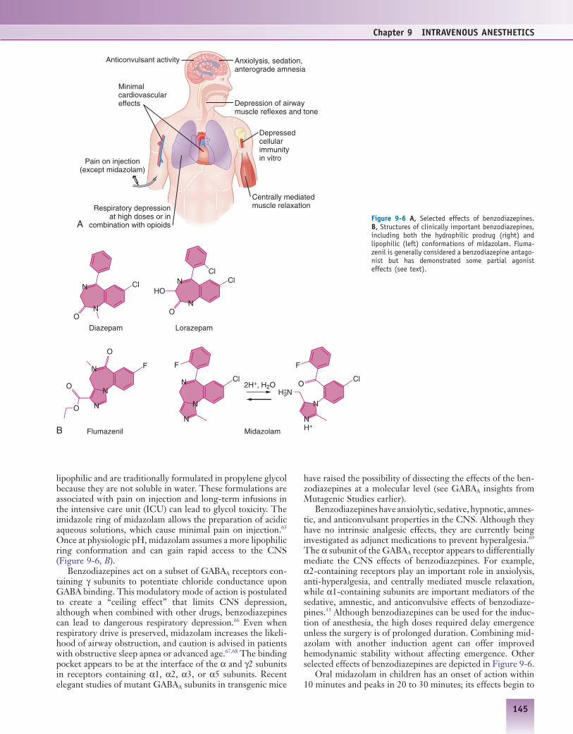
Benzodiazepines have anxiolytic, sedative, hypnotic, amnes-tic, and anticonvulsant properties in the CNS. Although they have no intrinsic analgesic effects, they are currently being investigated as adjunct medications to prevent hyperalgesia.69 The α subunit of the GABAA receptor appears to differentially mediate the CNS effects of benzodiazepines. For example, α2-containing receptors play an important role in anxiolysis, anti-hyperalgesia, and centrally mediated muscle relaxation, while α1-containing subunits are important mediators of the sedative, amnestic, and anticonvulsive effects of benzodiaze-pines.33 Although benzodiazepines can be used for the induc-tion of anesthesia, the high doses required delay emergence unless the surgery is of prolonged duration. Combining mid-azolam with another induction agent can offer improved hemodynamic stability without affecting emergence. Other selected effects of benzodiazepines are depicted in Figure 9-6.
Oral midazolam in children has an onset of action within 10 minutes and peaks in 20 to 30 minutes; its effects begin to
lipophilic and are traditionally formulated in propylene glycol because they are not soluble in water. These formulations are associated with pain on injection and long-term infusions in the intensive care unit (ICU) can lead to glycol toxicity. The imidazole ring of midazolam allows the preparation of acidic aqueous solutions, which cause minimal pain on injection.65 Once at physiologic pH, midazolam assumes a more lipophilic ring conformation and can gain rapid access to the CNS (Figure 9-6, B).
Benzodiazepines act on a subset of GABAA receptors con-taining γ subunits to potentiate chloride conductance upon GABA binding. This modulatory mode of action is postulated to create a “ceiling effect” that limits CNS depression, although when combined with other drugs, benzodiazepines can lead to dangerous respiratory depression.66 Even when respiratory drive is preserved, midazolam increases the likeli-hood of airway obstruction, and caution is advised in patients with obstructive sleep apnea or advanced age.67,68 The binding pocket appears to be at the interface of the α and γ2 subunits in receptors containing α1, α2, α3, or α5 subunits. Recent elegant studies of mutant GABAA subunits in transgenic mice
Figure 9-6 A, Selected effects of benzodiazepines. B, Structures of clinically important benzodiazepines, including both the hydrophilic prodrug (right) and lipophilic (left) conformations of midazolam. Fluma-zenil is generally considered a benzodiazepine antago-nist but has demonstrated some partial agonist effects (see text).
Diazepam Lorazepam
MidazolamFlumazenil
Respiratory depressionat high doses or in
combination with opioids
Anticonvulsant activity
Pain on injection(except midazolam)
Anxiolysis, sedation,anterograde amnesia
Depression of airwaymuscle reflexes and tone
Minimalcardiovasculareffects
Depressedcellularimmunityin vitro
Centrally mediatedmuscle relaxation
Cl NN
NH3N
N NH+
NN
O
HO
O
Cl Cl
Cl
F
O
F
2H+, H2O
A
B
Cl
N
N
N
F
O
O
O
N
N
+
Section II NERVOUS SYSTEM
146
Etomidate
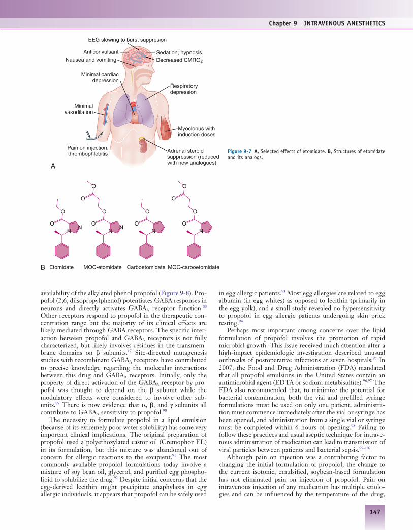
Etomidate is a rapidly acting intravenous agent that was introduced into clinical practice in the 1970s. Compared with other induction agents, it has minimal effects on the cardio-vascular system.79 Because of its association with nausea and prolonged suppression of adrenocortical synthesis of steroids, its main clinical use is for inductions in which hemodynamic stability is essential.80 The R(+) isomer has much greater hyp-notic effects (Figure 9-7), and it is formulated as a single enantiomer. Like propofol, etomidate interacts with GABAA receptors in a stereoselective manner.81 Its enhancement of GABA-mediated current is smaller on receptors containing the β1 subunit.82 Of all of the clinically used intravenous anesthetics, etomidate exhibits the greatest selectivity for GABAA receptors and has the fewest relevant interactions with other ion channels (see Table 9-1).
Following intravenous injection, etomidate is tightly bound to plasma proteins such as albumin. The uncharged drug is highly lipophilic so etomidate rapidly penetrates the blood-brain barrier; peak brain levels are achieved within 2 minutes of injection (see Figure 9-5, A). Etomidate is metabo-lized in the liver by ester hydrolysis to a pharmacologically inactive metabolite.
The effects of etomidate on the CNS are similar to those of propofol and the barbiturates. Induction doses are associ-ated with a high incidence of myoclonus, possibly via a loss of cortical inhibition during the transition from consciousness to unconsciousness. Although this myoclonic activity could be mistaken for generalized tonic-clonic seizures, etomidate has anticonvulsant activity in several experimental models. Epi-leptic attacks occur less frequently during etomidate anesthe-sia but it is likely that propofol and thiopental possess greater anticonvulsant effects; etomidate is therefore a viable option for electroconvulsive therapy.
Etomidate causes less depression of ventilation compared to the barbiturates (see Figure 9-7).83,84 Despite its favorable hemodynamic profile it should be noted that patients with high sympathetic tone such as those suffering from shock, intoxica-tion or drug withdrawal can have a precipitous drop in blood pressure even when etomidate is used to induce anesthesia.
In 1983, investigators reported increased mortality in ICU patients sedated for days with etomidate.85 The increased mortality was attributed to suppression of cortisol synthesis since etomidate is a potent inhibitor of the synthetic enzyme 11β-hydroxylase in the adrenal cortex. The original retro-spective study has been criticized for failing to control for the severity of illness and for the potential role played by concurrent administration of adjuncts to ICU sedation (e.g., opioids) in those patients. Randomized controlled trials in elective cardiac surgery and critically ill patients verified the adrenal suppression but did not show differences in clinical outcome.80,86 Nevertheless, a recent meta-analysis reported a weak association between etomidate use and mortality in criti-cally ill patients, and new etomidate analogs with rapid metab-olism are currently under development to avoid endocrine disturbance (see Emerging Developments).87
Propofol
The most important factor in increasing use of total intrave-nous anesthesia (TIVA) since the late 1980s has been the
dissipate 45 minutes after administration. These kinetics require some planning to get the child to the operating room within the window of efficacy. As shown in Figure 9-5, A, intravenous midazolam does not reach peak effect site con-centration until nearly 10 minutes after administration. When titrating midazolam, one must therefore be patient to avoid “stacking” the doses and oversedating the patient. The onset of intravenous diazepam is more than twice as rapid as that of midazolam, but its use is limited by an extremely long duration of action. Not only is the elimination half-life 10 times longer than that of midazolam, but about half of the parent drug is metabolized to the active compound desmeth-yldiazepam, which has an even longer elimination half-life. Both midazolam and diazepam are metabolized by CYP3A family members in the liver and are subject to interactions with erythromycin and antifungal medications. Diazepam is also metabolized by CYP1A2 and CYP2C19.
Premedication is currently the main role of benzodiaze-pines in anesthesia; a majority of U.S. practitioners surveyed in the 1990s used intravenous midazolam for adults (>70%) and oral midazolam for children (80%).70 For patients requir-ing long-term mechanical ventilation, combinations of an opioid and benzodiazepine have been the traditional method of sedation. Although midazolam is often considered a short-acting benzodiazepine, prolonged infusions in critically ill patients leads to markedly delayed awakening in part due to accumulation of its active metabolite 1-hydroxymidazolam (see Figure 9-5, B and C). For this reason, recent Society for Critical Care Medicine (SCCM) sedation guidelines recom-mend limiting midazolam use to 2 to 3 days.71 For longer periods of sedation, the benzodiazepine of choice is loraze-pam, but some formulations of lorazepam are formulated in a glycol-based vehicle and prolonged infusions lead to glycol toxicity. An emerging body of literature links benzodiazepine use to delirium in critically ill patients.72,73 Because delirium increases time on mechanical ventilation, length of stay, and morbidity and mortality, there has been a movement toward other sedatives in the ICU. Benzodiazepines also have some immunomodulatory effects on monocytes and T cells in vitro, but the clinical significance of these effects is not clear.74,75
An important and unique feature of the benzodiazepines compared to the other GABA-ergic sedative hypnotics is that a competitive antagonist for the reversal of benzodiazepine effects is available. As an intravenous rescue agent, flumazenil can rapidly reverse the CNS depression associated with ben-zodiazepine intoxication. Routine administration of flumaze-nil to patients presenting to the emergency room with the suspicion of overdose can lead to seizures via precipitating acute withdrawal in chronic benzodiazepine users.76 But, there is some evidence that flumazenil has anticonvulsant properties suggesting that this drug may have mixed or partial agonist effects on the GABAA receptor, even in the absence of benzodiazepine administration.77 In support of this phe-nomenon is the potential for high doses of flumazenil to potentiate the hypnosis of other positive GABA modulators such as propofol.78 Its use to augment recovery from general anesthesia is being explored with mixed results (see Emerging Developments). While flumazenil can be life saving in cases of benzodiazepine overdose, its short acting kinetic profile creates the possibility of resedation after the effects of fluma-zenil dissipate.
Chapter 9 INTRAVENOUS ANESTHETICS
147
in egg allergic patients.93 Most egg allergies are related to egg albumin (in egg whites) as opposed to lecithin (primarily in the egg yolk), and a small study revealed no hypersensitivity to propofol in egg allergic patients undergoing skin prick testing.94
Perhaps most important among concerns over the lipid formulation of propofol involves the promotion of rapid microbial growth. This issue received much attention after a high-impact epidemiologic investigation described unusual outbreaks of postoperative infections at seven hospitals.95 In 2007, the Food and Drug Administration (FDA) mandated that all propofol emulsions in the United States contain an antimicrobial agent (EDTA or sodium metabisulfite).96,97 The FDA also recommended that, to minimize the potential for bacterial contamination, both the vial and prefilled syringe formulations must be used on only one patient, administra-tion must commence immediately after the vial or syringe has been opened, and administration from a single vial or syringe must be completed within 6 hours of opening.98 Failing to follow these practices and usual aseptic technique for intrave-nous administration of medication can lead to transmission of viral particles between patients and bacterial sepsis.99-102
Although pain on injection was a contributing factor to changing the initial formulation of propofol, the change to the current isotonic, emulsified, soybean-based formulation has not eliminated pain on injection of propofol. Pain on intravenous injection of any medication has multiple etiolo-gies and can be influenced by the temperature of the drug,
availability of the alkylated phenol propofol (Figure 9-8). Pro-pofol (2,6, diisopropylphenol) potentiates GABA responses in neurons and directly activates GABAA receptor function.88 Other receptors respond to propofol in the therapeutic con-centration range but the majority of its clinical effects are likely mediated through GABA receptors. The specific inter-action between propofol and GABAA receptors is not fully characterized, but likely involves residues in the transmem-brane domains on β subunits.17 Site-directed mutagenesis studies with recombinant GABAA receptors have contributed to precise knowledge regarding the molecular interactions between this drug and GABAA receptors. Initially, only the property of direct activation of the GABAA receptor by pro-pofol was thought to depend on the β subunit while the modulatory effects were considered to involve other sub-units.89 There is now evidence that α, β, and γ subunits all contribute to GABAA sensitivity to propofol.90
The necessity to formulate propofol in a lipid emulsion (because of its extremely poor water solubility) has some very important clinical implications. The original preparation of propofol used a polyethoxylated castor oil (Cremophor EL) in its formulation, but this mixture was abandoned out of concern for allergic reactions to the excipient.91 The most commonly available propofol formulations today involve a mixture of soy bean oil, glycerol, and purified egg phospho-lipid to solubilize the drug.92 Despite initial concerns that the egg-derived lecithin might precipitate anaphylaxis in egg allergic individuals, it appears that propofol can be safely used
Figure 9-7 A, Selected effects of etomidate. B, Structures of etomidate and its analogs.
Pain on injection,thrombophlebitis
Respiratorydepression
Decreased CMRO2
Minimal cardiacdepression
Minimalvasodilation
Etomidate MOC-etomidate Carboetomidate MOC-carboetomidate
AnticonvulsantNausea and vomiting
Sedation, hypnosis
Adrenal steroidsuppression (reducedwith new analogues)
Myoclonus withinduction doses
A
B
NN
O
O
NN
O
O
O
O
NO
O
NO
O
O
O
EEG slowing to burst suppresion

Section II NERVOUS SYSTEM
148
the drug.107 Like most of the intravenous anesthetics, the offset of the hypnotic effect after bolus administration occurs mainly through redistribution of propofol from the brain to less well-perfused sites. Plasma concentrations for inducing unconsciousness are 2 to 2.5 µg/mL and for maintenance of anesthesia are 2 to 6 µg/mL. The elimination half-life of propofol is prolonged because of the slow mobilization from adipose tissue. Regardless, obesity should not be considered a contraindication to its use for induction or maintenance. Obesity does not drastically prolong recovery and dose adjust-ments based on lean body weight have been suggested.108 However, there is some controversy about this as other groups have demonstrated total body weight as the best predictor of propofol pharmacokinetics.109
Moderate hepatic or renal impairment has little effect on the duration of clinical effect. The dose requirement for pro-pofol is reduced in the elderly because of reduced metabolic clearance of drugs and reduced relative volume of the central compartment. Dosing is increased in pediatric populations because relative central compartment volume is larger and clearance and metabolism are increased.
Propofol has a number of advantages over other intrave-nous agents for induction and maintenance of anesthesia. It depresses airway reflexes, reduces nausea, does not induce adrenocortical suppression, and has a short context-sensitive half-time even after prolonged infusion (see Figure 9-5, C).110,111 Simulations of bolus kinetics indicate that it is second only to ketamine in speed of onset and offset (see Figure 9-5, A).
the site of administration, the size of the vein, the speed of injection, and the rate of infusion of the carrier fluid. Some common hyperosmolar formulations of diazepam and etomi-date also cause pain on injection and this pain is often reduced with formulations that are closer to blood in osmolarity.103,104 However, the modern formulation of propofol is not hyper-osmolar and has a pH in the physiologic range. It is thought that the pain on injection of propofol is related to its free aqueous concentration, because the pain on injection appears to be less when the propofol concentration is reduced in its current formulation.105 Coadministration of lidocaine (either as a pretreatment or mixed with propofol) can reduce the incidence of pain on injection, although other strategies have been suggested.106
The rapid effects of propofol on the brain make it a fre-quent choice for sedation in monitored anesthesia care (MAC) settings and for induction and maintenance of general anes-thesia. Its rapid metabolic clearance is useful in pediatric pro-cedures like magnetic resonance imaging (MRI), in the critical care arena for sedation during mechanical ventilation, and in neuroanesthesia to temporarily reduce cerebral metabolic rate (i.e., burst suppression or isoelectric EEG). Propofol also possesses unique antiemetic qualities that are beneficial for ambulatory procedures.
Propofol is metabolized via conjugation into inactive metabolites, however its clearance (total body clearance esti-mated at 25 mL/kg/min) exceeds that of hepatic blood flow and continues even during the anhepatic phase of liver trans-plantation; this argues for some extrahepatic metabolism of
Figure 9-8 A, Selected effects of propofol. B, Structures of propofol and the water-soluble prodrug fospropofol.
Bronchodilation,respiratory depression
Decreasedvascular
resistance
Decreased CMRO2
Sedation, hypnosis
Propofol Fospropofol
Anticonvulsant
Antiemesis
Depression of airwaymuscle reflexes and tone
Myocardialdepression
Rhabdomyolysis withinfusion syndrome (rare)
Hypertriglyceridemia withprolonged infusionsA
B
OH
O
O
O O–
O–P
Pain oninjection
EEG slowing to burst suppresion

Chapter 9 INTRAVENOUS ANESTHETICS
149
renal failure, and hemodynamic instability, known as propofol infusion syndrome. These complications are not common but patients should be monitored closely for propofol infusions greater than 48 hours. Although moderate-dose infusions (>40 µg/kg/min) can increase triglyceride levels in critically ill patients after just 3 days, low-dose propofol infusions (<33 µg/kg/min) showed no detectable increase in triglycer-ides after 2 weeks of constant infusion.125
It is difficult to imagine the practice of anesthesia without propofol. It is also essential outside of the operating room in settings such as the ICU, pediatric imaging procedures, and endoscopy suites. Propofol has numerous advantages (see Figure 9-8), but its depression of respiration makes it poten-tially dangerous in unskilled hands. Propofol appears to have greater relaxation effects on the pharyngeal musculature than thiopental and should be administered only by persons trained in the rescue of patients who experience deeper-than-intended sedation, including general anesthesia, (and who are not involved in the conduct of the procedure).110
Currently, no widely accepted method is available to anes-thesia providers in the United States that allows estimates of blood concentrations of intravenous drugs being infused for maintenance of anesthesia during TIVA. This presents a dis-advantage of intravenous anesthetics in comparison to inhaled anesthetics which are typically used in conjunction with mon-itors equipped with gas sampling for measurements of expired concentrations. Using computer-based models of drug phar-macokinetics, mathematical modeling of a drug’s disposition can provide a convenient way to estimate these blood concen-trations of intravenous agents.126 Target-controlled infusion devices (e.g., Diprifusor) have been used successfully in main-taining anesthesia for both inpatient and outpatient surgery.127 Despite obvious advantages of these devices in predicting blood concentrations some controversy exists in regard to which pharmacokinetic model should be used.128,129 Another impediment to widespread use of these devices has been the unfamiliarity of providers in targeting an actual blood con-centration range, as it is more common to think in terms of infusion rates rather than blood concentrations for infusions of other medications in the hospital.130 There has been some attempts to improve the adoption of these devices by target-ing depth of anesthesia using a processed EEG device rather than targeting a desired blood concentration (see Emerging Developments).131
Ketamine
Ketamine, an arylcyclohexylamine related to phencyclidine, was developed in the 1960s and approved for use in the United States in 1970. Early test subjects described a sense of disconnection from their environment, leading to the term “dissociative anesthesia.”132 Its combination of hypnosis and analgesia showed great promise as a complete anesthetic with minimal effects on cardiovascular function, respiratory drive, and airway reflexes. Concern over psychologic side effects such as hallucinations and emergence delirium limited its clinical use as a primary anesthetic agent, but there is an increasing body of evidence supporting the use of subanes-thetic doses for treatment of acute and chronic pain (see Emerging Developments).
Ketamine was originally produced as a racemic mixture and most commercial preparations continue to be a racemic
The primary disadvantage of propofol is its depressive effect on the cardiovascular system. Patients who are hypovolemic, debilitated, or reliant on high sympathetic tone to maintain blood pressure require careful titration of propofol to avoid severe hypotension. Animal models of hemorrhagic shock suggest that the induction dose should be reduced to between 10% and 20% of usual doses if given before fluid resuscitation and decreased 50% if given after resuscitation.112 Use of pro-pofol in patients with cardiac tamponade or critical aortic stenosis can result in hemodynamic collapse. Mixtures of low-dose ketamine infusions (10-20 µg/kg/min) with propofol infusions (100-200 µg/kg/min) have been used to mitigate the cardiovascular effects of both drugs, especially in pediatric anesthesia.113 However, there is no improvement in respiratory complications compared with using propofol alone.114
Early clinical trials in the late 1980s and early 1990s con-sistently reported faster time to eye-opening and other recov-ery criteria for propofol-based anesthetics compared with “traditional” regimens of thiopental and volatile agents such as enflurane or isoflurane. More recent studies comparing propofol-based techniques to newer inhaled agents with lower blood and tissue solubility (desflurane and sevoflurane) have either failed to show benefit or have shown only minimal benefits in speed of recovery.115 Propofol-based TIVA prob-ably confers advantages over volatile agents in anesthetic maintenance for patients at high risk for postoperative nausea and vomiting.111 It is beneficial not only for patients at high risk for malignant hyperthermia or postoperative nausea and vomiting but it also led to improved operating conditions for endoscopic sinus surgery and decreased pain and opioid con-sumption in gynecologic procedures.116,117
Anesthetic depth is an important consideration when com-paring propofol-based TIVA techniques with volatile anes-thesia. The concentration of expired anesthetic gases measured by modern anesthesia monitors correlates with the concentra-tion of inhaled agent in the CNS.118 Because propofol blood concentrations are not easily measured in the operating room, many practitioners choosing TIVA with propofol as their maintenance anesthetic attempt to control for individual vari-ability in drug clearance through the use of an EEG-based depth of anesthesia monitor becasue it has been proven to decrease anesthetic use and facilitate postoperative recov-ery.119 Favorable pharmacokinetic properties and effects on cerebral blood flow (CBF) and cerebral metabolic require-ment for oxygen (CMRO2) have made propofol popular for neuroanesthesia. In animal studies, reduction of CBF occurs even when mean arterial blood pressure is held constant, and propofol can reduce ICP even when cerebral perfusion pres-sure is fixed.120 Cerebral autoregulation also appears to be preserved in the setting of propofol anesthesia.121 In compari-son, the volatile agents tend to increase ICP and at high doses compromise cerebral autoregulation. Although propofol and thiopental have similar effects on brain physiology, propofol might suppress apoptosis in models of ischemia in vitro (see Chapter 8).122-124
The latest Society for Critical Care Medicine sedation guidelines recommended the use of propofol when rapid or frequent interruptions of sedation are required, such as serial neurologic evaluations.71 Awakening times were similar after 24-, 48-, 72-, and 96-hour constant rate infusions of propofol. Prolonged, high-dose infusions can lead to hypertriglyceride-mia or a constellation of metabolic acidosis, rhabdomyolysis,

Section II NERVOUS SYSTEM
150
preservation of respiratory drive, and maintenance of respira-tory muscle tone.140,141 It produces direct relaxation of airway smooth muscle ex vivo but may also cause bronchodilation indirectly through the modulation of vagal tone.142,143 Ket-amine has a number of other effects detailed in Figure 9-9.
Compared to other intravenous anesthetics, ketamine has relatively low plasma protein binding. This, in conjunction with its lipid solubility, allows rapid accumulation in the CNS. Intravenous bolus administration leads to peak effect site con-centration within a minute (see Figure 9-5, A). Peak effects for other routes of administration are 10 to 15 minutes for intramuscular injection and 15 to 30 minutes following oral administration. Hepatic cytochrome P450 enzymes (espe-cially CYP3A4) metabolize ketamine to norketamine by N-methylation in a perfusion-limited manner. Extensive first-pass metabolism leads to less than 20% bioavailability of orally administered ketamine. Norketamine is roughly one-third as potent as ketamine; its contribution to clinical effects appears relatively more significant in settings of long infusions or chronic use.
Although ketamine continues to play a major role in peri-operative care in developing nations, concerns over side effects have limited its use in modern operating rooms to special situations. Low doses are frequently used to augment regional anesthesia during caesarean sections when the regional block is inadequate. It can be used for inductions in patients with asthma or hypovolemia; in a randomized con-trolled trial of acutely ill patients requiring rapid sequence intubation it was as effective as etomidate.80 Ketamine is sometimes used in critical care in cases of status asthmaticus as an adjunctive bronchodilator, and as an analgesic for dress-ing changes in the burn ICU.
The role of ketamine in patients with neurologic injury is controversial. Its blockade of NMDA receptors could attenu-ate excitotoxicity, but animal studies have demonstrated
mix of the R and S enantiomers (see Chapter 2). S(+) ket-amine, a single enantiomer formulation that is available in some areas of Europe and South America, is more potent in most clinical and experimental settings (Figure 9-9).
Despite more than 40 years of clinical use, the key site or sites of ketamine action have not been fully elucidated. The diverse effects of ketamine are likely mediated by a variety of receptors and signaling pathways. Landmark studies in the 1980s identified ketamine as an NMDA receptor antagonist in the spinal cord and brain.133,134 Ketamine binds uncompeti-tively within the ion channel pore in a use-dependent fashion, but there is some evidence for allosteric binding as well.39,135 Experimental data support a role for NMDA receptors in mediating analgesia, but their role in ketamine anesthesia is unclear.46,47 Ketamine also inhibits nicotinic acetylcholine receptors at clinically relevant concentrations, which could contribute to analgesia but does not appear to influence its sedative properties.136 Ketamine has some local anesthetic properties on Na+ channels and binds µ- and κ-opioid recep-tors.137 Although blunted responses to ketamine have been noted in µ-opioid receptor knockout mice, the limited effect of naloxone on ketamine actions in humans suggests a limited role for these receptors. Finally, knockout of the HCN1 receptor in mice abolishes the hypnotic effect of ketamine and propofol but not of etomidate.138 Further research is needed to confirm the significance of this pathway in humans.
In the brain, ketamine has a unique profile combining depressed consciousness with increased sympathetic tone. This sympathetic activation leads to increases in cardiac output in patients with normal catecholamine stores, which can make ketamine a useful induction agent in patients with hypovolemia. In patients with shock and depleted catechol-amine reserves, the inability to mount a sympathetic surge unmasks a direct myocardial depressant effect of ketamine.139 Respiratory effects of ketamine include bronchodilation,
Figure 9-9 A, Selected effects of ketamine. B, Structures of S(+) ketamine and its parent drug phencyclidine. Ketamine is usually supplied as a racemic mixture but the more active S(+) isomer is available in some countries.
Phencyclidine S(+)-Ketamine
Bronchodilationwith preservedrespiratory drive
Increased vascularresistance from
sympathetic outflow
Dissociative sedation,possible emergence
delirium
Supraspinal analgesia
Spinal analgesia
Nystagmus
Increased salivation
Indirect myocardialstimulation (predominant),direct myocardial depression
Cl
NN
H
O
A
B
Chapter 9 INTRAVENOUS ANESTHETICS
151
with cardiac disease there is a potential benefit of decreased myocardial demand in response to painful stimuli. A recent meta-analysis found decreased mortality in vascular surgery patients, but with increased bradycardia and hypotension.153 Bradycardia can be dangerous in the setting of heart block and dexmedetomidine should be used with caution in this setting. Significant hypotension is more likely in patients with high sympathetic tone such as diabetics, the elderly, and those with hypovolemia. Selected other effects are shown in Figure 9-10.
Dexmedetomidine is metabolized in the liver primarily by glucuronidation with a small portion undergoing hydroxyl-ation by CYP2A6. The distribution half-life is 6 to 8 minutes and its lipophilicity leads to a high volume of distribution. The context-sensitive half-times range from 25 to 120 minutes for a 1-hour infusion to 87 to 250 minutes for infusions greater than 6 hours (Figure 9-11).154-156
Dexmedetomidine sedation is increasingly used in the ICU and is associated with less delirium than benzodiazepine seda-tion.157,158 Dexmedetomidine may reduce delirium in part because its sedation closely mimics physiologic sleep in adults and children.159,160 The ability to extubate patients without weaning dexmedetomidine can facilitate earlier extubation in those patients who become agitated when sedation is held. In the United States, dexmedetomidine is approved for infusions up to 24 hours, but much longer infusions have been reported in the literature. There are concerns for withdrawal phenom-ena for prolonged infusions but no clear withdrawal syndrome has been reported.
neurotoxic effects on the developing brain.144 The tradeoffs are presently unclear but recent German guidelines have out-lined a role for ketamine in brain injured patients whose PaCO2 is being controlled with mechanical ventilation.145 The sympathomimetic properties of ketamine allow sedation with preserved or augmented blood pressure to improve cere-bral perfusion. In general, however, ketamine has traditionally been avoided in neuroanesthesia (see Chapter 8).
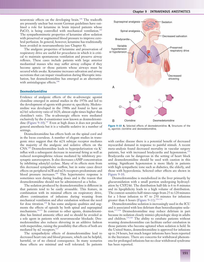
The analgesic properties of ketamine and preservation of respiratory drive are useful for procedures in which it is criti-cal to maintain spontaneous ventilation and preserve airway reflexes. These cases include patients with large anterior mediastinal masses who may suffer airway collapse if they become apneic or those patients whose airways must be secured while awake. Ketamine increases salivation and airway secretions that can impair visualization during fiberoptic intu-bation, but dexmedetomidine has emerged as an alternative with antisialagogue effects.146
Dexmedetomidine
Evidence of analgesic effects of the α-adrenergic agonist clonidine emerged in animal studies in the 1970s and led to the development of agents with greater α2 specificity. Medeto-midine was developed in the 1980s and found to have an α2/α1 selectivity ratio of 1620, almost eight times higher than clonidine’s ratio. The α-adrenergic effects were mediated exclusively by the d-enantiomer now known as dexmedetomi-dine (Figure 9-10).147 Even at high doses it does not produce general anesthesia but it is a valuable sedative in a number of settings.
Dexmedetomidine has effects both on the spinal cord and in the locus coeruleus. A number of elegant studies in trans-genic mice suggest that the α2A subtype receptor mediates the majority of the analgesic and sedative effects on the CNS.148 Dexmedetomidine leads to hyperpolarization via K+ influx with a subsequent decrease in neuronal firing as well as reduced norepinephrine release mediated by inhibitory pre-synaptic autoreceptors. It also decreases cAMP concentration by inhibiting adenylyl cyclase. Many of its effects stem from this decreased sympathetic outflow, but in some cases direct effects on peripheral α2B and α2A receptors predominate and blood pressure increases.149 This hypertensive response is sometimes seen during loading doses and is the reason that dexmedetomidine should not be administered as a bolus.
The sedation produced by dexmedetomidine is different in that patients tend to be easily arousable. This feature, in combination with its minimal effects on respiratory drive, allows continuation of infusions during weaning from mechanical ventilation and after extubation without the need for dose titration.150 It has some analgesic qualities and aug-ments the effects of opioids by both spinal and supraspinal mechanisms.54,151 In contrast to other agents, dexmedetomi-dine has limited amnestic effect and so should be avoided as a sole agent in patients with neuromuscular blockade. Dex-medetomidine also reduces shivering in an additive fashion with meperidine, raising the possibility that effects of both are mediated by α2 receptors.152
The sympatholytic effects of dexmedetomidine lead to decreased heart rate and blood pressure, which can be helpful, harmful, or of no clinical consequence. In many scenarios these effects are minimal and well tolerated. In patients
Figure 9-10 A, Selected effects of dexmedetomidine. B, Structures of the α2 agonists clonidine and dexmedetomidine.
Clonidine Dexmedetomidine
Variablehypertension
or hypotension
Preservedrespiratory drive
Supraspinal analgesia
Spinal analgesia
Sedation
Decreased salivationBradycardia
Decreasedshivering
A
B
N
NN
Cl Cl
NN

Section II NERVOUS SYSTEM
152
intravenously administered drugs.164,165 Commonly referred to as closed-loop anesthesia delivery systems (CLADS), these devices are typically associated with feedback controls based on predicting depth of hypnosis via EEG analysis or other brain monitor (e.g., auditory evoked potentials).166 Recently, a Canadian group has developed an automated system that titrates dose based on three separate parameters: pain, muscle relaxation, and depth of hypnosis (measured via EEG analy-sis).167 Several studies have shown some benefit to CLADS over manual infusion rates and open-loop target controlled infusions, but there is some controversy regarding their eco-nomic efficiency and the choice of physiologic parameters to which dosage is titrated.131,166,168,169 Controversy surrounding the supervision of these automated devices for use in endo-scopic and other office-based procedures will also necessitate discussions regarding the safety of traditional anesthesia drugs in the hands of nonanesthesiologists.
Novel Sedatives
The rapid growth in outpatient and office-based procedures has stimulated the development of several new drugs. One example is fospropofol, a water-soluble prodrug of propofol. Circulating phosphatases metabolize the drug into propofol, formaldehyde, and a phosphate group (Figure 9-12, A).170 It was envisioned during the drug development process that the slower onset of drug effect associated with the metabolism of the propofol prodrug would allow safe bolus administration in the setting of procedural sedation. However, the FDA product labeling ultimately recommended that fospropofol should only be administered by persons trained in general anesthesia as unanticipated transitions to general anesthesia remain a possibility. The formaldehyde levels measured after routine fospropofol administration are indistinguishable from baseline levels.171 After administration of the drug, formaldehyde is rapidly metabolized to formate. Although high formate levels can lead to metabolic acidosis, there has been no reported formate toxicity from fospropofol. Other supposed advantages from this alternative formulation of propofol are decreased risk of bacteremia, decreased risk of hyperlipidemia, and decreased pain on injection, although pruritus and perineal paresthesias are reported side effects of its administration.172
Other intravenous anesthetics have been designed to mimic the rapid, end-organ independent metabolism of drugs such as remifentanil. The benzodiazepine CNS-7056, also known as remimazolam, undergoes ester hydrolysis to form a carboxylic acid metabolite which is 300 times less potent (see Figure 9-12, B).173 It may offer sedation with less postproce-dure cognitive effects than midazolam and is now being inves-tigated in humans.
A similar strategy has been applied to modify etomidate and reduce its adrenal suppression effects. Investigators have developed an ultrarapid metabolizing etomidate named methoxycarbonyletomidate (MOC-etomidate), which is broken down via nonspecific esterases (see Figure 9-12, C).174 The in vitro half-life of MOC-etomidate is approximately one-tenth that of its parent compound etomidate. The rapid metabolism of MOC-etomidate into a carboxylic acid metab-olite, which has low affinity for 11β-hydroxylase, results in less adrenocortical suppression than its parent compound.175 This compound, as well as carboetomidate (see Figure 9-7), a pyrrole analog of etomidate specifically designed not to
Controversy over the use of propofol by non-anesthesiologists has prompted a number of studies of dex-medetomidine as an alternative in sedation practice.161 A small clinical trial suggested it was comparable to propofol but had longer recovery times; conventional doses up to 1 µg/kg/hr often require supplementation with other drugs. One group reported success of dexmedetomidine as a sole agent in over 97% of pediatric MRI scans by escalating the loading dose to 3 µg/kg and the infusion to 2 µg/kg/hr.162
The clinical applications of dexmedetomidine continue to expand. Although initially approved by regulatory agencies only for ICU sedation, dexmedetomidine was recently approved for monitored anesthesia care (MAC) sedation in the United States. The antisialagogue effect of dexmedetomi-dine represents an advantage over ketamine for awake fiber-optic intubations.146 It has also been administered intranasally for premedication of children.163 The specific and distinctive mechanism of action of dexmedetomidine provides an impor-tant alternative amid ongoing uncertainty about short- and long-term cognitive effects of more established anesthetics in both young and old patients.73
EMERGING DEVELOPMENTS
Ongoing efforts to improve the safety of intravenous anes-thetics include both new systems to titrate existing drugs and new compounds with the potential for improved safety profiles.
High-Tech Delivery Systems
The development of a computer-aided device that dynamically adjusts anesthetic administration based on measured physio-logic parameters has been explored for both inhaled164 and
Figure 9-11 Context-sensitive half-times of dexmedetomidine reported in adults and neonates based on various pharmacokinetic models. (Modified from Dyck JB, Maze M, Haack C, et al. Computer-controlled infusion of intra-venous dexmedetomidine hydrochloride in adult human volunteers. Anesthe-siology. 1993;78:821-828; Potts AL, Warman GR, Anderson BJ. Dexmedetomidine disposition in children: a population analysis. Paediatr Anaesth. 2008;18:722-730; and Lin L, Guo X, Zhang MZ, et al. Pharmacokinetics of dexmedetomidine in Chinese post-surgical intensive care unit patients. Acta Anaesthesiol Scand. 2011;55:359-367.)
0 2 4 6 8 10 12
Dyck 1993 Adult Lin 2011 AdultPotts 2008 Neonate Potts 2008 Adult
Infusion duration (hr)
Dex
med
etom
idin
e co
ntex
t-se
nsiti
veha
lf-tim
e (m
in)
0
50
100
150
200
250
300
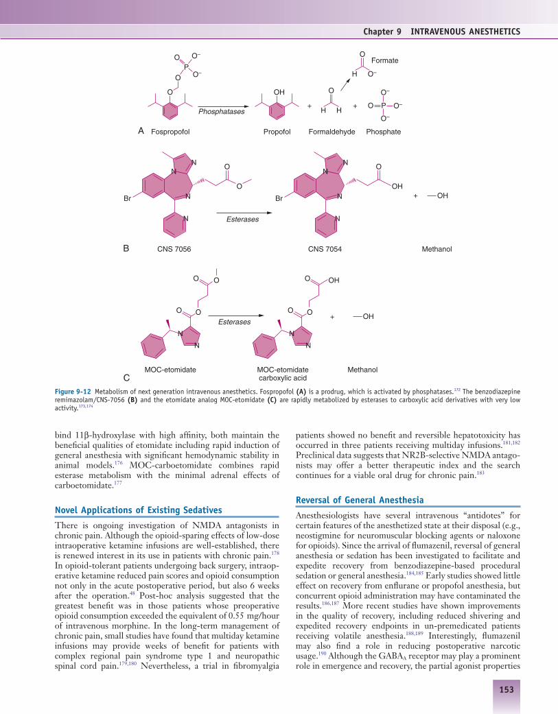
Chapter 9 INTRAVENOUS ANESTHETICS
153
patients showed no benefit and reversible hepatotoxicity has occurred in three patients receiving multiday infusions.181,182 Preclinical data suggests that NR2B-selective NMDA antago-nists may offer a better therapeutic index and the search continues for a viable oral drug for chronic pain.183
Reversal of General Anesthesia
Anesthesiologists have several intravenous “antidotes” for certain features of the anesthetized state at their disposal (e.g., neostigmine for neuromuscular blocking agents or naloxone for opioids). Since the arrival of flumazenil, reversal of general anesthesia or sedation has been investigated to facilitate and expedite recovery from benzodiazepine-based procedural sedation or general anesthesia.184,185 Early studies showed little effect on recovery from enflurane or propofol anesthesia, but concurrent opioid administration may have contaminated the results.186,187 More recent studies have shown improvements in the quality of recovery, including reduced shivering and expedited recovery endpoints in un-premedicated patients receiving volatile anesthesia.188,189 Interestingly, flumazenil may also find a role in reducing postoperative narcotic usage.190 Although the GABAA receptor may play a prominent role in emergence and recovery, the partial agonist properties
bind 11β-hydroxylase with high affinity, both maintain the beneficial qualities of etomidate including rapid induction of general anesthesia with significant hemodynamic stability in animal models.176 MOC-carboetomidate combines rapid esterase metabolism with the minimal adrenal effects of carboetomidate.177
Novel Applications of Existing Sedatives
There is ongoing investigation of NMDA antagonists in chronic pain. Although the opioid-sparing effects of low-dose intraoperative ketamine infusions are well-established, there is renewed interest in its use in patients with chronic pain.178 In opioid-tolerant patients undergoing back surgery, intraop-erative ketamine reduced pain scores and opioid consumption not only in the acute postoperative period, but also 6 weeks after the operation.48 Post-hoc analysis suggested that the greatest benefit was in those patients whose preoperative opioid consumption exceeded the equivalent of 0.55 mg/hour of intravenous morphine. In the long-term management of chronic pain, small studies have found that multiday ketamine infusions may provide weeks of benefit for patients with complex regional pain syndrome type 1 and neuropathic spinal cord pain.179,180 Nevertheless, a trial in fibromyalgia
Figure 9-12 Metabolism of next generation intravenous anesthetics. Fospropofol (A) is a prodrug, which is activated by phosphatases.172 The benzodiazepine remimazolam/CNS-7056 (B) and the etomidate analog MOC-etomidate (C) are rapidly metabolized by esterases to carboxylic acid derivatives with very low activity.173,174
O
A
B
C
O–
O OH O
O
O
O
PO
O–
Fospropofol
Phosphatases+ +
Propofol Formaldehyde Phosphate
Formate
P
O–
O–
O–H H
O
H O–
Br
N
N
NN
O
O
Br
N
N
NN
OHOH
CNS 7056
Esterases
Esterases
+
OH+
CNS 7054 Methanol
OO
O
N
N
OH
OO
O
N
N
MOC-etomidate MOC-etomidatecarboxylic acid
Methanol
Section II NERVOUS SYSTEM
154
of this drug may make it difficult to study as a pure reversal agent of general anesthesia. Studies investigating the role of dopamine and nicotine in anesthesia recovery are also promising.191,192
KEY POINTS
• Intravenous anesthetics are used both to induce and maintain general anesthesia in the operating room, for sedation and anxiolysis preoperatively, for procedures outside the operating room, and in the ICU.
• Potentiation of the effect of the inhibitory neurotransmitter GABA at GABAA receptors is the most important mechanism of action for barbiturates, benzodiazepines, etomidate, and propofol.
• Ongoing molecular studies are dissecting not only the specific binding pockets for intravenous anesthetics but also the receptor subunit combinations that differentially mediate effects such as sedation, anxiolysis, and amnesia.
• Etomidate plays an important role in the induction of anesthesia in patients with significant cardiovascular comorbidities such as congestive heart failure or hemorrhagic shock.
• Propofol-based TIVA has advantages over anesthetic maintenance with volatile anesthetics in some patient populations.
• Ketamine blocks glutamate signaling at NMDA-type glutamate receptors, but its clinical effects are broader and more profound than those of more specific NMDA receptor antagonists.
• Subanesthetic doses of ketamine are useful adjuvants for chronic pain patients and its combination of low cost, analgesia, and preserved respiratory drive make it an important anesthetic, especially in developing nations.
• Dexmedetomidine is a highly specific α2 adrenergic receptor agonist that produces sedation with preserved respiratory drive. Dexmedetomidine has seen an increasing role in perioperative and ICU sedation.
• The combination of intravenous anesthetics can be tailored for each patient and procedure to optimize the key aspects of the anesthetic and minimize toxicity.
Anis NA, Berry SC, Burton NR, Lodge D. The dissociative anaes-thetics, ketamine and phencyclidine, selectively reduce excitation of central mammalian neurones by N-methyl-aspartate. Br J Pharmacol. 1983;79(2):565-575. Representative of several seminal publications by Lodge demonstrating blockade of NMDA action in isolated spinal cord cells as well as after intravenous administra-tion in intact animals. (Ref. 133)
Belelli D, Lambert JJ, Peters JA, Wafford K, Whiting PJ. The interaction of the general anesthetic etomidate with the gamma-aminobutyric acid type A receptor is influenced by a single amino acid. Proc Natl Acad Sci U S A. 1997;94(20):11031-11036. A pio-neering demonstration of manipulating the sensitivity of GABAA receptors to intravenous anesthetics by specific amino-acid sub-stitutions. (Ref. 31)
Cummings GC, Dixon J, Kay NH, et al. Dose requirements of ICI 35,868 (propofol, ‘Diprivan’) in a new formulation for induction of anaesthesia. Anaesthesia. 1984;39(12):1168-1171. First demon-
stration of propofol’s clinical usage in humans with its new soy-based emulsion. (Ref. 92)
Glen JB. Animal studies of the anaesthetic activity of ICI 35 868. Br J Anaesth. 1980;52(8):731-742. Glen’s original characterization of the phenolic compound now known as propofol. Glen would later go on to prepare propofol in the soy-based emulsion suitable for human anesthesia after concerns of adverse reactions to its origi-nal carrier solution (Cremaphor EL) surfaced. (Ref. 91)
Loftus RW, Yeager MP, Clark JA, et al. Intraoperative ketamine reduces perioperative opiate consumption in opiate-dependent patients with chronic back pain undergoing back surgery. Anes-thesiology. 2010;113(3):639-646. In a population of opioid-tolerant patients, sub-anesthetic ketamine infusion decreased pain and opioid consumption not only immediately postoperatively but also 6 weeks after surgery. (Ref. 48)
Lundy JS. The barbiturates as anesthetics, hypnotics and antispas-modics. Anesthesia & Analgesia. 1929;8(1-6):360-364. After verify-ing that barbiturates reduce local anesthetic toxicity in dogs, Lundy outlined their use as part of a balanced anesthetic in over 1000 patients. The lessons gleaned from his progression from oral premedication to sole anesthetic laid the foundation for his later work with thiopental. (Ref. 55)
Pandharipande PP, Pun BT, Herr DL, et al. Effect of sedation with dexmedetomidine vs lorazepam on acute brain dysfunction in mechanically ventilated patients: the MENDS randomized con-trolled trial. JAMA. 2007;298(22):2644-2653. First of the large clinical trials showing a reduction in delirium with dexmedetomi-dine compared to benzodiazepine sedation. (Ref. 157)
Unwin N. Refined structure of the nicotinic acetylcholine receptor at 4A resolution. J Mol Biol. 2005;346(4):967-989. Our current best understanding of the structure of the GABAA receptor is based on homology to this high resolution structure. (Ref. 13)
Vickery RG, Sheridan BC, Segal IS, Maze M. Anesthetic and hemo-dynamic effects of the stereoisomers of medetomidine, an alpha 2-adrenergic agonist, in halothane-anesthetized dogs. Anesth Analg. 1988;67(7):611-615. Demonstrated not only that the racemic compound medetomidine reduces MAC for halothane but also that its effects were exclusively mediated by the D-enantiomer now marketed as dexmedetomidine. (Ref. 47)
Key References
References1. Bergman NA. Early intravenous anesthesia: an eyewitness account.
Anesthesiology. 1990;72(1):185-186.2. Adams RC. Intravenous anesthesia. Anesthesiology. 1947;8(5):489-
496.3. Hatcher RA, Eggleston C. A contribution to the pharmacology of
Novocain. J Pharmacol Exp Ther. 1916;8(Jul):385-405.4. Dundee JW, McIlroy PD. The history of barbiturates. Anaesthesia.
1982;37(7):726-734.5. Hilgenberg JC. Intraoperative awareness during high-dose
fentanyl—oxygen anesthesia. Anesthesiology. 1981;54(4):341-343.6. Lundy J. Balanced anesthesia. Minn Med. 1926;(9):399.7. Alkire MT, Hudetz AG, Tononi G. Consciousness and anesthesia.
Science. 2008;322(5903):876-880.8. Franks NP. General anaesthesia: from molecular targets to neuronal
pathways of sleep and arousal. Nat Rev Neurosci. 2008;9(5):370-386.
9. Sieghart W, Sperk G. Subunit composition, distribution and func-tion of GABA(A) receptor subtypes. Curr Top Med Chem. 2002;2(8):795-816.
10. Antkowiak B, Kirschfeld K. [Neural mechanisms of anesthesia.] Anasthesiol Intensivmed Notfallmed Schmerzther. 2000;35(12):731-743.
11. Collins JG, Kendig JJ, Mason P. Anesthetic actions within the spinal cord: contributions to the state of general anesthesia. Trends Neuro-sci. 1995;18(12):549-553.
12. McGaugh JL, Izquierdo I. The contribution of pharmacology to research on the mechanisms of memory formation. Trends Pharmacol Sci. 2000;21(6):208-210.
13. Unwin N. Refined structure of the nicotinic acetylcholine receptor at 4A resolution. J Mol Biol. 2005;346(4):967-989.

Chapter 9 INTRAVENOUS ANESTHETICS
155
GABA(A) receptor alpha1 subtype. Nat Neurosci. 2000;3(6):587-592.
38. Morris HV, Dawson GR, Reynolds DS, Atack JR, Stephens DN. Both alpha2 and alpha3 GABAA receptor subtypes mediate the anxiolytic properties of benzodiazepine site ligands in the condi-tioned emotional response paradigm. Eur J Neurosci. 2006;23(9):2495-2504.
39. Traynelis SF, Wollmuth LP, McBain CJ, et al. Glutamate receptor ion channels: structure, regulation, and function. Pharmacol Rev. 2010;62(3):405-496.
40. Harris RA, Mihic SJ, Dildy-Mayfield JE, Machu TK. Actions of anesthetics on ligand-gated ion channels: role of receptor subunit composition. FASEB J. 1995;9(14):1454-1462.
41. Duguid IC, Smart TG. Presynaptic NMDA Receptors. In: Van Dongen AM, ed. Biology of the NMDA Receptor. Boca Raton, FL: CRC Press; 2009 [chapter 14].
42. Grasshoff C, Drexler B, Rudolph U, Antkowiak B. Anaesthetic drugs: linking molecular actions to clinical effects. Curr Pharm Des. 2006;12(28):3665-3679.
43. Paoletti P, Neyton J. NMDA receptor subunits: function and phar-macology. Curr Opin Pharmacol. 2007;7(1):39-47.
44. Nicoll RA, Malenka RC. Expression mechanisms underlying NMDA receptor-dependent long-term potentiation. Ann N Y Acad Sci. 1999;868:515-525.
45. Daw NW, Stein PS, Fox K. The role of NMDA receptors in infor-mation processing. Annu Rev Neurosci. 1993;16:207-222.
46. South SM, Kohno T, Kaspar BK, et al. A conditional deletion of the NR1 subunit of the NMDA receptor in adult spinal cord dorsal horn reduces NMDA currents and injury-induced pain. J Neurosci. 2003;23(12):5031-5040.
47. Garraway SM, Xu Q, Inturrisi CE. siRNA-mediated knockdown of the NR1 subunit gene of the NMDA receptor attenuates formalin-induced pain behaviors in adult rats. J Pain. 2009;10(4):380-390.
48. Loftus RW, Yeager MP, Clark JA, et al. Intraoperative ketamine reduces perioperative opiate consumption in opiate-dependent patients with chronic back pain undergoing back surgery. Anesthe-siology. 2010;113(3):639-646.
49. Dickinson R, Peterson BK, Banks P, et al. Competitive inhibition at the glycine site of the N-methyl-D-aspartate receptor by the anesthetics xenon and isoflurane: evidence from molecular model-ing and electrophysiology. Anesthesiology. 2007;107(5):756-767.
50. Grasshoff C, Antkowiak B. Propofol and sevoflurane depress spinal neurons in vitro via different molecular targets. Anesthesiology. 2004;101(5):1167-1176.
51. Ratnakumari L, Hemmings HC Jr. Effects of propofol on sodium channel-dependent sodium influx and glutamate release in rat cere-brocortical synaptosomes. Anesthesiology. 1997;86(2):428-439.
52. Frenkel C, Urban BW. Molecular actions of racemic ketamine on human CNS sodium channels. Br J Anaesth. 1992;69(3):292-297.
53. Ying SW, Abbas SY, Harrison NL, Goldstein PA. Propofol block of I(h) contributes to the suppression of neuronal excitability and rhythmic burst firing in thalamocortical neurons. Eur J Neurosci. 2006;23(2):465-480.
54. Guo TZ, Jiang JY, Buttermann AE, Maze M. Dexmedetomidine injection into the locus ceruleus produces antinociception. Anesthe-siology. 1996;84(4):873-881.
55. Lundy JS. The barbiturates as anesthetics, hypnotics and antispas-modics. AnesthAnalg. 1929;8(1-6):360-364.
56. Gibson WR, Doran WJ, Wood WC, Swanson EE. Pharmacology of stereoisomers of 1-methyl-5-(1-methyl-2-pentynyl)-5-allyl-bar-bituric acid. J Pharmacol Exp Ther. 1959;125(1):23-27.
57. Brunner H, Ittner K-P, Lunz D, Schmatloch S, Schmidt T, Zabel M. Highly enriched mixtures of methohexital stereoisomers by palladium-catalyzed allylation and their anaesthetic activity. Eur J Organic Chem 2003;2003:855-862.
58. Tomlin SL, Jenkins A, Lieb WR, Franks NP. Preparation of barbi-turate optical isomers and their effects on GABA(A) receptors. Anes-thesiology. 1999;90(6):1714-1722.
59. Bieda MC, Su H, Maciver MB. Anesthetics discriminate between tonic and phasic gamma-aminobutyric acid receptors on hippocam-pal CA1 neurons. Anesth Analg. 2009;108(2):484-490.
60. Bennetts FE. Thiopentone anaesthesia at Pearl Harbor. Br J Anaesth. 1995;75(3):366-368.
61. Hughes MA, Glass PS, Jacobs JR. Context-sensitive half-time in multicompartment pharmacokinetic models for intravenous anes-thetic drugs. Anesthesiology. 1992;76(3):334-341.
14. Simon J, Wakimoto H, Fujita N, Lalande M, Barnard EA. Analysis of the set of GABA(A) receptor genes in the human genome. J Biol Chem. 2004;279(40):41422-41435.
15. Mody I, Pearce RA. Diversity of inhibitory neurotransmission through GABA(A) receptors. Trends Neurosci. 2004;27(9):569-575.
16. Sieghart W, Schuster A. Affinity of various ligands for benzodiaz-epine receptors in rat cerebellum and hippocampus. Biochem Phar-macol. 1984;33(24):4033-4038.
17. Richardson JE, Garcia PS, O’Toole KK, Derry JM, Bell SV, Jenkins A. A conserved tyrosine in the beta2 subunit M4 segment is a determinant of gamma-aminobutyric acid type A receptor sensitiv-ity to propofol. Anesthesiology. 2007;107(3):412-418.
18. Jurd R, Arras M, Lambert S, et al. General anesthetic actions in vivo strongly attenuated by a point mutation in the GABA(A) recep-tor beta3 subunit. FASEB J. 2003;17(2):250-252.
19. Zeller A, Arras M, Jurd R, Rudolph U. Identification of a molecular target mediating the general anesthetic actions of pentobarbital. Mol Pharmacol. 2007;71(3):852-859.
20. Laurie DJ, Seeburg PH, Wisden W. The distribution of 13 GABAA receptor subunit mRNAs in the rat brain. II. Olfactory bulb and cerebellum. J Neurosci. 1992;12(3):1063-1076.
21. Wisden W, Laurie DJ, Monyer H, Seeburg PH. The distribution of 13 GABAA receptor subunit mRNAs in the rat brain. I. Telen-cephalon, diencephalon, mesencephalon. J Neurosci. 1992;12(3):1040-1062.
22. Reynolds DS, Rosahl TW, Cirone J, et al. Sedation and anesthesia mediated by distinct GABA(A) receptor isoforms. J Neurosci. 2003;23(24):8608-8617.
23. O’Meara GF, Newman RJ, Fradley RL, Dawson GR, Reynolds DS. The GABA-A beta3 subunit mediates anaesthesia induced by etomi-date. Neuroreport. 2004;15(10):1653-1656.
24. Ching S, Cimenser A, Purdon PL, Brown EN, Kopell NJ. Thala-mocortical model for a propofol-induced alpha-rhythm associated with loss of consciousness. Proc Natl Acad Sci U S A. 2010;107(52):22665-22670.
25. Talavera JA, Esser SK, Amzica F, Hill S, Antognini JF. Modeling the GABAergic action of etomidate on the thalamocortical system. Anesth Analg. 2009;108(1):160-167.
26. Sigel E, Baur R, Kellenberger S, Malherbe P. Point mutations affecting antagonist affinity and agonist dependent gating of GABAA receptor channels. EMBO J. 1992;11(6):2017-2023.
27. Kleingoor C, Wieland HA, Korpi ER, Seeburg PH, Kettenmann H. Current potentiation by diazepam but not GABA sensitivity is determined by a single histidine residue. Neuroreport. 1993;4(2):187-190.
28. Amin J, Weiss DS. GABAA receptor needs two homologous domains of the beta-subunit for activation by GABA but not by pentobarbital. Nature. 1993;366(6455):565-569.
29. Smith GB, Olsen RW. Functional domains of GABAA receptors. Trends Pharmacol Sci. 1995;16(5):162-168.
30. Mihic SJ, Ye Q, Wick MJ, et al. Sites of alcohol and volatile anaes-thetic action on GABA(A) and glycine receptors. Nature. 1997;389(6649):385-389.
31. Belelli D, Lambert JJ, Peters JA, Wafford K, Whiting PJ. The interaction of the general anesthetic etomidate with the gamma-aminobutyric acid type A receptor is influenced by a single amino acid. Proc Natl Acad Sci U S A. 1997;94(20):11031-11036.
32. Siegwart R, Jurd R, Rudolph U. Molecular determinants for the action of general anesthetics at recombinant alpha(2)beta(3)gamma(2)gamma-aminobutyric acid(A) receptors. J Neurochem. 2002;80(1):140-148.
33. Rudolph U, Mohler H. Analysis of GABAA receptor function and dissection of the pharmacology of benzodiazepines and general anesthetics through mouse genetics. Annu Rev Pharmacol Toxicol. 2004;44:475-498.
34. Rudolph U, Crestani F, Benke D, et al. Benzodiazepine actions mediated by specific gamma-aminobutyric acid(A) receptor sub-types. Nature. 1999;401(6755):796-800.
35. Low K, Crestani F, Keist R, et al. Molecular and neuronal substrate for the selective attenuation of anxiety. Science. 2000;290(5489):131-134.
36. Crestani F, Keist R, Fritschy JM, et al. Trace fear conditioning involves hippocampal alpha5 GABA(A) receptors. Proc Natl Acad Sci U S A. 2002;99(13):8980-8985.
37. McKernan RM, Rosahl TW, Reynolds DS, et al. Sedative but not anxiolytic properties of benzodiazepines are mediated by the

Section II NERVOUS SYSTEM
156
87. Albert SG, Ariyan S, Rather A. The effect of etomidate on adrenal function in critical illness: a systematic review. Intensive Care Med. 2011;37(6):901-910.
88. Orser BA, McAdam LC, Roder S, MacDonald JF. General anaes-thetics and their effects on GABA(A) receptor desensitization. Toxicol Lett. 1998;100-101:217-224.
89. Sanna E, Mascia MP, Klein RL, Whiting PJ, Biggio G, Harris RA. Actions of the general anesthetic propofol on recombinant human GABAA receptors: influence of receptor subunits. J Pharmacol Exp Ther. 1995;274(1):353-360.
90. Jones MV, Harrison NL, Pritchett DB, Hales TG. Modulation of the GABAA receptor by propofol is independent of the gamma subunit. J Pharmacol Exp Ther. 1995;274(2):962-968.
91. Glen JB. Animal studies of the anaesthetic activity of ICI 35 868. Br J Anaesth. 1980;52(8):731-742.
92. Cummings GC, Dixon J, Kay NH, et al. Dose requirements of ICI 35,868 (propofol, ‘Diprivan’) in a new formulation for induction of anaesthesia. Anaesthesia. 1984;39(12):1168-1171.
93. Dewachter P, Mouton-Faivre C, Castells MC, Hepner DL. Anes-thesia in the patient with multiple drug allergies: are all allergies the same? Curr Opin Anaesthesiol. 2011;24(3):320-325.
94. Murphy A, Campbell DE, Baines D, Mehr S. Allergic reactions to propofol in egg-allergic children. Anesth Analg. 2011;113(1):140-144.
95. Bennett SN, McNeil MM, Bland LA, et al. Postoperative infections traced to contamination of an intravenous anesthetic, propofol. N Engl J Med. 1995;333(3):147-154.
96. Jansson JR, Fukada T, Ozaki M, Kimura S. Propofol EDTA and reduced incidence of infection. Anaesth Intensive Care. 2006;34(3):362-368.
97. Thompson KA, Goodale DB. The recent development of propofol (DIPRIVAN). Intensive Care Med. 2000;26 Suppl 4:S400-S404.
98. FDA. Information for Healthcare Professionals: propofol (marketed as Diprivan and as generic products). 2007; http://www.fda.gov/Drugs/DrugSafety/PostmarketDrugSafetyInformationforPatients andProviders/ucm125817.htm. Accessed December 16, 2011.
99. Germain JM, Carbonne A, Thiers V, et al. Patient-to-patient trans-mission of hepatitis C virus through the use of multidose vials during general anesthesia. Infect Control Hosp Epidemiol. 2005;26(9):789-792.
100. Fischer GE, Schaefer MK, Labus BJ, et al. Hepatitis C virus infections from unsafe injection practices at an endoscopy clinic in Las Vegas, Nevada, 2007-2008. Clin Infect Dis. 2010;51(3):267-273.
101. Henry B, Plante-Jenkins C, Ostrowska K. An outbreak of Serratia marcescens associated with the anesthetic agent propofol. Am J Infect Control. 2001;29(5):312-315.
102. Muller AE, Huisman I, Roos PJ, et al. Outbreak of severe sepsis due to contaminated propofol: lessons to learn. J Hosp Infect. 2010;76(3):225-230.
103. Klement W, Arndt JO. Pain on I.V. injection of some anaesthetic agents is evoked by the unphysiological osmolality or pH of their formulations. Br J Anaesth. 1991;66(2):189-195.
104. Bretschneider H. Osmolalities of commercially supplied drugs often used in anesthesia. Anesth Analg. 1987;66(4):361-362.
105. Klement W, Arndt JO. Pain on injection of propofol: effects of concentration and diluent. Br J Anaesth. 1991;67(3):281-284.
106. Tan CH, Onsiong MK. Pain on injection of propofol. Anaesthesia. 1998;53(5):468-476.
107. Veroli P, O’Kelly B, Bertrand F, Trouvin JH, Farinotti R, Ecoffey C. Extrahepatic metabolism of propofol in man during the anhe-patic phase of orthotopic liver transplantation. Br J Anaesth. 1992;68(2):183-186.
108. Ingrande J, Brodsky JB, Lemmens HJ. Lean body weight scalar for the anesthetic induction dose of propofol in morbidly obese sub-jects. Anesth Analg. 2011;113(1):57-62.
109. Cortinez LI, Anderson BJ, Penna A, et al. Influence of obesity on propofol pharmacokinetics: derivation of a pharmacokinetic model. Br J Anaesth. 2010;105(4):448-456.
110. McKeating K, Bali IM, Dundee JW. The effects of thiopentone and propofol on upper airway integrity. Anaesthesia. 1988;43(8):638-640.
111. Apfel CC, Korttila K, Abdalla M, et al. A factorial trial of six inter-ventions for the prevention of postoperative nausea and vomiting. N Engl J Med. 2004;350(24):2441-2451.
62. Russo H, Bressolle F. Pharmacodynamics and pharmacokinetics of thiopental. Clin Pharmacokinet. 1998;35(2):95-134.
63. Mustajoki P, Heinonen J. General anesthesia in “inducible” por-phyrias. Anesthesiology. 1980;53(1):15-20.
64. Martin IL. The benzodiazepines and their receptors: 25 years of progress. Neuropharmacology. 1987;26(7B):957-970.
65. Gerecke M. Chemical structure and properties of midazolam com-pared with other benzodiazepines. Br J Clin Pharmacol. 1983;16 Suppl 1:11S-16S.
66. Bailey PL, Pace NL, Ashburn MA, Moll JW, East KA, Stanley TH. Frequent hypoxemia and apnea after sedation with midazolam and fentanyl. Anesthesiology. 1990;73(5):826-830.
67. Gueye PN, Lofaso F, Borron SW, et al. Mechanism of respiratory insufficiency in pure or mixed drug-induced coma involving benzo-diazepines. J Toxicol Clin Toxicol. 2002;40(1):35-47.
68. Sun GC, Hsu MC, Chia YY, Chen PY, Shaw FZ. Effects of age and gender on intravenous midazolam premedication: a randomized double-blind study. Br J Anaesth. 2008;101(5):632-639.
69. Shih A, Miletic V, Miletic G, Smith LJ. Midazolam administration reverses thermal hyperalgesia and prevents gamma-aminobutyric acid transporter loss in a rodent model of neuropathic pain. Anesth Analg. 2008;106(4):1296-1302.
70. Kain ZN, Mayes LC, Bell C, Weisman S, Hofstadter MB, Rimar S. Premedication in the United States: a status report. Anesth Analg. 1997;84(2):427-432.
71. Jacobi J, Fraser GL, Coursin DB, et al. Clinical practice guidelines for the sustained use of sedatives and analgesics in the critically ill adult. Crit Care Med. 2002;30(1):119-141.
72. Pisani MA, Murphy TE, Araujo KL, Slattum P, Van Ness PH, Inouye SK. Benzodiazepine and opioid use and the duration of intensive care unit delirium in an older population. Crit Care Med. 2009;37(1):177-183.
73. Hughes CG, Pandharipande PP. Review articles: the effects of peri-operative and intensive care unit sedation on brain organ dysfunc-tion. Anesth Analg. 2011;112(5):1212-1217.
74. Taupin V, Jayais P, Descamps-Latscha B, et al. Benzodiazepine anes-thesia in humans modulates the interleukin-1 beta, tumor necrosis factor-alpha and interleukin-6 responses of blood monocytes. J Neuroimmunol. 1991;35(1-3):13-19.
75. Wei M, Li L, Meng R, et al. Suppressive effect of diazepam on IFN-gamma production by human T cells. Int Immunopharmacol. 2010;10(3):267-271.
76. Spivey WH. Flumazenil and seizures: analysis of 43 cases. Clin Ther. 1992;14(2):292-305.
77. Scollo-Lavizzari G. The clinical anti-convulsant effects of flumaze-nil, a benzodiazepine antagonist. Eur J Anaesthesiol Suppl. 1988;2:129-138.
78. Adachi YU, Watanabe K, Higuchi H, Satoh T. Flumazenil reduces the hypnotic dose of propofol in male patients under spinal anes-thesia. J Anesth. 2002;16(1):9-12.
79. Ray DC, McKeown DW. Effect of induction agent on vasopressor and steroid use, and outcome in patients with septic shock. Crit Care. 2007;11(3):R56.
80. Jabre P, Combes X, Lapostolle F, et al. Etomidate versus ketamine for rapid sequence intubation in acutely ill patients: a multicentre randomised controlled trial. Lancet. 2009;374(9686):293-300.
81. Tomlin SL, Jenkins A, Lieb WR, Franks NP. Stereoselective effects of etomidate optical isomers on gamma-aminobutyric acid type A receptors and animals. Anesthesiology. 1998;88(3):708-717.
82. Hill-Venning C, Belelli D, Peters JA, Lambert JJ. Subunit-dependent interaction of the general anaesthetic etomidate with the gamma-aminobutyric acid type A receptor. Br J Pharmacol. 1997;120(5):749-756.
83. McCollum JS, Dundee JW. Comparison of induction characteristics of four intravenous anaesthetic agents. Anaesthesia. 1986;41(10):995-1000.
84. Choi SD, Spaulding BC, Gross JB, Apfelbaum JL. Comparison of the ventilatory effects of etomidate and methohexital. Anesthesiology. 1985;62(4):442-447.
85. Ledingham IM, Watt I. Influence of sedation on mortality in criti-cally ill multiple trauma patients. Lancet. 1983;1(8336):1270.
86. Morel J, Salard M, Castelain C, et al. Haemodynamic consequences of etomidate administration in elective cardiac surgery: a random-ized double-blinded study. Br J Anaesth. 2011;107(4):503-509.
Chapter 9 INTRAVENOUS ANESTHETICS
157
137. Hirota K, Okawa H, Appadu BL, Grandy DK, Devi LA, Lambert DG. Stereoselective interaction of ketamine with recombinant mu, kappa, and delta opioid receptors expressed in Chinese hamster ovary cells. Anesthesiology. 1999;90(1):174-182.
138. Chen X, Shu S, Bayliss DA. HCN1 channel subunits are a molecu-lar substrate for hypnotic actions of ketamine. J Neurosci. 2009;29(3):600-609.
139. Gelissen HP, Epema AH, Henning RH, Krijnen HJ, Hennis PJ, den Hertog A. Inotropic effects of propofol, thiopental, midazolam, etomidate, and ketamine on isolated human atrial muscle. Anesthe-siology. 1996;84(2):397-403.
140. Hemmingsen C, Nielsen PK, Odorico J. Ketamine in the treatment of bronchospasm during mechanical ventilation. Am J Emerg Med. 1994;12(4):417-420.
141. Drummond GB. Comparison of sedation with midazolam and ket-amine: effects on airway muscle activity. Br J Anaesth. 1996;76(5):663-667.
142. Gateau O, Bourgain JL, Gaudy JH, Benveniste J. Effects of ket-amine on isolated human bronchial preparations. Br J Anaesth. 1989;63(6):692-695.
143. Brown RH, Wagner EM. Mechanisms of bronchoprotection by anesthetic induction agents: propofol versus ketamine. Anesthesiol-ogy. 1999;90(3):822-828.
144. Paule MG, Li M, Allen RR, et al. Ketamine anesthesia during the first week of life can cause long-lasting cognitive deficits in rhesus monkeys. Neurotoxicol Teratol. 2011;33(2):220-230.
145. Martin J, Heymann A, Basell K, et al. Evidence and consensus-based German guidelines for the management of analgesia, sedation and delirium in intensive care–short version. Ger Med Sci. 2010;8:Doc02.
146. Abdelmalak B, Makary L, Hoban J, Doyle DJ. Dexmedetomidine as sole sedative for awake intubation in management of the critical airway. J Clin Anesth. 2007;19(5):370-373.
147. Vickery RG, Sheridan BC, Segal IS, Maze M. Anesthetic and hemo-dynamic effects of the stereoisomers of medetomidine, an alpha 2-adrenergic agonist, in halothane-anesthetized dogs. Anesth Analg. 1988;67(7):611-615.
148. Fairbanks CA, Stone LS, Wilcox GL. Pharmacological profiles of alpha 2 adrenergic receptor agonists identified using genetically altered mice and isobolographic analysis. Pharmacol Ther. 2009;123(2):224-238.
149. Seyrek M, Halici Z, Yildiz O, Ulusoy HB. Interaction between dexmedetomidine and alpha-adrenergic receptors: emphasis on vas-cular actions. J Cardiothorac Vasc Anesth. 2011;25(5):856-862.
150. Herr DL, Sum-Ping ST, England M. ICU sedation after coronary artery bypass graft surgery: dexmedetomidine-based versus propofol-based sedation regimens. J Cardiothorac Vasc Anesth. 2003;17(5):576-584.
151. Sullivan AF, Kalso EA, McQuay HJ, Dickenson AH. The antinoci-ceptive actions of dexmedetomidine on dorsal horn neuronal responses in the anaesthetized rat. Eur J Pharmacol. 1992;215(1):127-133.
152. Doufas AG, Lin CM, Suleman MI, et al. Dexmedetomidine and meperidine additively reduce the shivering threshold in humans. Stroke. 2003;34(5):1218-1223.
153. Wijeysundera DN, Bender JS, Beattie WS. Alpha-2 adrenergic agonists for the prevention of cardiac complications among patients undergoing surgery. Cochrane Database Syst Rev. 2009(4):CD004126.
154. Dyck JB, Maze M, Haack C, Azarnoff DL, Vuorilehto L, Shafer SL. Computer-controlled infusion of intravenous dexmedetomidine hydrochloride in adult human volunteers. Anesthesiology. 1993;78(5):821-828.
155. Potts AL, Warman GR, Anderson BJ. Dexmedetomidine disposi-tion in children: a population analysis. Paediatr Anaesth. 2008;18(8):722-730.
156. Lin L, Guo X, Zhang MZ, Qu CJ, Sun Y, Bai J. Pharmacokinetics of dexmedetomidine in Chinese post-surgical intensive care unit patients. Acta Anaesthesiol Scand. 2011;55(3):359-367.
157. Pandharipande PP, Pun BT, Herr DL, et al. Effect of sedation with dexmedetomidine vs lorazepam on acute brain dysfunction in mechanically ventilated patients: the MENDS randomized con-trolled trial. JAMA. 2007;298(22):2644-2653.
158. Riker RR, Shehabi Y, Bokesch PM, et al. Dexmedetomidine vs midazolam for sedation of critically ill patients: a randomized trial. JAMA. 2009;301(5):489-499.
112. Shafer SL. Shock values. Anesthesiology. 2004;101(3):567-568.113. Weatherall A, Venclovas R. Experience with a propofol-ketamine
mixture for sedation during pediatric orthopedic surgery. Paediatr Anaesth. 2010;20(11):1009-1016.
114. Hui TW, Short TG, Hong W, Suen T, Gin T, Plummer J. Additive interactions between propofol and ketamine when used for anesthe-sia induction in female patients. Anesthesiology. 1995;82(3):641-648.
115. Gupta A, Stierer T, Zuckerman R, Sakima N, Parker SD, Fleisher LA. Comparison of recovery profile after ambulatory anesthesia with propofol, isoflurane, sevoflurane and desflurane: a systematic review. Anesth Analg. 2004;98(3):632-641.
116. Eberhart LH, Folz BJ, Wulf H, Geldner G. Intravenous anesthesia provides optimal surgical conditions during microscopic and endo-scopic sinus surgery. Laryngoscope. 2003;113(8):1369-1373.
117. Cheng SS, Yeh J, Flood P. Anesthesia matters: patients anesthetized with propofol have less postoperative pain than those anesthetized with isoflurane. Anesth Analg. 2008;106(1):264-269.
118. Katoh T, Suguro Y, Nakajima R, Kazama T, Ikeda K. Blood con-centrations of sevoflurane and isoflurane on recovery from anaes-thesia. Br J Anaesth. 1992;69(3):259-262.
119. Punjasawadwong Y, Boonjeungmonkol N, Phongchiewboon A. Bispectral index for improving anaesthetic delivery and postopera-tive recovery. Cochrane Database Syst Rev. 2007(4):CD003843.
120. Artru AA, Shapira Y, Bowdle TA. Electroencephalogram, cerebral metabolic, and vascular responses to propofol anesthesia in dogs. J Neurosurg Anesthesiol. 1992;4(2):99-109.
121. Fox J, Gelb AW, Enns J, Murkin JM, Farrar JK, Manninen PH. The responsiveness of cerebral blood flow to changes in arterial carbon dioxide is maintained during propofol-nitrous oxide anesthesia in humans. Anesthesiology. 1992;77(3):453-456.
122. Hans P, Bonhomme V. Why we still use intravenous drugs as the basic regimen for neurosurgical anaesthesia. Curr Opin Anaesthesiol. 2006;19(5):498-503.
123. Sagara Y, Hendler S, Khoh-Reiter S, et al. Propofol hemisuccinate protects neuronal cells from oxidative injury. J Neurochem. 1999;73(6):2524-2530.
124. Iijima T, Mishima T, Akagawa K, Iwao Y. Neuroprotective effect of propofol on necrosis and apoptosis following oxygen-glucose deprivation–relationship between mitochondrial membrane poten-tial and mode of death. Brain Res. 2006;1099(1):25-32.
125. Carrasco G, Cabre L, Sobrepere G, et al. Synergistic sedation with propofol and midazolam in intensive care patients after coronary artery bypass grafting. Crit Care Med. 1998;26(5):844-851.
126. Roberts FL, Dixon J, Lewis GT, Tackley RM, Prys-Roberts C. Induction and maintenance of propofol anaesthesia. A manual infu-sion scheme. Anaesthesia. 1988;43(Suppl):14-17.
127. Coates D. ‘Diprifusor’ for general and day-case surgery. Anaesthesia. 1998;53 Suppl 1:46-48.
128. Servin FS. TCI compared with manually controlled infusion of propofol: a multicentre study. Anaesthesia. 1998;53 Suppl 1:82-86.
129. Enlund M. TCI: Target controlled infusion, or totally confused infusion? Call for an optimised population based pharmacokinetic model for propofol. Ups J Med Sci. 2008;113(2):161-170.
130. Egan TD. Target-controlled drug delivery: progress toward an intravenous “vaporizer” and automated anesthetic administration. Anesthesiology. 2003;99(5):1214-1219.
131. Absalom AR, Kenny GN. Closed-loop control of propofol anaes-thesia using bispectral index: performance assessment in patients receiving computer-controlled propofol and manually controlled remifentanil infusions for minor surgery. Br J Anaesth. 2003;90(6):737-741.
132. Domino EF. Taming the ketamine tiger. 1965. Anesthesiology. 2010;113(3):678-684.
133. Anis NA, Berry SC, Burton NR, Lodge D. The dissociative anaes-thetics, ketamine and phencyclidine, selectively reduce excitation of central mammalian neurones by N-methyl-aspartate. Br J Pharma-col. 1983;79(2):565-575.
134. Thomson AM, West DC, Lodge D. An N-methylaspartate receptor-mediated synapse in rat cerebral cortex: a site of action of ketamine? Nature. 1985;313(6002):479-481.
135. Orser BA, Pennefather PS, MacDonald JF. Multiple mechanisms of ketamine blockade of N-methyl-D-aspartate receptors. Anesthesiol-ogy. 1997;86(4):903-917.
136. Udesky JO, Spence NZ, Achiel R, Lee C, Flood P. The role of nicotinic inhibition in ketamine-induced behavior. Anesth Analg. 2005;101(2):407-411.
Section II NERVOUS SYSTEM
158
177. Pejo E, Cotten JF, Kelly EW, et al. In vivo and in vitro pharmaco-logical studies of methoxycarbonyl-carboetomidate. Anesth Analg. 2011.
178. Laskowski K, Stirling A, McKay WP, Lim HJ. A systematic review of intravenous ketamine for postoperative analgesia. Can J Anaesth. 2011;58(10):911-923.
179. Sigtermans MJ, van Hilten JJ, Bauer MC, et al. Ketamine produces effective and long-term pain relief in patients with Complex Regional Pain Syndrome Type 1. Pain. 2009;145(3):304-311.
180. Amr YM. Multi-day low dose ketamine infusion as adjuvant to oral gabapentin in spinal cord injury related chronic pain: a prospective, randomized, double blind trial. Pain Physician. 2010;13(3):245-249.
181. Noppers I, Niesters M, Swartjes M, et al. Absence of long-term analgesic effect from a short-term S-ketamine infusion on fibromy-algia pain: a randomized, prospective, double blind, active placebo-controlled trial. Eur J Pain. 2011;15(9):942-949.
182. Noppers IM, Niesters M, Aarts LP, et al. Drug-induced liver injury following a repeated course of ketamine treatment for chronic pain in CRPS type 1 patients: a report of 3 cases. Pain. 2011;152(9):2173-2178.
183. Swartjes M, Morariu A, Niesters M, Aarts L, Dahan A. Nonselec-tive and NR2B-selective N-methyl-D-aspartic acid receptor antag-onists produce antinociception and long-term relief of allodynia in acute and neuropathic pain. Anesthesiology. 2011;115(1):165-174.
184. Ricou B, Forster A, Bruckner A, Chastonay P, Gemperle M. Clinical evaluation of a specific benzodiazepine antagonist (RO 15-1788). Studies in elderly patients after regional anaesthesia under benzo-diazepine sedation. Br J Anaesth. 1986;58(9):1005-1011.
185. Nilsson A, Persson MP, Hartvig P. Effects of flumazenil on post-operative recovery after total intravenous anesthesia with midazolam and alfentanil. Eur J Anaesthesiol Suppl. 1988;2:251-256.
186. Schwieger IM, Szlam F, Hug Jr CC. Absence of agonistic or antago-nistic effect of flumazenil (Ro 15-1788) in dogs anesthetized with enflurane, isoflurane, or fentanyl-enflurane. Anesthesiology. 1989;70(3):477-480.
187. Fan SZ, Liu CC, Yu HY, Chao CC, Lin SM. Lack of effect of flumazenil on the reversal of propofol anaesthesia. Acta Anaesthesiol Scand. 1995;39(3):299-301.
188. Weinbroum AA, Geller E. Flumazenil improves cognitive and neu-romotor emergence and attenuates shivering after halothane-, enflurane- and isoflurane-based anesthesia. Can J Anaesth. 2001;48(10):963-972.
189. Karakosta A, Andreotti B, Chapsa C, Pouliou A, Anastasiou E. Flumazenil expedites recovery from sevoflurane/remifentanil anaes-thesia when administered to healthy unpremedicated patients. Eur J Anaesthesiol. 2010;27(11):955-959.
190. Holtman JR, Jr., Sloan JW, Jing X, Wala EP. Modification of mor-phine analgesia and tolerance by flumazenil in male and female rats. Eur J Pharmacol. 2003;470(3):149-156.
191. Solt K, Cotten JF, Cimenser A, Wong KF, Chemali JJ, Brown EN. Methylphenidate actively induces emergence from general anesthe-sia. Anesthesiology. 2011;115(4):791-803.
192. Alkire MT, McReynolds JR, Hahn EL, Trivedi AN. Thalamic microinjection of nicotine reverses sevoflurane-induced loss of righting reflex in the rat. Anesthesiology. 2007;107(2):264-272.
159. Huupponen E, Maksimow A, Lapinlampi P, et al. Electroencepha-logram spindle activity during dexmedetomidine sedation and physiological sleep. Acta Anaesthesiol Scand. 2008;52(2):289-294.
160. Mason KP, O’Mahony E, Zurakowski D, Libenson MH. Effects of dexmedetomidine sedation on the EEG in children. Paediatr Anaesth. 2009;19(12):1175-1183.
161. Mason KP. Sedation trends in the 21st century: the transition to dexmedetomidine for radiological imaging studies. Paediatr Anaesth. 2010;20(3):265-272.
162. Mason KP, Zurakowski D, Zgleszewski SE, et al. High dose dex-medetomidine as the sole sedative for pediatric MRI. Paediatr Anaesth. 2008;18(5):403-411.
163. Yuen VM, Hui TW, Irwin MG, Yuen MK. A comparison of intra-nasal dexmedetomidine and oral midazolam for premedication in pediatric anesthesia: a double-blinded randomized controlled trial. Anesth Analg. 2008;106(6):1715-1721.
164. Locher S, Stadler KS, Boehlen T, et al. A new closed-loop control system for isoflurane using bispectral index outperforms manual control. Anesthesiology. 2004;101(3):591-602.
165. Struys MM, Mortier EP, De Smet T. Closed loops in anaesthesia. Best Pract Res Clin Anaesthesiol. 2006;20(1):211-220.
166. Mi WD, Sakai T, Kudo T, Kudo M, Matsuki A. Performance of bispectral index and auditory evoked potential monitors in detecting loss of consciousness during anaesthetic induction with propofol with and without fentanyl. Eur J Anaesthesiol. 2004;21(10):807-811.
167. Hemmerling TM, Charabati S, Zaouter C, Minardi C, Mathieu PA. A randomized controlled trial demonstrates that a novel closed-loop propofol system performs better hypnosis control than manual administration. Can J Anaesth. 2010;57(8):725-735.
168. Liu N, Chazot T, Hamada S, et al. Closed-loop coadministration of propofol and remifentanil guided by bispectral index: a random-ized multicenter study. Anesth Analg. 2011;112(3):546-557.
169. Suttner S, Boldt J, Schmidt C, Piper S, Kumle B. Cost analysis of target-controlled infusion-based anesthesia compared with standard anesthesia regimens. Anesth Analg. 1999;88(1):77-82.
170. Gan TJ. Pharmacokinetic and pharmacodynamic characteristics of medications used for moderate sedation. Clin Pharmacokinet. 2006;45(9):855-869.
171. Fechner J, Schwilden H, Schuttler J. Pharmacokinetics and phar-macodynamics of GPI 15715 or fospropofol (Aquavan injection)—a water-soluble propofol prodrug. Handb Exp Pharmacol. 2008;182:253-266.
172. Pergolizzi Jr JV, Gan TJ, Plavin S, Labhsetwar S, Taylor R. Perspec-tives on the role of fospropofol in the monitored anesthesia care setting. Anesthesiol Res Pract. 2011;2011:458920.
173. Kilpatrick GJ, McIntyre MS, Cox RF, et al. CNS 7056: a novel ultra-short-acting Benzodiazepine. Anesthesiology. 2007;107(1):60-66.
174. Cotten JF, Husain SS, Forman SA, et al. Methoxycarbonyl-etomidate: a novel rapidly metabolized and ultra-short-acting etomidate analogue that does not produce prolonged adrenocortical suppression. Anesthesiology. 2009;111(2):240-249.
175. Ge RL, Pejo E, Haburcak M, Husain SS, Forman SA, Raines DE. Pharmacological studies of methoxycarbonyl etomidate’s carboxylic acid metabolite. Anesth Analg. 2011.
176. Cotten JF, Forman SA, Laha JK, et al. Carboetomidate: a pyrrole analog of etomidate designed not to suppress adrenocortical func-tion. Anesthesiology. 2010;112(3):637-644.